Click image to see more details
-

-
-
-
-
+3
Product Info Summary
| SKU: | M00150-2 |
|---|---|
| Size: | 100 μg/vial |
| Reactive Species: | Human, Mouse, Rat |
| Host: | Mouse |
| Application: | Flow Cytometry, IF, IHC, ICC, WB |
Customers Who Bought This Also Bought

Product info
Product Name
Anti-SHP2/PTPN11 Antibody Picoband® (monoclonal, 2E6)
SKU/Catalog Number
M00150-2
Size
100 μg/vial
Form
Lyophilized
Description
Boster Bio Anti-SHP2/PTPN11 Antibody Picoband® (monoclonal, 2E6) catalog # M00150-2. Tested in Flow Cytometry, IF, IHC, ICC, WB applications. This antibody reacts with Human, Mouse, Rat. The brand Picoband indicates this is a premium antibody that guarantees superior quality, high affinity, and strong signals with minimal background in Western blot applications. Only our best-performing antibodies are designated as Picoband, ensuring unmatched performance.
Storage & Handling
Store at -20˚C for one year from date of receipt. After reconstitution, at 4˚C for one month. It can also be aliquotted and stored frozen at -20˚C for six months. Avoid repeated freeze-thaw cycles.
Cite This Product
Anti-SHP2/PTPN11 Antibody Picoband® (monoclonal, 2E6) (Boster Biological Technology, Pleasanton CA, USA, Catalog # M00150-2)
Host
Mouse
Contents
Each vial contains 4mg Trehalose, 0.9mg NaCl, 0.2mg Na2HPO4, 0.05mg NaN3.
Clonality
Monoclonal
Clone Number
Clone: 2E6
Isotype
Mouse IgG2b
Immunogen
A synthetic peptide corresponding to a sequence at the N-terminus of human SHP2, identical to the related mouse and rat sequences.
Cross-reactivity
No cross-reactivity with other proteins.
Reactive Species
M00150-2 is reactive to PTPN11 in Human, Mouse, Rat
Observed Molecular Weight
68 kDa
Calculated molecular weight
68.0 kDa
Background of PTPN11
PTPN11 (Tyrosine-protein phosphatase non-receptor type 11), also known as protein-tyrosine phosphatase 1D (PTP-1D), protein-tyrosine phosphatase 2C (PTP-2C), TYROSINE PHOSPHATASE SHP2 (SHP2), BPTP3, SH-PTP2, SHP-2, SH-PTP3, is an enzyme that in humans is encoded by the PTPN11 gene. PTPN11 is a member of the protein tyrosine phosphatase (PTP) family. The open reading frame consists of 1,779 nucleotides potentially encoding a protein of 593 amino acids with a predicted molecular mass of 68 kD. PTPs are known to be signaling molecules that regulate a variety of cellular processes including cell growth, differentiation, mitotic cycle, and oncogenic transformation. This PTP contains two tandem Src homology-2 domains, which function as phospho-tyrosine binding domains and mediate the interaction of this PTP with its substrates. This PTP is widely expressed in most tissues and plays a regulatory role in various cell signaling events that are important for a diversity of cell functions, such as mitogenic activation, metabolic control, transcription regulation, and cell migration. Mutations in this gene are a cause of Noonan syndrome as well as acute myeloid leukemia.
Antibody Validation
Boster validates all antibodies on WB, IHC, ICC, Immunofluorescence, and ELISA with known positive control and negative samples to ensure specificity and high affinity, including thorough antibody incubations.
Application & Images
Applications
M00150-2 is guaranteed for Flow Cytometry, IF, IHC, ICC, WB Boster Guarantee
Recommend Dilution
| Application | Dilution | Species |
|---|---|---|
| Western blot | 0.1-0.5μg/ml | |
| Immunohistochemistry (Paraffin-embedded Section) | 0.5-1μg/ml | |
| Immunocytochemistry/Immunofluorescence | 2μg/ml | |
| Flow Cytometry (Fixed) | 1-3μg/1x106 cells |
Tested application
Suggested blocking solution with 5% non-fat milk or BSA; (*)Recommended protein loading: 20-40 µg per lane
Use TE buffer pH 9.0 for antigen retrieval; (*) citrate buffer pH 6.0 is an alternative.
Validation Images & Assay Conditions
Click image to see more details
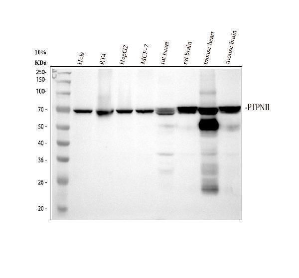
Western blot analysis of SHP2/PTPN11 using anti-SHP2/PTPN11 antibody (M00150-2).
Electrophoresis was performed on a 10% SDS-PAGE gel at 80V (Stacking gel) / 120V (Resolving gel) for 2 hours. The sample well of each lane was loaded with 30 ug of sample under reducing conditions.
Lane 1: human Hela whole cell lysates,
Lane 2: human RT4 whole cell lysates,
Lane 3: human HepG2 whole cell lysates.
Lane 4: human MCF-7 whole cell lysates,
Lane 5: rat heart tissue lysates,
Lane 6: rat brain tissue lysates,
Lane 7: mouse heart tissue lysates,
Lane 8: mouse brain tissue lysates.
After Electrophoresis, proteins were transferred to a Nitrocellulose membrane at 150mA for 50-90 minutes. Blocked the membrane with 5% Non-fat Milk/ TBS for 1.5 hour at RT. The membrane was incubated with mouse anti-SHP2/PTPN11 antigen affinity purified monoclonal antibody (Catalog # M00150-2) at 0.5 μg/mL overnight at 4°C, then washed with TBS-0.1%Tween 3 times with 5 minutes each and probed with a goat anti-mouse IgG-HRP secondary antibody at a dilution of 1:10000 for 1.5 hour at RT. The signal is developed using an ECL Plus Western Blotting Substrate (Catalog # AR1196-200) with Tanon 5200 system. A specific band was detected for SHP2/PTPN11 at approximately 68 kDa. The expected band size for SHP2/PTPN11 is at 68 kDa.

Click image to see more details
IHC analysis of SHP2/PTPN11 using anti SHP2/PTPN11 antibody (M00150-2).
SHP2/PTPN11 was detected in paraffin-embedded section of human colon cancer tissue. Heat mediated antigen retrieval was performed in EDTA buffer (pH8.0, epitope retrieval solution). The tissue section was blocked with 10% goat serum. The tissue section was then incubated with 1μg/ml mouse anti-SHP2/PTPN11 Antibody (M00150-2) overnight at 4°C. Biotinylated goat anti-mouse IgG was used as secondary antibody and incubated for 30 minutes at 37°C. The tissue section was developed using Strepavidin-Biotin-Complex (SABC) (Catalog # SA1021) with DAB as the chromogen.

Click image to see more details
IHC analysis of SHP2/PTPN11 using anti SHP2/PTPN11 antibody (M00150-2).
SHP2/PTPN11 was detected in paraffin-embedded section of human tonsil tissue. Heat mediated antigen retrieval was performed in EDTA buffer (pH8.0, epitope retrieval solution). The tissue section was blocked with 10% goat serum. The tissue section was then incubated with 1μg/ml mouse anti-SHP2/PTPN11 Antibody (M00150-2) overnight at 4°C. Biotinylated goat anti-mouse IgG was used as secondary antibody and incubated for 30 minutes at 37°C. The tissue section was developed using Strepavidin-Biotin-Complex (SABC) (Catalog # SA1021) with DAB as the chromogen.

Click image to see more details
IHC analysis of SHP2/PTPN11 using anti SHP2/PTPN11 antibody (M00150-2).
SHP2/PTPN11 was detected in paraffin-embedded section of mouse brain tissue. Heat mediated antigen retrieval was performed in EDTA buffer (pH8.0, epitope retrieval solution). The tissue section was blocked with 10% goat serum. The tissue section was then incubated with 1μg/ml mouse anti-SHP2/PTPN11 Antibody (M00150-2) overnight at 4°C. Biotinylated goat anti-mouse IgG was used as secondary antibody and incubated for 30 minutes at 37°C. The tissue section was developed using Strepavidin-Biotin-Complex (SABC) (Catalog # SA1021) with DAB as the chromogen.

Click image to see more details
IHC analysis of SHP2/PTPN11 using anti SHP2/PTPN11 antibody (M00150-2).
SHP2/PTPN11 was detected in paraffin-embedded section of rat brain tissue. Heat mediated antigen retrieval was performed in EDTA buffer (pH8.0, epitope retrieval solution). The tissue section was blocked with 10% goat serum. The tissue section was then incubated with 1μg/ml mouse anti-SHP2/PTPN11 Antibody (M00150-2) overnight at 4°C. Biotinylated goat anti-mouse IgG was used as secondary antibody and incubated for 30 minutes at 37°C. The tissue section was developed using Strepavidin-Biotin-Complex (SABC) (Catalog # SA1021) with DAB as the chromogen.

Click image to see more details
IF analysis of SHP2/PTPN11 using anti-SHP2/PTPN11 antibody (M00150-2).
SHP2/PTPN11 was detected in immunocytochemical section of U251 cells. Enzyme antigen retrieval was performed using IHC enzyme antigen retrieval reagent (AR0022) for 15 mins. The cells were blocked with 10% goat serum. And then incubated with 2μg/mL mouse anti-SHP2/PTPN11 Antibody (M00150-2) overnight at 4°C. DyLight®488 Conjugated Goat Anti-Mouse IgG (BA1126) was used as secondary antibody at 1:100 dilution and incubated for 30 minutes at 37°C. The section was counterstained with DAPI. Visualize using a fluorescence microscope and filter sets appropriate for the label used.

Click image to see more details
Flow Cytometry analysis of A549 cells using anti-SHP2/PTPN11 antibody (M00150-2).
Overlay histogram showing A549 cells stained with M00150-2 (Blue line). To facilitate intracellular staining, cells were fixed with 4% paraformaldehyde and permeabilized with permeabilization buffer. The cells were blocked with 10% normal goat serum. And then incubated with mouse anti-SHP2/PTPN11 Antibody (M00150-2, 1μg/1x106 cells) for 30 min at 20°C. DyLight®488 conjugated goat anti-mouse IgG (BA1126, 5-10μg/1x106 cells) was used as secondary antibody for 30 minutes at 20°C. Isotype control antibody (Green line) was mouse IgG (1μg/1x106) used under the same conditions. Unlabelled sample (Red line) was also used as a control.
Specific Publications For Anti-SHP2/PTPN11 Antibody Picoband® (monoclonal, 2E6) (M00150-2)
Loading publications
Recommended Resources
Here are featured tools and databases that you might find useful.
- Boster's Pathways Library
- Protein Databases
- Bioscience Research Protocol Resources
- Data Processing & Analysis Software
- Photo Editing Software
- Scientific Literature Resources
- Research Paper Management Tools
- Molecular Biology Software
- Primer Design Tools
- Bioinformatics Tools
- Phylogenetic Tree Analysis
Customer Reviews
Have you used Anti-SHP2/PTPN11 Antibody Picoband® (monoclonal, 2E6)?
Share your experimental results or join a short interview to earn up to $1,000 in product credits or other rewards.
0 Reviews For Anti-SHP2/PTPN11 Antibody Picoband® (monoclonal, 2E6)
Customer Q&As
Have a question?
Find answers in Q&As, reviews.
Can't find your answer?
Submit your question
