Click image to see more details
-

-
-
-
-
+2
Product Info Summary
| SKU: | M05696 |
|---|---|
| Size: | 0.1 mg |
| Reactive Species: | Human |
| Host: | Mouse |
| Application: | Flow Cytometry, IP, WB |
Customers Who Bought This Also Bought

Product info
Product Name
Anti-SIT Purified SIT1 Monoclonal Antibody
SKU/Catalog Number
M05696
Size
0.1 mg
Form
Liquid
Description
Boster Bio Anti-SIT Purified SIT1 Monoclonal Antibody (Catalog# M05696). Tested in Flow Cytometry, IP, WB application(s). This antibody reacts with Human.
Storage & Handling
Store at 2-8°C. Do not freeze.
Cite This Product
Anti-SIT Purified SIT1 Monoclonal Antibody (Boster Biological Technology, Pleasanton CA, USA, Catalog # M05696)
Host
Mouse
Contents
Phosphate buffered saline (PBS), pH 7.4, 15 mM sodium azide
Clonality
Monoclonal
Clone Number
SIT-01
Isotype
Mouse IgG1
Immunogen
Bacterially produced recombinant intracellular fragment of human SIT. The antibody SIT-01 reacts with an intracellular epitope of SHP2-interacting transmembrane adaptor protein (SIT) expressed exclusively in lymphoid organs.
Cross-reactivity
This antibody weakly cross-reacts with murine SIT.
Reactive Species
M05696 is reactive to SIT1 in Human
Observed Molecular Weight
42 kDa
Calculated molecular weight
21.1 kDa
Background of SIT1
SIT (SHP2-interacting transmembrane adaptor protein) is expressed exclusively in lymphoid organs and acts either as a positive or as a negative regulatory element in T cell activation and in T cell development. Binding to Grb2 plays a pivotal role in signal transduction. Hubener et al. (2001) determined that the SIT gene contains 5 exons and spans 1.8 kb of genomic DNA. The SIT promoter demonstrated strong transcriptional activity and potential binding sites for both ubiquitous and lymphoid-specific transcription factors.
Antibody Validation
Boster validates all antibodies on WB, IHC, ICC, Immunofluorescence, and ELISA with known positive control and negative samples to ensure specificity and high affinity, including thorough antibody incubations.
Application & Images
Applications
M05696 is guaranteed for Flow Cytometry, IP, WB Boster Guarantee
Recommend Dilution
| Application | Dilution | Species |
|---|---|---|
| Flow cytometry: 1-5 μg/ml | intracellular staining. | |
| Western blotting: 1-2 μg/ml | reducing conditions. |
Validation Images & Assay Conditions
Click image to see more details
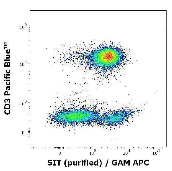
Flow cytometry multicolor intracellular staining of human peripheral whole blood stained using anti-SIT (SIT-01) purified antibody (concentration in sample 9 µg/ml, GAM APC) and anti-human CD3 (UCHT1) Pacific Blue™ antibody (20 µl reagent / 100 µl of peripheral whole blood).

Click image to see more details
Separation of human CD3 negative SIT positive lymphocytes (red-filled) from CD3 negative SIT negative lymphocytes (black-dashed) in flow cytometry analysis (intracellular staining) of peripheral whole blood stained using anti-SIT (SIT-01) purified antibody (concentration in sample 9 µg/ml, GAM APC).

Click image to see more details
Flow cytometry intracellular staining pattern of human peripheral whole blood using anti-SIT (SIT-01) purified antibody (concentration in sample 9 µg/ml, GAM APC).

Click image to see more details
Western blotting analysis of human SIT using mouse monoclonal antibody SIT-01 on lysates of Molt-4 and HEK-293T cells under reducing and non-reducing conditions. Nitrocellulose membrane was probed with 2 µg/ml of mouse anti-SIT monoclonal antibody followed by IRDye800-conjugated anti-mouse secondary antibody. SIT was detected around 36 kDa.

Click image to see more details
Anti-SIT Purified (clone SIT-01) works in WB application.
Western blotting analysis was performed on whole cell extracts (RIPA lysis buffer) of Ramos and Jurkat cell lines, mixed and heated (100°C, 5 min) with reducing (2-mercaptoethanol) SDS-loading buffer. Samples were resolved using 10% SDS-PAGE gel.
Nitrocellulose membrane blot was probed simultaneously with mouse IgG1 monoclonal antibody SIT-01 (2 µg/ml), and rat IgG2a anti-tubulin monoclonal antibody YOL1/34 (1 µg/ml) used as the loading control. Subclass-specific secondary antibodies IRDye 800CW Goat-anti-Rat IgG (green) and IRDye 680LT Goat-anti-Mouse IgG (red) were used for multiplex fluorescent Western blot detection.
SIT was detected at ~32 kDa in tested cell lines.

Click image to see more details
Anti-SIT Purified (clone SIT-01) specificity verification by WB.
The specificity of SIT-01 antibody was assessed by comparing binding signals in HEK293T cells overexpressing the target SIT protein to wild type cells (control) with low level of endogenous protein expression.
Western blotting analysis was performed on whole cell extracts (urea lysis buffer) of transfected and control cells, mixed and heated (100°C, 5 min) with reducing (2-mercaptoethanol) SDS-loading buffer. Samples were resolved using 10% SDS-PAGE gel.
Nitrocellulose membrane blot was probed simultaneously with mouse IgG1 monoclonal antibody SIT-01 (2 µg/ml), and rat IgG2a anti-tubulin monoclonal antibody YOL1/34 (1 µg/ml) used as the loading control. Subclass-specific secondary antibodies IRDye 800CW Goat-anti-Rat IgG (green) and IRDye 680LT Goat-anti-Mouse IgG (red) were used for multiplex fluorescent Western blot detection.
Specific Publications For Anti-SIT Purified SIT1 Monoclonal Antibody (M05696)
Loading publications
Recommended Resources
Here are featured tools and databases that you might find useful.
- Boster's Pathways Library
- Protein Databases
- Bioscience Research Protocol Resources
- Data Processing & Analysis Software
- Photo Editing Software
- Scientific Literature Resources
- Research Paper Management Tools
- Molecular Biology Software
- Primer Design Tools
- Bioinformatics Tools
- Phylogenetic Tree Analysis
Customer Reviews
Have you used Anti-SIT Purified SIT1 Monoclonal Antibody?
Share your experimental results or join a short interview to earn up to $1,000 in product credits or other rewards.
0 Reviews For Anti-SIT Purified SIT1 Monoclonal Antibody
Customer Q&As
Have a question?
Find answers in Q&As, reviews.
Can't find your answer?
Submit your question
