Click image to see more details
Product Info Summary
| SKU: | PB9439 |
|---|---|
| Size: | 100μg/vial |
| Reactive Species: | Human, Mouse, Rat |
| Host: | Rabbit |
| Application: | IHC, WB |
Customers Who Bought This Also Bought

Product info
Product Name
Anti-SLUG/SNAI2 Antibody Picoband®
SKU/Catalog Number
PB9439
Size
100μg/vial
Form
Lyophilized
Description
Boster Bio Anti-SLUG/SNAI2 Antibody Picoband® catalog # PB9439. Tested in IHC, WB applications. This antibody reacts with Human, Mouse, Rat. The brand Picoband indicates this is a premium antibody that guarantees superior quality, high affinity, and strong signals with minimal background in Western blot applications. Only our best-performing antibodies are designated as Picoband, ensuring unmatched performance.
Storage & Handling
Store at -20˚C for one year from date of receipt. After reconstitution, at 4˚C for one month. It can also be aliquotted and stored frozen at -20˚C for six months. Avoid repeated freeze-thaw cycles.
Cite This Product
Anti-SLUG/SNAI2 Antibody Picoband® (Boster Biological Technology, Pleasanton CA, USA, Catalog # PB9439)
Host
Rabbit
Contents
Each vial contains 4 mg Trehalose, 0.9 mg NaCl and 0.2 mg Na2HPO4.
Clonality
Polyclonal
Immunogen
A synthetic peptide corresponding to a sequence in the middle region of human SLUG, identical to the related mouse and rat sequences.
Cross-reactivity
No cross reactivity with other proteins
Reactive Species
PB9439 is reactive to SNAI2 in Human, Mouse, Rat
Observed Molecular Weight
30 kDa
Calculated molecular weight
30.0 kDa
Background of SNAI2
SLUG is also known as SNAI2. This gene encodes a member of the Snail family of C2H2-type zinc finger transcription factors. The encoded protein acts as a transcriptional repressor that binds to E-box motifs and is also likely to repress E-cadherin transcription in breast carcinoma. This protein is involved in epithelial-mesenchymal transitions and has antiapoptotic activity. Mutations in this gene may be associated with sporatic cases of neural tube defects.
Antibody Validation
Boster validates all antibodies on WB, IHC, ICC, Immunofluorescence, and ELISA with known positive control and negative samples to ensure specificity and high affinity, including thorough antibody incubations.
Application & Images
Applications
PB9439 is guaranteed for IHC, WB Boster Guarantee
Recommend Dilution
| Application | Dilution | Species |
|---|---|---|
| Western blot | 0.1-0.5μg/ml | Human, Mouse, Rat |
| Immunohistochemistry (Paraffin-embedded Section) | 2-5μg/ml | Rat |
Tested application
Suggested blocking solution with 5% non-fat milk or BSA; (*)Recommended protein loading: 20-40 µg per lane
Use TE buffer pH 9.0 for antigen retrieval; (*) citrate buffer pH 6.0 is an alternative.
Validation Images & Assay Conditions
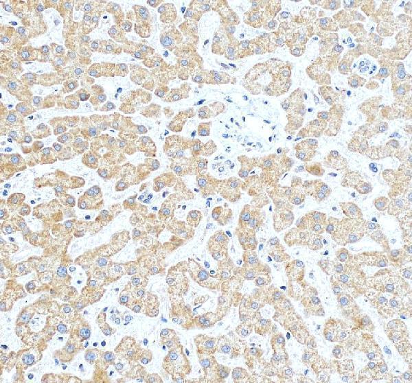
Click image to see more details
IHC analysis of SNAI2 using anti-SNAI2 antibody (PB9439).
SNAI2 was detected in a paraffin-embedded section of human liver tissue. Heat mediated antigen retrieval was performed in EDTA buffer (pH 8.0, epitope retrieval solution). The tissue section was blocked with 10% goat serum. The tissue section was then incubated with 2 μg/ml rabbit anti-SNAI2 Antibody (PB9439) overnight at 4°C. Peroxidase Conjugated Goat Anti-rabbit IgG was used as secondary antibody and incubated for 30 minutes at 37°C. The tissue section was developed using HRP Conjugated Rabbit IgG Super Vision Assay Kit (Catalog # SV0002) with DAB as the chromogen.

Click image to see more details
EMP3 affects the malignant phenotype of glioblastoma by promoting the EMT process ( A )-( D ). Correlation analysis of VIM , FOS , SNAI2 , TWIST1 and EMP3 using TCGA data ( E )-( H ). These related EMT markers were tested by qRT-PCR after the knockdown of EMP3 for 96 h cultured with siRNA. On the other hand, proteins of EMT markers VIM, SNAI2, FOS, TWIST1 were investigated by western blot after EMP3 siRNA transient transfection ( I )-( M ), Actin as the internal parameter. Cells were divided into three groups, NC (negtive control) group, siRNA EMP3-1 and siRNA EMP3-2 group. U87 cells were lysis after siRNA incubation for 72 h. The efficiency of trasfection of siRNA EMP3 was detected by WB ( N ). All of the full-length blots/gels of western blot are presented in Supplementary Figure C-H. * p < 0.05, ** P < 0.01, *** P < 0.001,**** p < 0.0001
Index in PubMed under a CC BY license. PMID: 38229014

Click image to see more details
Western blot analysis of SNAI2 using anti-SNAI2 antibody (PB9439).
Electrophoresis was performed on a 5-20% SDS-PAGE gel at 70V (Stacking gel) / 90V (Resolving gel) for 2-3 hours. The sample well of each lane was loaded with 30 ug of sample under reducing conditions.
Lane 1: human HepG2 whole cell lysates,
Lane 2: human Hela whole cell lysates,
Lane 3: human A431 whole cell lysates,
Lane 4: rat lung tissue lysates,
Lane 5: mouse lung tissue lysates,
Lane 6: mouse NIH/3T3 whole cell lysates.
After electrophoresis, proteins were transferred to a nitrocellulose membrane at 150 mA for 50-90 minutes. Blocked the membrane with 5% non-fat milk/TBS for 1.5 hour at RT. The membrane was incubated with rabbit anti-SNAI2 antigen affinity purified polyclonal antibody (Catalog # PB9439) at 0.5 μg/mL overnight at 4°C, then washed with TBS-0.1%Tween 3 times with 5 minutes each and probed with a goat anti-rabbit IgG-HRP secondary antibody at a dilution of 1:5000 for 1.5 hour at RT. The signal is developed using an Enhanced Chemiluminescent detection (ECL) kit (Catalog # EK1002) with Tanon 5200 system. A specific band was detected for SNAI2 at approximately 30 kDa. The expected band size for SNAI2 is at 30 kDa.

Click image to see more details
IHC analysis of SNAI2 using anti-SNAI2 antibody (PB9439).
SNAI2 was detected in a paraffin-embedded section of rat colon tissue. Heat mediated antigen retrieval was performed in EDTA buffer (pH 8.0, epitope retrieval solution). The tissue section was blocked with 10% goat serum. The tissue section was then incubated with 2 μg/ml rabbit anti-SNAI2 Antibody (PB9439) overnight at 4°C. Peroxidase Conjugated Goat Anti-rabbit IgG was used as secondary antibody and incubated for 30 minutes at 37°C. The tissue section was developed using HRP Conjugated Rabbit IgG Super Vision Assay Kit (Catalog # SV0002) with DAB as the chromogen.
Specific Publications For Anti-SLUG/SNAI2 Antibody Picoband® (PB9439)
Loading publications
Recommended Resources
Here are featured tools and databases that you might find useful.
- Boster's Pathways Library
- Protein Databases
- Bioscience Research Protocol Resources
- Data Processing & Analysis Software
- Photo Editing Software
- Scientific Literature Resources
- Research Paper Management Tools
- Molecular Biology Software
- Primer Design Tools
- Bioinformatics Tools
- Phylogenetic Tree Analysis
Customer Reviews
Have you used Anti-SLUG/SNAI2 Antibody Picoband®?
Share your experimental results or join a short interview to earn up to $1,000 in product credits or other rewards.
0 Reviews For Anti-SLUG/SNAI2 Antibody Picoband®
Customer Q&As
Have a question?
Find answers in Q&As, reviews.
Can't find your answer?
Submit your question
