Click image to see more details
-

-
-
-
-
+7
Product Info Summary
| SKU: | A07969 |
|---|---|
| Size: | 100 µg/vial |
| Reactive Species: | Human, Mouse, Rat |
| Host: | Rabbit |
| Application: | ELISA, IF, IHC, WB |
Customers Who Bought This Also Bought

Product info
Product Name
Anti-SPA17 Antibody Picoband®
SKU/Catalog Number
A07969
Size
100 µg/vial
Form
Lyophilized
Description
Boster Bio Anti-SPA17 Antibody Picoband® catalog # A00955. Tested in WB, IHC, IF, ELISA applications. This antibody reacts with Human, Mouse, Rat. The brand Picoband indicates this is a premium antibody that guarantees superior quality, high affinity, and strong signals with minimal background in Western blot applications. Only our best-performing antibodies are designated as Picoband, ensuring unmatched performance.
Storage & Handling
At -20°C for one year from date of receipt. After reconstitution, at 4°C for one month. It can also be aliquotted and stored frozen at -20°C for six months. Avoid repeated freezing and thawing.
Cite This Product
Anti-SPA17 Antibody Picoband® (Boster Biological Technology, Pleasanton CA, USA, Catalog # A07969)
Host
Rabbit
Contents
Each vial contains 4 mg Trehalose, 0.9 mg NaCl, 0.2 mg Na2HPO4.
Clonality
Polyclonal
Isotype
IgG
Immunogen
E.coli-derived human SPA17 recombinant protein (Position: M1-K136). Human SPA17 shares 75.7% amino acid (aa) sequence identity with mouse SPA17.
Cross-reactivity
No cross reactivity with other proteins.
Reactive Species
A07969 is reactive to SPA17 in Human, Mouse, Rat
Observed Molecular Weight
22 kDa
Calculated molecular weight
17.4 kDa
Background of SPA17
Sperm surface protein Sp17 is a protein that in humans is encoded by the SPA17 gene. This gene encodes a protein present at the cell surface. The N-terminus has sequence similarity to human cAMP-dependent protein kinase A (PKA) type II alpha regulatory subunit (RIIa) while the C-terminus has an IQ calmodulin-binding motif. The central portion of the protein has carbohydrate binding motifs and likely functions in cell-cell adhesion. The protein was initially characterized by its involvement in the binding of sperm to the zona pellucida of the oocyte. Recent studies indicate that it is also involved in additional cell-cell adhesion functions such as immune cell migration and metastasis. A retrotransposed pseudogene is present on chromosome 10q22.
Antibody Validation
Boster validates all antibodies on WB, IHC, ICC, Immunofluorescence, and ELISA with known positive control and negative samples to ensure specificity and high affinity, including thorough antibody incubations.
Application & Images
Applications
A07969 is guaranteed for ELISA, IF, IHC, WB Boster Guarantee
Recommend Dilution
| Application | Dilution | Species |
|---|---|---|
| Western blot | 0.25-0.5 μg/ml | Mouse, Rat |
| Immunohistochemistry(Paraffin-embedded Section) | 2-5 μg/ml | Human, Mouse, Rat |
| Immunofluorescence | 5 μg/ml | Human |
| ELISA | 0.1-0.5 μg/ml | - |
Tested application
Suggested blocking solution with 5% non-fat milk or BSA; (*)Recommended protein loading: 20-40 µg per lane
Use TE buffer pH 9.0 for antigen retrieval; (*) citrate buffer pH 6.0 is an alternative.
Validation Images & Assay Conditions
Click image to see more details
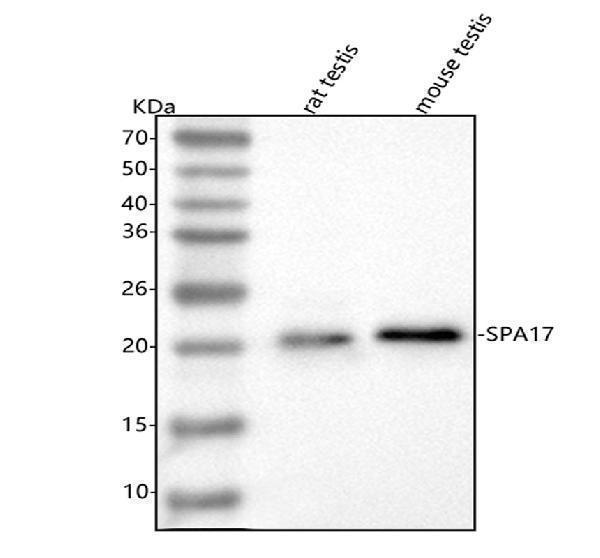
Western blot analysis of SPA17 using anti-SPA17 antibody (A07969).
Electrophoresis was performed on a 5-20% SDS-PAGE gel at 70V (Stacking gel) / 90V (Resolving gel) for 2-3 hours. The sample well of each lane was loaded with 30 ug of sample under reducing conditions.
Lane 1: rat testis tissue lysates,
Lane 2: mouse testis tissue lysates.
After electrophoresis, proteins were transferred to a nitrocellulose membrane at 150 mA for 50-90 minutes. Blocked the membrane with 5% non-fat milk/TBS for 1.5 hour at RT. The membrane was incubated with rabbit anti-SPA17 antigen affinity purified polyclonal antibody (Catalog # A07969) at 0.5 μg/mL overnight at 4°C, then washed with TBS-0.1%Tween 3 times with 5 minutes each and probed with a goat anti-rabbit IgG-HRP secondary antibody at a dilution of 1:5000 for 1.5 hour at RT. The signal is developed using an Enhanced Chemiluminescent detection (ECL) kit (Catalog # EK1002) with Tanon 5200 system. A specific band was detected for SPA17 at approximately 22 kDa. The expected band size for SPA17 is at 17 kDa.

Click image to see more details
IHC analysis of SPA17 using anti-SPA17 antibody (A07969).
SPA17 was detected in a paraffin-embedded section of human placenta tissue. Heat mediated antigen retrieval was performed in EDTA buffer (pH 8.0, epitope retrieval solution). The tissue section was blocked with 10% goat serum. The tissue section was then incubated with 2 μg/ml rabbit anti-SPA17 Antibody (A07969) overnight at 4°C. Peroxidase Conjugated Goat Anti-rabbit IgG was used as secondary antibody and incubated for 30 minutes at 37°C. The tissue section was developed using HRP Conjugated Rabbit IgG Super Vision Assay Kit (Catalog # SV0002) with DAB as the chromogen.

Click image to see more details
IHC analysis of SPA17 using anti-SPA17 antibody (A07969).
SPA17 was detected in a paraffin-embedded section of human rectum adenocarcinoma tissue. Heat mediated antigen retrieval was performed in EDTA buffer (pH 8.0, epitope retrieval solution). The tissue section was blocked with 10% goat serum. The tissue section was then incubated with 2 μg/ml rabbit anti-SPA17 Antibody (A07969) overnight at 4°C. Peroxidase Conjugated Goat Anti-rabbit IgG was used as secondary antibody and incubated for 30 minutes at 37°C. The tissue section was developed using HRP Conjugated Rabbit IgG Super Vision Assay Kit (Catalog # SV0002) with DAB as the chromogen.

Click image to see more details
IHC analysis of SPA17 using anti-SPA17 antibody (A07969).
SPA17 was detected in a paraffin-embedded section of mouse testis tissue. Heat mediated antigen retrieval was performed in EDTA buffer (pH 8.0, epitope retrieval solution). The tissue section was blocked with 10% goat serum. The tissue section was then incubated with 2 μg/ml rabbit anti-SPA17 Antibody (A07969) overnight at 4°C. Peroxidase Conjugated Goat Anti-rabbit IgG was used as secondary antibody and incubated for 30 minutes at 37°C. The tissue section was developed using HRP Conjugated Rabbit IgG Super Vision Assay Kit (Catalog # SV0002) with DAB as the chromogen.

Click image to see more details
IHC analysis of SPA17 using anti-SPA17 antibody (A07969).
SPA17 was detected in a paraffin-embedded section of rat testis tissue. Heat mediated antigen retrieval was performed in EDTA buffer (pH 8.0, epitope retrieval solution). The tissue section was blocked with 10% goat serum. The tissue section was then incubated with 2 μg/ml rabbit anti-SPA17 Antibody (A07969) overnight at 4°C. Peroxidase Conjugated Goat Anti-rabbit IgG was used as secondary antibody and incubated for 30 minutes at 37°C. The tissue section was developed using HRP Conjugated Rabbit IgG Super Vision Assay Kit (Catalog # SV0002) with DAB as the chromogen.

Click image to see more details
IF analysis of SPA17 using anti-SPA17 antibody (A07969).
SPA17 was detected in a paraffin-embedded section of human cervical cancer tissue. Heat mediated antigen retrieval was performed in EDTA buffer (pH 8.0, epitope retrieval solution). The tissue section was blocked with 10% goat serum. The tissue section was then incubated with 5 μg/mL rabbit anti-SPA17 Antibody (A07969) overnight at 4°C. FITC Conjugated Goat Anti-Rabbit IgG (BA1105) was used as secondary antibody at 1:500 dilution and incubated for 30 minutes at 37°C. The section was counterstained with DAPI. Visualize using a fluorescence microscope and filter sets appropriate for the label used.

Click image to see more details
IF analysis of SPA17 using anti-SPA17 antibody (A07969).
SPA17 was detected in a paraffin-embedded section of human ovarian tissue. Heat mediated antigen retrieval was performed in EDTA buffer (pH 8.0, epitope retrieval solution). The tissue section was blocked with 10% goat serum. The tissue section was then incubated with 5 μg/mL rabbit anti-SPA17 Antibody (A07969) overnight at 4°C. FITC Conjugated Goat Anti-Rabbit IgG (BA1105) was used as secondary antibody at 1:500 dilution and incubated for 30 minutes at 37°C. The section was counterstained with DAPI. Visualize using a fluorescence microscope and filter sets appropriate for the label used.
Specific Publications For Anti-SPA17 Antibody Picoband® (A07969)
Loading publications
Recommended Resources
Here are featured tools and databases that you might find useful.
- Boster's Pathways Library
- Protein Databases
- Bioscience Research Protocol Resources
- Data Processing & Analysis Software
- Photo Editing Software
- Scientific Literature Resources
- Research Paper Management Tools
- Molecular Biology Software
- Primer Design Tools
- Bioinformatics Tools
- Phylogenetic Tree Analysis
Customer Reviews
Have you used Anti-SPA17 Antibody Picoband®?
Share your experimental results or join a short interview to earn up to $1,000 in product credits or other rewards.
0 Reviews For Anti-SPA17 Antibody Picoband®
Customer Q&As
Have a question?
Find answers in Q&As, reviews.
Can't find your answer?
Submit your question
