Click image to see more details
Product Info Summary
| SKU: | M01315 |
|---|---|
| Size: | 100 μl/vial |
| Reactive Species: | Human |
| Host: | Rabbit |
| Application: | IP, IHC, WB |
Customers Who Bought This Also Bought

Product info
Product Name
Anti-TCF7 Antibody (Monoclonal, 31T28)
SKU/Catalog Number
M01315
Size
100 μl/vial
Form
Liquid
Description
Boster Bio Anti-TCF7 Antibody (Monoclonal, 31T28) catalog # M01315. Tested in WB, IHC, IP applications. This antibody reacts with Human.
Storage & Handling
Store at -20°C for one year. For short term storage and frequent use, store at 4°C for up to one month. Avoid repeated freeze-thaw cycles.
Cite This Product
Anti-TCF7 Antibody (Monoclonal, 31T28) (Boster Biological Technology, Pleasanton CA, USA, Catalog # M01315)
Host
Rabbit
Contents
Rabbit IgG in stabilizing components, phosphate buffered saline, pH 7.4, 150mM NaCl, 0.02% sodium azide and 50% glycerol.
This antibody is supplied in a stabilized formulation.
Compatibility with conjugation reactions depends on the chemistry of the conjugation method used.
For conjugation methods that are not compatible with the stabilizing components present in this formulation, a carrier-free antibody format is required.
Clonality
Monoclonal
Clone Number
31T28
Immunogen
Recombinant protein within human TCF7 aa 3-298.
Reactive Species
M01315 is reactive to TCF7 in Human
Observed Molecular Weight
42 kDa
Calculated molecular weight
41.6 kDa
Background of TCF7
Transcription factor 7 is a protein that in humans is encoded by the TCF7 gene. It is mapped to 5q31.1. This gene encodes a member of the T-cell factor/lymphoid enhancer-binding factor family of high mobility group (HMG) box transcriptional activators. This gene is expressed predominantly in T-cells and plays a critical role in natural killer cell and innate lymphoid cell development. The encoded protein forms a complex with beta-catenin and activates transcription through a Wnt/beta-catenin signaling pathway. Mice with a knockout of this gene are viable and fertile, but display a block in T-lymphocyte differentiation. Alternative splicing results in multiple transcript variants. Naturally-occurring isoforms lacking the N-terminal beta-catenin interaction domain may act as dominant negative regulators of Wnt signaling.
Antibody Validation
Boster validates all antibodies on WB, IHC, ICC, Immunofluorescence, and ELISA with known positive control and negative samples to ensure specificity and high affinity, including thorough antibody incubations.
Application & Images
Applications
M01315 is guaranteed for IP, IHC, WB Boster Guarantee
Recommend Dilution
| Application | Dilution | Species |
|---|---|---|
| Western blot | 1:500-2000 | |
| Immunohistochemistry | 1:50-200 | |
| ImmunoPrecipitation | 1:50 |
Tested application
Suggested blocking solution with 5% non-fat milk or BSA; (*)Recommended protein loading: 20-40 µg per lane
Validation Images & Assay Conditions
Click image to see more details
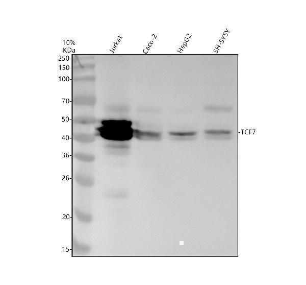
Western blot analysis of TCF7 using anti-TCF7 antibody (M01315).
Electrophoresis was performed on a 10% SDS-PAGE gel at 80V (Stacking gel) / 120V (Resolving gel) for 2 hours. The sample well of each lane was loaded with 30 ug of sample under reducing conditions.
Lane 1: human Jurkat whole cell lysates,
Lane 2: human Caco-2 whole cell lysates,
Lane 3: human HepG2 whole cell lysates,
Lane 4: human SH-SY5Y whole cell lysates.
After electrophoresis, proteins were transferred to a nitrocellulose membrane at 150 mA for 50-90 minutes. Blocked the membrane with 5% non-fat milk/TBS for 1.5 hour at RT. The membrane was incubated with rabbit anti-TCF7 antigen affinity purified monoclonal antibody (M01315) at 1:500 overnight at 4°C, then washed with TBS-0.1%Tween 3 times with 5 minutes each and probed with a goat anti-rabbit IgG-HRP secondary antibody at a dilution of 1:5000 for 1.5 hour at RT. The signal is developed using an ECL Plus Western Blotting Substrate (Catalog # AR1196-200) with Tanon 5200 system. A specific band was detected for TCF7 at approximately 42 kDa. The expected band size for TCF7 is at 42 kDa.
Specific Publications For Anti-TCF7 Antibody (Monoclonal, 31T28) (M01315)
Loading publications
Recommended Resources
Here are featured tools and databases that you might find useful.
- Boster's Pathways Library
- Protein Databases
- Bioscience Research Protocol Resources
- Data Processing & Analysis Software
- Photo Editing Software
- Scientific Literature Resources
- Research Paper Management Tools
- Molecular Biology Software
- Primer Design Tools
- Bioinformatics Tools
- Phylogenetic Tree Analysis
Customer Reviews
Have you used Anti-TCF7 Antibody (Monoclonal, 31T28)?
Share your experimental results or join a short interview to earn up to $1,000 in product credits or other rewards.
0 Reviews For Anti-TCF7 Antibody (Monoclonal, 31T28)
Customer Q&As
Have a question?
Find answers in Q&As, reviews.
Can't find your answer?
Submit your question
