Click image to see more details
Product Info Summary
| SKU: | A00657-5 |
|---|---|
| Size: | 100 μl/vial |
| Reactive Species: | Human, Mouse, Rat |
| Host: | Rabbit |
| Application: | IF, IHC, WB |
Customers Who Bought This Also Bought

Product info
Product Name
Anti-TIM3/HAVCR2 Antibody
SKU/Catalog Number
A00657-5
Size
100 μl/vial
Form
Liquid
Description
Boster Bio Anti-TIM3/HAVCR2 Antibody catalog # A00657-5. Tested in WB, IHC, IF applications. This antibody reacts with Human, Mouse, Rat.
Storage & Handling
12 months from date of receipt,-20℃ as supplied. 6 months 2 to 8℃ after reconstitution. Avoid repeated freezing and thawing.
Cite This Product
Anti-TIM3/HAVCR2 Antibody (Boster Biological Technology, Pleasanton CA, USA, Catalog # A00657-5)
Host
Rabbit
Contents
500 μg/ml antibody with PBS, 0.02% NaN3, 1 mg stabilizing protein and 50% glycerol
*This antibody is supplied in a stabilized formulation.
Compatibility with conjugation reactions depends on the chemistry of the conjugation method used.
For conjugation methods that are not compatible with the stabilizing components present in this formulation, a carrier-free antibody format is required.
Clonality
Polyclonal
Immunogen
E.coli-derived human TIM3/HAVCR2 recombinant protein (Position: M1-L140).
Reactive Species
A00657-5 is reactive to HAVCR2 in Human, Mouse, Rat
Observed Molecular Weight
70 kDa
Calculated molecular weight
33.4 kDa
Background of HAVCR2
The protein encoded by this gene belongs to the immunoglobulin superfamily, and TIM family of proteins. CD4-positive T helper lymphocytes can be divided into types 1 (Th1) and 2 (Th2) on the basis of their cytokine secretion patterns. Th1 cells are involved in cell-mediated immunity to intracellular pathogens and delayed-type hypersensitivity reactions, whereas, Th2 cells are involved in the control of extracellular helminthic infections and the promotion of atopic and allergic diseases. This protein is a Th1-specific cell surface protein that regulates macrophage activation, and inhibits Th1-mediated auto- and alloimmune responses, and promotes immunological tolerance.
Antibody Validation
Boster validates all antibodies on WB, IHC, ICC, Immunofluorescence, and ELISA with known positive control and negative samples to ensure specificity and high affinity, including thorough antibody incubations.
Application & Images
Applications
A00657-5 is guaranteed for IF, IHC, WB Boster Guarantee
Recommend Dilution
| Application | Dilution | Species |
|---|---|---|
| Western blot | 1:500-2000 | |
| Immunohistochemistry | 1:50-400 | |
| Immunofluorescence | 1:50-400 |
Tested application
Suggested blocking solution with 5% non-fat milk or BSA; (*)Recommended protein loading: 20-40 µg per lane
Validation Images & Assay Conditions
Click image to see more details
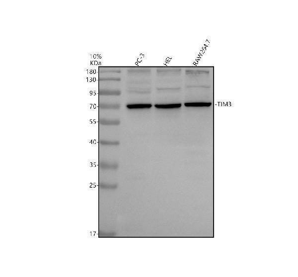
Western blot analysis of TIM3/HAVCR2 using anti-TIM3/HAVCR2 antibody (A00657-5).
Electrophoresis was performed on a 10% SDS-PAGE gel at 80V (Stacking gel) / 120V (Resolving gel) for 2 hours. The sample well of each lane was loaded with 30 ug of sample under reducing conditions.
Lane 1: human PC-3 whole cell lysates,
Lane 2: human HEL whole cell lysates,
Lane 3: mouse RAW264.7 whole cell lysates.
After electrophoresis, proteins were transferred to a nitrocellulose membrane at 150 mA for 50-90 minutes. Blocked the membrane with 5% non-fat milk/TBS for 1.5 hour at RT. The membrane was incubated with rabbit anti-TIM3/HAVCR2 antigen affinity purified polyclonal antibody (A00657-5) at 1:1000 overnight at 4°C, then washed with TBS-0.1%Tween 3 times with 5 minutes each and probed with a goat anti-rabbit IgG-HRP secondary antibody at a dilution of 1:5000 for 1.5 hour at RT. The signal is developed using an ECL Plus Western Blotting Substrate (Catalog # AR1196-200) with Tanon 5200 system. A specific band was detected for TIM3/HAVCR2 at approximately 70 kDa. The expected band size for TIM3/HAVCR2 is at 33 kDa.

Click image to see more details
The knockdown of MFSD12 inhibited the proliferation, migration, and invasion of LIHC cells, as well as the TIM-3/Galectin-9 signaling pathway. (A, B) RT-qPCR and Western blot validation of MFSD12 silencing efficiency using siRNAs (si-MFSD12–1 to −4) with GAPDH as loading control. (C) CCK-8 cell viability assay showing reduced HEP 3B2.1–7 cells proliferation after MFSD12 knockdown (si-MFSD12-3). (D) Transwell assay revealed a reduction in the migratory and invasive capabilities of HEP 3B2.1–7 cells following the knockdown of MFSD12. (E) Immunoblot analysis of EMT markers and TIM-3 axis components showing up-regulation of E-cadherin and down-regulation of Vimentin, MMP-2, MMP-9, HAVCR2 (TIM-3) and LGALS9 in si-MFSD12-treated cells. * P < 0.05, ** P < 0.01, *** P < 0.001. CTRL, control untreated; si-NC, negative control siRNA; si-MFSD12, MFSD12-targeting siRNA; E-cadherin, epithelial cadherin; MMP-2/9, matrix metalloproteinase-2/9; HAVCR2, hepatitis A virus cellular receptor 2 (TIM-3); LGALS9, lectin galactoside-binding soluble 9 (Galectin-9).
Index in PubMed under a CC BY license. PMID: 41194934

Click image to see more details
The knockdown of CHMP4A inhibited the proliferation, migration, and invasion of LIHC cells, as well as the TIM-3/Galectin-9 signaling pathway. (A, B) RT-qPCR and Western blot validation of CHMP4A silencing efficiency using siRNAs (si-CHMP4A-1 to -4) with GAPDH as a loading control. (C) CCK-8 cell viability assay showing reduced proliferation of HEP 3B2.1–7 cells after CHMP4A knockdown (si-CHMP4A-2). (D) Transwell assay revealing a reduction in the migratory and invasive capabilities of HEP 3B2.1–7 cells following the knockdown of CHMP4A. (E) Immunoblot analysis of EMT markers and TIM-3 axis components showing upregulation of E-cadherin and downregulation of Vimentin, MMP-2, and MMP-9 in si-CHMP4A-treated cells. (F) Immunoblot analysis of TIM-3 axis components showing downregulation of HAVCR2 (TIM-3) and LGALS9 in si-CHMP4A-treated cells. ** P <0.01, *** P <0.001. CTRL, control untreated; si-NC, negative control siRNA; si-CHMP4A, CHMP4A-targeting siRNA; E-cadherin, epithelial cadherin; MMP-2/9, matrix metalloproteinase-2/9; HAVCR2, hepatitis A virus cellular receptor 2 (TIM-3); LGALS9, lectin galactoside-binding soluble 9 (Galectin-9).
Index in PubMed under a CC BY license. PMID: 41112257
Specific Publications For Anti-TIM3/HAVCR2 Antibody (A00657-5)
Loading publications
Recommended Resources
Here are featured tools and databases that you might find useful.
- Boster's Pathways Library
- Protein Databases
- Bioscience Research Protocol Resources
- Data Processing & Analysis Software
- Photo Editing Software
- Scientific Literature Resources
- Research Paper Management Tools
- Molecular Biology Software
- Primer Design Tools
- Bioinformatics Tools
- Phylogenetic Tree Analysis
Customer Reviews
Have you used Anti-TIM3/HAVCR2 Antibody?
Share your experimental results or join a short interview to earn up to $1,000 in product credits or other rewards.
0 Reviews For Anti-TIM3/HAVCR2 Antibody
Customer Q&As
Have a question?
Find answers in Q&As, reviews.
Can't find your answer?
Submit your question
