Click image to see more details
Product Info Summary
| SKU: | A10501 |
|---|---|
| Size: | 100ug |
| Reactive Species: | Human |
| Host: | Rabbit |
| Application: | ELISA, IHC, WB |
Customers Who Bought This Also Bought

Product info
Product Name
Anti-TMBIM1/Recs1 Antibody
SKU/Catalog Number
A10501
Size
100ug
Form
Liquid (sterile filtered)
Description
Boster Bio Anti-TMBIM1/Recs1 Antibody (Catalog # A10501). Tested in ELISA, IHC, WB applications. This antibody reacts with Human.
Storage & Handling
Store vial at -20°C prior to opening. Aliquot contents and freeze at -20°C or below for extended storage. Avoid cycles of freezing and thawing. Centrifuge product if not completely clear after standing at room temperature. This product is stable for several weeks at 4°C as an undiluted liquid. Dilute only prior to immediate use. Expiration date is one (1) year from date of opening. (Ship on dry ice.)
Cite This Product
Anti-TMBIM1/Recs1 Antibody (Boster Biological Technology, Pleasanton CA, USA, Catalog # A10501)
Host
Rabbit
Contents
0.02 M Potassium Phosphate, 0.15 M Sodium Chloride, pH 7.2, 0.01% (w/v) Sodium Azide
Clonality
Polyclonal
Isotype
IgG
Immunogen
This protein A purified antibody was prepared from whole rabbit serum produced by repeated immunizations with a recombinant protein corresponding to the amino terminus of human TMBIM1 protein.
Reactive Species
A10501 is reactive to TMBIM1 in Human
Calculated molecular weight
34.6 kDa
Background of TMBIM1
This antibody is suitable for Cancer, Immunology and Nuclear Signaling research. TMBIM1 (Transmembrane BAX inhibitor motif-containing protein 1) is a member of the 7 TMS (7 transmembrane domains) family of receptors known to mediate the activation of various transcription factors. TMBIM1 is identified as a novel modulator of NF-kB activation. A drosophila homolog of this protein (dNMDA1 with 40% homology to hTMBIM1) is reported to be unregulated during aging and oxidative stress.
Antibody Validation
Boster validates all antibodies on WB, IHC, ICC, Immunofluorescence, and ELISA with known positive control and negative samples to ensure specificity and high affinity, including thorough antibody incubations.
Application & Images
Applications
A10501 is guaranteed for ELISA, IHC, WB Boster Guarantee
Recommend Dilution
| Application | Dilution | Species |
|---|---|---|
| ELISA: 1:10 | 000 | |
| WB: 1:500 - 1:2 | 000 | |
| This protein A purified antibody has been tested for use in ELISA | Immunohistochemistry | and western blotting. Specific conditions for reactivity should be optimized by the end user. Expect a band approximately 35 kDa in size corresponding to TMBIM-1 by western blotting in the appropriate cell lysate or extract. To date this antibody has shown the ability to recognize over-expressed TMBIM1 but not endogenous protein. |
Validation Images & Assay Conditions
Click image to see more details
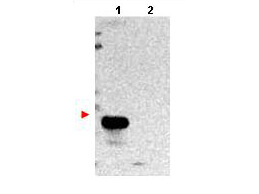
Western blot using Boster's protein A purified anti-TMBIM1 antibody shows detection of exogenous TMBIM1 in lysates from HeLa cells transfected with pcDNA3-hTMBIM1 (lane 1). No staining is observed in lysates from mock transformed HeLa cells (lane 2). To date this antibody has shown the ability to recognize over expressed TMBIM1 ~35kDa, but not endogenous protein. The membrane was probed with the primary antibody at a 1:1,000 dilution at 4° C, overnight. Personal Communication from Srinivasa Srinivasula, CCR-NCI, Bethesda, MD.

Click image to see more details
Immunohistochemistry of Rabbit anti-TMBIM1 antibody. Tissue: human tonsil. Fixation: formalin fixed paraffin embedded. Antigen retrieval: not required. Primary antibody: anti-TMBIM1 antibody at 10 µg/mL for 1 h at RT. Secondary antibody: Peroxidase rabbit secondary antibody at 1:10,000 for 45 min at RT. Staining: TMBIM1 as precipitated red signal with hematoxylin purple nuclear counterstain.

Click image to see more details
Immunohistochemistry of Rabbit anti-TMBIM1 antibody. Tissue: human prostate. Fixation: formalin fixed paraffin embedded. Antigen retrieval: not required. Primary antibody: anti-TMBIM1 antibody at 10 µg/mL for 1 h at RT. Secondary antibody: Peroxidase rabbit secondary antibody at 1:10,000 for 45 min at RT. Staining: TMBIM1 as precipitated red signal with hematoxylin purple nuclear counterstain.
Specific Publications For Anti-TMBIM1/Recs1 Antibody (A10501)
Loading publications
Recommended Resources
Here are featured tools and databases that you might find useful.
- Boster's Pathways Library
- Protein Databases
- Bioscience Research Protocol Resources
- Data Processing & Analysis Software
- Photo Editing Software
- Scientific Literature Resources
- Research Paper Management Tools
- Molecular Biology Software
- Primer Design Tools
- Bioinformatics Tools
- Phylogenetic Tree Analysis
Customer Reviews
Have you used Anti-TMBIM1/Recs1 Antibody?
Share your experimental results or join a short interview to earn up to $1,000 in product credits or other rewards.
0 Reviews For Anti-TMBIM1/Recs1 Antibody
Customer Q&As
Have a question?
Find answers in Q&As, reviews.
Can't find your answer?
Submit your question
