Click image to see more details
Product Info Summary
| SKU: | A02502 |
|---|---|
| Size: | 100ug |
| Reactive Species: | Human, Mouse |
| Host: | Rabbit |
| Application: | ELISA, IP, WB |
Customers Who Bought This Also Bought

Product info
Product Name
Anti-TrkCT1 NTRK3 Antibody
SKU/Catalog Number
A02502
Size
100ug
Form
Liquid (sterile filtered)
Description
Boster Bio Anti-TrkCT1 NTRK3 Antibody (Catalog # A02502). Tested in ELISA, IP, WB applications. This antibody reacts with Human, Mouse.
Storage & Handling
Store vial at -20°C prior to opening. Aliquot contents and freeze at -20°C or below for extended storage. Avoid cycles of freezing and thawing. Centrifuge product if not completely clear after standing at room temperature. This product is stable for several weeks at 4°C as an undiluted liquid. Dilute only prior to immediate use. Expiration date is one (1) year from date of opening. (Ship on dry ice.)
Cite This Product
Anti-TrkCT1 NTRK3 Antibody (Boster Biological Technology, Pleasanton CA, USA, Catalog # A02502)
Host
Rabbit
Contents
0.02 M Potassium Phosphate, 0.15 M Sodium Chloride, pH 7.2, 0.01% (w/v) Sodium Azide
Clonality
Polyclonal
Isotype
IgG
Immunogen
This affinity purified antibody was prepared from whole rabbit serum produced by repeated immunizations with a synthetic peptide corresponding to amino acids near the carboxyl terminus of mouse TrkCT1 protein.
Reactive Species
A02502 is reactive to NTRK3 in Human, Mouse
Observed Molecular Weight
42 kDa
Calculated molecular weight
92.8 kDa
Background of NTRK3
This antibody is suitable for Cancer, Immunology and Nuclear Signaling research. TrkCT1, also named neurotrophic tyrosine kinase receptor type 3 (Ntrk3) non-catalytic isoform 2, is a non-catalytic isoform of TrkC, the high affinity receptor for Neurotrophin-3 (NT-3). This isoform lacks the kinase domain that is responsible for signaling by the full-length isoform. TrkCT1 is the product of an alternative splicing of Ntrk3 that leaves the extracellular and transmembrane domains intact but includes a shorter intracellular domain encoded by exons 13b and 14b. Recent studies indicate that the short cytoplasmic tail binds the scaffold protein Tamalin in a ligand-dependent manner and further activates the Arf6–Rac1 signaling pathway. Isoform 3 transcripts are readily detected early during embryogenesis and are expressed predominantly in adult brain and gonads.
Antibody Validation
Boster validates all antibodies on WB, IHC, ICC, Immunofluorescence, and ELISA with known positive control and negative samples to ensure specificity and high affinity, including thorough antibody incubations.
Application & Images
Applications
A02502 is guaranteed for ELISA, IP, WB Boster Guarantee
Recommend Dilution
| Application | Dilution | Species |
|---|---|---|
| ELISA: 1:5 | 000 - 1:20 | 000 |
| WB: 1:1 | 000 - 1:10 | 000 |
| This affinity purified antibody has been tested for use in ELISA | IP | and western blotting. Specific conditions for reactivity should be optimized by the end user. While the predicted molecular weight for TrkCT1 is approximately 68 kDa, the protein has been reported to migrate at about 95 kDa on western blots. The molecular weight noted for TrkCT1 depends somewhat upon the cell lysate or extract used for western blotting, and this variation may be due to post-translational modifications of the protein. |
Validation Images & Assay Conditions
Click image to see more details
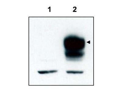
Western blot using Boster's affinity purified anti-TrkCT1 to detect over-expressed TrkCT1 in HEK293 cells (Lane 2, arrowhead). Lane 1 is a non-transfected control. Cell extracts were resolved by electrophoresis and transferred to nitrocellulose. The membrane was probed with the primary antibody at a 1:3,000 dilution. Personal Communication, V. Coppola, CCR-NCI, Frederick, MD.

Click image to see more details
Mouse cortex lysate was immunoprecipitated with anti-TrkCT1 antibody and further blotted with affinity purified anti-TrkCT1. Lane 1 is wild-type cortex lysate, Lane 2 is Tamalin knock-out cortex lysate, and Lane 3 is TrkCT1 knock-out cortex lysate. The membrane was probed with the primary antibody at a 1:6,000 dilution. Personal Communication, V. Coppola, CCR-NCI, Frederick, MD.

Click image to see more details
Western blot using Boster's affinity purified anti-TrkCT1 to detect endogenous TrkCT1 in mouse cortex lysate (Lane 1). Lane 2 is TrkCT1 knock-out cortex lysate. Cell extracts were resolved by electrophoresis and transferred to nitrocellulose. The membrane was probed with the primary antibody at a 1:6,000 dilution. Personal Communication, V. Coppola, CCRNCI, Frederick, MD.
Specific Publications For Anti-TrkCT1 NTRK3 Antibody (A02502)
Loading publications
Recommended Resources
Here are featured tools and databases that you might find useful.
- Boster's Pathways Library
- Protein Databases
- Bioscience Research Protocol Resources
- Data Processing & Analysis Software
- Photo Editing Software
- Scientific Literature Resources
- Research Paper Management Tools
- Molecular Biology Software
- Primer Design Tools
- Bioinformatics Tools
- Phylogenetic Tree Analysis
Customer Reviews
Have you used Anti-TrkCT1 NTRK3 Antibody?
Share your experimental results or join a short interview to earn up to $1,000 in product credits or other rewards.
0 Reviews For Anti-TrkCT1 NTRK3 Antibody
Customer Q&As
Have a question?
Find answers in Q&As, reviews.
Can't find your answer?
Submit your question
