Click image to see more details
Product Info Summary
| SKU: | A02810 |
|---|---|
| Size: | 500ug |
| Reactive Species: | Human |
| Host: | Rabbit |
| Application: | ELISA, IHC, WB |
Customers Who Bought This Also Bought

Product info
Product Name
Anti-Ubiquitin Activating Enzyme E1 UBA1 Antibody
SKU/Catalog Number
A02810
Size
500ug
Form
Lyophilized
Description
Boster Bio Anti-Ubiquitin Activating Enzyme E1 UBA1 Antibody (Catalog # A02810). Tested in IHC, WB applications. This antibody reacts with Human.
Storage & Handling
Store vial at 4°C prior to restoration. For extended storage aliquot contents and freeze at -20°C or below. Avoid cycles of freezing and thawing. Centrifuge product if not completely clear after standing at room temperature. This product is stable for several weeks at 4°C as an undiluted liquid. Dilute only prior to immediate use. Expiration date is one (1) year from date of opening. (Ship at ambient temperature.)
Cite This Product
Anti-Ubiquitin Activating Enzyme E1 UBA1 Antibody (Boster Biological Technology, Pleasanton CA, USA, Catalog # A02810)
Host
Rabbit
Contents
0.02 M Potassium Phosphate, 0.15 M Sodium Chloride, pH 7.2, 0.01% (w/v) Sodium Azide
Clonality
Polyclonal
Isotype
IgG
Immunogen
Anti-Ubiquitin Activating Enzyme E1 antibody was prepared from whole rabbit serum produced by repeated immunizations with a recombinant protein corresponding to full length Human Ubiquitin Activating Enzyme E1.
Cross-reactivity
beta-Actin antibody is human, mouse, rat, rabbit, chicken, zebrafish and drosophila reactive.
Reactive Species
A02810 is reactive to UBA1 in Human
Observed Molecular Weight
68 kDa
Calculated molecular weight
117.8 kDa
Background of UBA1
Ubiquitin Activating Enzyme (E1), also known as A1S9 and UBE1, is responsible for the first step in ubiquitin-protein isopeptide bond formation. E1 catalyzes the activation of the C-terminal carboxyl group of ubiquitin by forming a high-energy thioester bond in an ATP-dependent manner. UBE1 is monomeric and contains two active sites within the E1 molecule, allowing it to bind two ubiquitin moieties at a time, with a new ubiquitin forming an adenylate intermediate as the previous one is transferred to the thiol site. Alternative splicing results in 2 transcript variants encoding the same protein, but with different 5' UTR. Isoform 1 has a different 5' noncoding exon compared to isoform 2. Both variants encode the same protein.
Antibody Validation
Boster validates all antibodies on WB, IHC, ICC, Immunofluorescence, and ELISA with known positive control and negative samples to ensure specificity and high affinity, including thorough antibody incubations.
Application & Images
Applications
A02810 is guaranteed for ELISA, IHC, WB Boster Guarantee
Recommend Dilution
| Application | Dilution | Species |
|---|---|---|
| ELISA: 1:2 | 000 - 1:10 | 000 |
| WB: 1:1 | 000 - 1:5 | 000 |
| This purified antibody has been tested for use in ELISA | immunohistochemistry and western blot. Specific conditions for reactivity should be optimized by the end user. Expect a band at ~118 kDa in size corresponding to UBE1 by western blotting in the appropriate cell lysate or extract. |
Validation Images & Assay Conditions
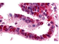
Click image to see more details
Boster's Affinity Purified anti-Ubiquitin Activating Enzyme antibody was used at a 10 µg/ml to detect UBE1 in a variety of tissues including adrenal, breast, colon (epithelium), kidney, liver, lung (respiratory epithelium), ovary (oocyte and endothelium), pancreas (islet and exocrine), placenta, prostate (epithelium), skin (epithelium), spleen (lymphocytes), stomach (chief), testis, thymus, tonsil, and uterus (glandular, stroma). In many cells a punctate nuclear staining was observed. Other cells showed both cytoplasmic and nuclear staining. This image shows UBE1 staining of human lung tissue. Tissue was formalin-fixed and paraffin embedded. Personal Communication, Tina Roush, LifeSpan Biosciences, Seattle, WA.

Click image to see more details
Western blot using Boster's purified anti-Ubiquitin Activating Enzyme (E1) antibody shows detection of a band at ~118 kDa corresponding to UBE1 (lane 1 800 nm channel). Approximately 35µg of an A431 whole cell lysate was separated on a 4-20% Tris-Glycine gel by SDS-PAGE and transferred onto nitrocellulose. After blocking the membrane was probed with the primary antibody diluted to 1:1,000. Incubation was for 2 h at room temperature followed by washes and reaction with a 1:10,000 dilution of IRDye™800 conjugated Gt-a-Rabbit IgG [H&L] MX10 for 45 min at room temperature. Molecular weight markers are shown in lane 2 (700 nm channel). IRDye™800 fluorescence image was captured using the Odyssey® Infrared Imaging System developed by LI-COR. IRDye is a trademark of LI-COR, Inc. Other detection systems will yield similar results.
Specific Publications For Anti-Ubiquitin Activating Enzyme E1 UBA1 Antibody (A02810)
Loading publications
Recommended Resources
Here are featured tools and databases that you might find useful.
- Boster's Pathways Library
- Protein Databases
- Bioscience Research Protocol Resources
- Data Processing & Analysis Software
- Photo Editing Software
- Scientific Literature Resources
- Research Paper Management Tools
- Molecular Biology Software
- Primer Design Tools
- Bioinformatics Tools
- Phylogenetic Tree Analysis
Customer Reviews
Have you used Anti-Ubiquitin Activating Enzyme E1 UBA1 Antibody?
Share your experimental results or join a short interview to earn up to $1,000 in product credits or other rewards.
0 Reviews For Anti-Ubiquitin Activating Enzyme E1 UBA1 Antibody
Customer Q&As
Have a question?
Find answers in Q&As, reviews.
Can't find your answer?
Submit your question
