Click image to see more details
-

-
-
-
-
+4
Product Info Summary
| SKU: | A04802-2 |
|---|---|
| Size: | 100 μg/vial |
| Reactive Species: | Human, Mouse, Rat |
| Host: | Rabbit |
| Application: | ELISA, Flow Cytometry, IHC, WB |
Customers Who Bought This Also Bought

Product info
Product Name
Anti-VDAC3 Antibody Picoband®
SKU/Catalog Number
A04802-2
Size
100 μg/vial
Form
Lyophilized
Description
Boster Bio Anti-VDAC3 Antibody Picoband® catalog # A04802-2. Tested in ELISA, Flow Cytometry, IHC, WB applications. This antibody reacts with Human, Mouse, Rat. The brand Picoband indicates this is a premium antibody that guarantees superior quality, high affinity, and strong signals with minimal background in Western blot applications. Only our best-performing antibodies are designated as Picoband, ensuring unmatched performance.
Storage & Handling
Store at -20˚C for one year from date of receipt. After reconstitution, at 4˚C for one month. It can also be aliquotted and stored frozen at -20˚C for six months. Avoid repeated freeze-thaw cycles.
Cite This Product
Anti-VDAC3 Antibody Picoband® (Boster Biological Technology, Pleasanton CA, USA, Catalog # A04802-2)
Host
Rabbit
Contents
Each vial contains 4mg Trehalose, 0.9mg NaCl, 0.2mg Na2HPO4, 0.01mg NaN3.
Clonality
Polyclonal
Isotype
Rabbit IgG
Immunogen
E.coli-derived human VDAC3 recombinant protein (Position: T83-E147).
Cross-reactivity
No cross-reactivity with other proteins.
Reactive Species
A04802-2 is reactive to VDAC3 in Human, Mouse, Rat
Observed Molecular Weight
31 kDa
Calculated molecular weight
30.7 kDa
Background of VDAC3
Voltage-dependent anion-selective channel protein 3 (VDAC3) is a protein that in humans is encoded by the VDAC3 gene on chromosome 8. This gene encodes a voltage-dependent anion channel (VDAC), and belongs to the mitochondrial porin family. VDACs are small, integral membrane proteins that traverse the outer mitochondrial membrane and conduct ATP and other small metabolites. They are known to bind several kinases of intermediary metabolism, thought to be involved in translocation of adenine nucleotides, and are hypothesized to form part of the mitochondrial permeability transition pore, which results in the release of cytochrome c at the onset of apoptotic cell death. Alternatively transcript variants encoding different isoforms have been described for this gene.
Antibody Validation
Boster validates all antibodies on WB, IHC, ICC, Immunofluorescence, and ELISA with known positive control and negative samples to ensure specificity and high affinity, including thorough antibody incubations.
Application & Images
Applications
A04802-2 is guaranteed for ELISA, Flow Cytometry, IHC, WB Boster Guarantee
Recommend Dilution
| Application | Dilution | Species |
|---|---|---|
| Western blot | 0.25-0.5μg/ml | Human, Mouse, Rat |
| Immunohistochemistry (Paraffin-embedded Section) | 2-5μg/ml | Human, Mouse, Rat |
| Flow Cytometry (Fixed) | 1-3μg/1x106 cells | Human |
| ELISA | 0.1-0.5μg/ml | - |
Tested application
Suggested blocking solution with 5% non-fat milk or BSA; (*)Recommended protein loading: 20-40 µg per lane
Use TE buffer pH 9.0 for antigen retrieval; (*) citrate buffer pH 6.0 is an alternative.
Validation Images & Assay Conditions
Click image to see more details
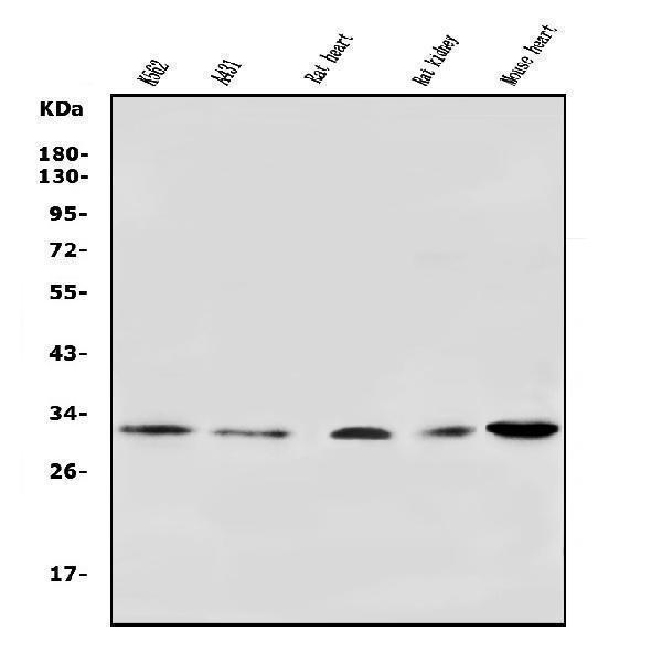
Western blot analysis of VDAC3 using anti-VDAC3 antibody (A04802-2).
Electrophoresis was performed on a 5-20% SDS-PAGE gel at 70V (Stacking gel) / 90V (Resolving gel) for 2-3 hours. The sample well of each lane was loaded with 50ug of sample under reducing conditions.
Lane 1: human K562 whole cell lysates,
Lane 2: human A431 whole cell lysates,
Lane 3: rat heart tissue lysates,
Lane 4: rat kidney tissue lysates,
Lane 5: mouse heart tissue lysates.
After Electrophoresis, proteins were transferred to a Nitrocellulose membrane at 150mA for 50-90 minutes. Blocked the membrane with 5% Non-fat Milk/ TBS for 1.5 hour at RT. The membrane was incubated with rabbit anti-VDAC3 antigen affinity purified polyclonal antibody (Catalog # A04802-2) at 0.5 μg/mL overnight at 4°C, then washed with TBS-0.1%Tween 3 times with 5 minutes each and probed with a goat anti-rabbit IgG-HRP secondary antibody at a dilution of 1:5000 for 1.5 hour at RT. The signal is developed using an Enhanced Chemiluminescent detection (ECL) kit (Catalog # EK1002) with Tanon 5200 system. A specific band was detected for VDAC3 at approximately 31KD. The expected band size for VDAC3 is at 31KD.

Click image to see more details
KEGG pathway clustering analysis of different comparison groups, the PPI network of differentially lactylated proteins in the pathways of interest, and verification of Vdac3 protein lactylation modifications. (A) Heatmap of lactylation sites for 12 key proteins. Each row represents a differentially modification site; each column represents a sample. Red indicates a high-level modification, blue indicates a low-level modification, and grey indicates sites that cannot be quantified in the corresponding samples. (B) The horizontal axis depicts different comparison groups, whereas the vertical axis indicates the enriched KEGG pathways. The colored blocks represent the functional descriptions of differentially expressed modified proteins enriched in each comparison group. Blue signifies high enrichment significance, whereas blue-white signifies low enrichment significance. * p < 0.05, ** p < 0.01, and *** p < 0.001. (C) PPI network of differentially lactylated proteins involved in the 4-VO group and sham group in the five pathways of interest. (D) PPI network in the 4-VO group and 4-VO + EA group. The color of the outer circle indicates the corresponding KEGG pathway of the protein, whereas the shape within the circle denotes the upregulation or downregulation of the modification sites contained within the protein. A diamond shape signifies proteins with upregulated modification sites, a downward arrow indicates proteins with downregulated modification sites, and a circle signifies proteins with upregulated and downregulated modification sites. The red box indicates the Vdac3 protein. (E) The IP experiment verified the lactylation modification level of Vdac3. (F) Quantification was performed using ImageJ software, and the values are expressed as the mean with the corresponding standard error from three replicate experiments ( n = 3, compared with the sham group, ## p < 0.01; compared with the 4-VO group, ** p < 0.01). 4-VO, four-vessel occlusion; EA, electroacupuncture; KEGG, Kyoto Encyclopedia of Genes and Genomes; PPI, protein–protein interaction; IP, immunoprecipitation; Vdac3, mitochondrial voltage-dependent anion channel protein3.
Index in PubMed under a CC BY license. PMID: 40963935

Click image to see more details
IHC analysis of VDAC3 using anti-VDAC3 antibody (A04802-2).
VDAC3 was detected in paraffin-embedded section of human testis cancer tissue. Heat mediated antigen retrieval was performed in EDTA buffer (pH8.0, epitope retrieval solution). The tissue section was blocked with 10% goat serum. The tissue section was then incubated with 2μg/ml rabbit anti-VDAC3 Antibody (A04802-2) overnight at 4°C. Biotinylated goat anti-rabbit IgG was used as secondary antibody and incubated for 30 minutes at 37°C. The tissue section was developed using Strepavidin-Biotin-Complex (SABC) (Catalog # SA1022) with DAB as the chromogen.

Click image to see more details
IHC analysis of VDAC3 using anti-VDAC3 antibody (A04802-2).
VDAC3 was detected in paraffin-embedded section of human breast cancer tissue. Heat mediated antigen retrieval was performed in EDTA buffer (pH8.0, epitope retrieval solution). The tissue section was blocked with 10% goat serum. The tissue section was then incubated with 2μg/ml rabbit anti-VDAC3 Antibody (A04802-2) overnight at 4°C. Biotinylated goat anti-rabbit IgG was used as secondary antibody and incubated for 30 minutes at 37°C. The tissue section was developed using Strepavidin-Biotin-Complex (SABC) (Catalog # SA1022) with DAB as the chromogen.

Click image to see more details
IHC analysis of VDAC3 using anti-VDAC3 antibody (A04802-2).
VDAC3 was detected in paraffin-embedded section of human melanoma tissue. Heat mediated antigen retrieval was performed in EDTA buffer (pH8.0, epitope retrieval solution). The tissue section was blocked with 10% goat serum. The tissue section was then incubated with 2μg/ml rabbit anti-VDAC3 Antibody (A04802-2) overnight at 4°C. Biotinylated goat anti-rabbit IgG was used as secondary antibody and incubated for 30 minutes at 37°C. The tissue section was developed using Strepavidin-Biotin-Complex (SABC) (Catalog # SA1022) with DAB as the chromogen.

Click image to see more details
IHC analysis of VDAC3 using anti-VDAC3 antibody (A04802-2).
VDAC3 was detected in paraffin-embedded section of mouse testis tissue. Heat mediated antigen retrieval was performed in EDTA buffer (pH8.0, epitope retrieval solution). The tissue section was blocked with 10% goat serum. The tissue section was then incubated with 2μg/ml rabbit anti-VDAC3 Antibody (A04802-2) overnight at 4°C. Biotinylated goat anti-rabbit IgG was used as secondary antibody and incubated for 30 minutes at 37°C. The tissue section was developed using Strepavidin-Biotin-Complex (SABC) (Catalog # SA1022) with DAB as the chromogen.

Click image to see more details
IHC analysis of VDAC3 using anti-VDAC3 antibody (A04802-2).
VDAC3 was detected in paraffin-embedded section of rat testis tissue. Heat mediated antigen retrieval was performed in EDTA buffer (pH8.0, epitope retrieval solution). The tissue section was blocked with 10% goat serum. The tissue section was then incubated with 2μg/ml rabbit anti-VDAC3 Antibody (A04802-2) overnight at 4°C. Biotinylated goat anti-rabbit IgG was used as secondary antibody and incubated for 30 minutes at 37°C. The tissue section was developed using Strepavidin-Biotin-Complex (SABC) (Catalog # SA1022) with DAB as the chromogen.

Click image to see more details
Flow Cytometry analysis of U20S cells using anti-VDAC3 antibody (A04802-2).
Overlay histogram showing U20S cells stained with A04802-2 (Blue line). To facilitate intracellular staining, cells were fixed with 4% paraformaldehyde and permeabilized with permeabilization buffer. The cells were blocked with 10% normal goat serum. And then incubated with rabbit anti-VDAC3 Antibody (A04802-2, 1μg/1x106 cells) for 30 min at 20°C. DyLight®488 conjugated goat anti-rabbit IgG (BA1127, 5-10μg/1x106 cells) was used as secondary antibody for 30 minutes at 20°C. Isotype control antibody (Green line) was rabbit IgG (1μg/1x106) used under the same conditions. Unlabelled sample without incubation with primary antibody and secondary antibody (Red line) was used as a blank control.
Specific Publications For Anti-VDAC3 Antibody Picoband® (A04802-2)
Loading publications
Recommended Resources
Here are featured tools and databases that you might find useful.
- Boster's Pathways Library
- Protein Databases
- Bioscience Research Protocol Resources
- Data Processing & Analysis Software
- Photo Editing Software
- Scientific Literature Resources
- Research Paper Management Tools
- Molecular Biology Software
- Primer Design Tools
- Bioinformatics Tools
- Phylogenetic Tree Analysis
Customer Reviews
Have you used Anti-VDAC3 Antibody Picoband®?
Share your experimental results or join a short interview to earn up to $1,000 in product credits or other rewards.
0 Reviews For Anti-VDAC3 Antibody Picoband®
Customer Q&As
Have a question?
Find answers in Q&As, reviews.
Can't find your answer?
Submit your question
