Click image to see more details
-

-
-
-
-
+4
Product Info Summary
| SKU: | A02672-2 |
|---|---|
| Size: | 100 μg/vial |
| Reactive Species: | Human, Mouse, Rat |
| Host: | Rabbit |
| Application: | ELISA, IP, IF, IHC, ICC, WB |
Customers Who Bought This Also Bought

Product info
Product Name
Anti-WWTR1 Antibody Picoband®
SKU/Catalog Number
A02672-2
Size
100 μg/vial
Form
Lyophilized
Description
Boster Bio Anti-WWTR1 Antibody Picoband® catalog # A02672-2. Tested in WB, IHC, ICC/IF, IP, ELISA applications. This antibody reacts with Human, Mouse, Rat. The brand Picoband indicates this is a premium antibody that guarantees superior quality, high affinity, and strong signals with minimal background in Western blot applications. Only our best-performing antibodies are designated as Picoband, ensuring unmatched performance.
Storage & Handling
Store at -20˚C for one year from date of receipt. After reconstitution, at 4˚C for one month. It can also be aliquotted and stored frozen at -20˚C for six months. Avoid repeated freeze-thaw cycles.
Cite This Product
Anti-WWTR1 Antibody Picoband® (Boster Biological Technology, Pleasanton CA, USA, Catalog # A02672-2)
Host
Rabbit
Contents
Each vial contains 4 mg Trehalose, 0.9 mg NaCl, 0.2 mg Na2HPO4.
Clonality
Polyclonal
Isotype
Rabbit IgG
Immunogen
E.coli-derived human WWTR1 recombinant protein (Position: M1-F396).
Reactive Species
A02672-2 is reactive to WWTR1 in Human, Mouse, Rat
Observed Molecular Weight
50 kDa
Calculated molecular weight
44.1 kDa
Background of WWTR1
WW domain-containing transcription regulator protein 1 (WWTR1), also known as Transcriptional coactivator with PDZ-binding motif (TAZ), is a protein that in humans is encoded by the WWTR1 gene. WWTR1 acts as a transcriptional coregulator and has no effect on transcription alone. When in complex with transcription factor binding partners, WWTR1 helps promote gene expression in pathways associated with development, cell growth and survival, and inhibiting apoptosis. Aberrant WWTR1 function has been implicated for its role in driving cancers. WWTR1 is often referred to as TAZ due to its initial characterization with the name TAZ. However, WWTR1 (TAZ) is not to be confused with the protein tafazzin, which originally held the official gene symbol TAZ, and is now TAFAZZIN.
Antibody Validation
Boster validates all antibodies on WB, IHC, ICC, Immunofluorescence, and ELISA with known positive control and negative samples to ensure specificity and high affinity, including thorough antibody incubations.
Application & Images
Applications
A02672-2 is guaranteed for ELISA, IP, IF, IHC, ICC, WB Boster Guarantee
Assay Dilutions Recommendation
The recommendations below provide a starting point for assay optimization. The actual working concentration varies and should be decided by the user.
Western blot, 0.25-0.5 μg/ml, Human
Immunohistochemistry (Paraffin-embedded Section), 2-5 μg/ml, Human, Mouse, Rat
Immunocytochemistry/Immunofluorescence, 5 μg/ml, Human
Immunoprecipitation, 0.5-2 μg/ml, Human
ELISA, 0.1-0.5 μg/ml, -
Positive Control
WB: human A431 whole cell, human U251 whole cell, human A549 whole cell
IHC: human ovarian cancer tissue, mouse brain tissue, rat kidney tissue, rat brain tissue
ICC/IF: U251 cell
IP: Hela cell
Validation Images & Assay Conditions
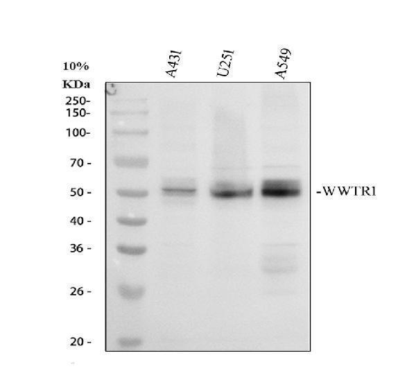
Click image to see more details
Western blot analysis of WWTR1 using anti-WWTR1 antibody (A02672-2).
Electrophoresis was performed on a 10% SDS-PAGE gel at 80V (Stacking gel) / 120V (Resolving gel) for 2 hours. The sample well of each lane was loaded with 30 ug of sample under reducing conditions.
Lane 1: human A431 whole cell lysates,
Lane 2: human U251 whole cell lysates,
Lane 3: human A549 whole cell lysates.
After electrophoresis, proteins were transferred to a nitrocellulose membrane at 150 mA for 50-90 minutes. Blocked the membrane with 5% non-fat milk/TBS for 1.5 hour at RT. The membrane was incubated with rabbit anti-WWTR1 antigen affinity purified polyclonal antibody (A02672-2) at 0.5 μg/mL overnight at 4°C, then washed with TBS-0.1%Tween 3 times with 5 minutes each and probed with a goat anti-rabbit IgG-HRP secondary antibody at a dilution of 1:5000 for 1.5 hour at RT. The signal is developed using an ECL Plus Western Blotting Substrate (Catalog # AR1196-200) with Tanon 5200 system. A specific band was detected for WWTR1 at approximately 50 kDa. The expected band size for WWTR1 is at 44 kDa.

Click image to see more details
IHC analysis of WWTR1 using anti-WWTR1 antibody (A02672-2).
WWTR1 was detected in a paraffin-embedded section of human ovarian cancer tissue. Heat mediated antigen retrieval was performed in EDTA buffer (pH 8.0, epitope retrieval solution). The tissue section was blocked with 10% goat serum. The tissue section was then incubated with 2 μg/ml rabbit anti-WWTR1 Antibody (A02672-2) overnight at 4°C. Peroxidase Conjugated Goat Anti-rabbit IgG was used as secondary antibody and incubated for 30 minutes at 37°C. The tissue section was developed using HRP Conjugated Rabbit IgG Super Vision Assay Kit (Catalog # SV0002) with DAB as the chromogen.

Click image to see more details
IHC analysis of WWTR1 using anti-WWTR1 antibody (A02672-2).
WWTR1 was detected in a paraffin-embedded section of mouse brain tissue. Heat mediated antigen retrieval was performed in EDTA buffer (pH 8.0, epitope retrieval solution). The tissue section was blocked with 10% goat serum. The tissue section was then incubated with 2 μg/ml rabbit anti-WWTR1 Antibody (A02672-2) overnight at 4°C. Peroxidase Conjugated Goat Anti-rabbit IgG was used as secondary antibody and incubated for 30 minutes at 37°C. The tissue section was developed using HRP Conjugated Rabbit IgG Super Vision Assay Kit (Catalog # SV0002) with DAB as the chromogen.

Click image to see more details
IHC analysis of WWTR1 using anti-WWTR1 antibody (A02672-2).
WWTR1 was detected in a paraffin-embedded section of rat kidney tissue. Heat mediated antigen retrieval was performed in EDTA buffer (pH 8.0, epitope retrieval solution). The tissue section was blocked with 10% goat serum. The tissue section was then incubated with 2 μg/ml rabbit anti-WWTR1 Antibody (A02672-2) overnight at 4°C. Peroxidase Conjugated Goat Anti-rabbit IgG was used as secondary antibody and incubated for 30 minutes at 37°C. The tissue section was developed using HRP Conjugated Rabbit IgG Super Vision Assay Kit (Catalog # SV0002) with DAB as the chromogen.

Click image to see more details
IHC analysis of WWTR1 using anti-WWTR1 antibody (A02672-2).
WWTR1 was detected in a paraffin-embedded section of rat brain tissue. Heat mediated antigen retrieval was performed in EDTA buffer (pH 8.0, epitope retrieval solution). The tissue section was blocked with 10% goat serum. The tissue section was then incubated with 2 μg/ml rabbit anti-WWTR1 Antibody (A02672-2) overnight at 4°C. Peroxidase Conjugated Goat Anti-rabbit IgG was used as secondary antibody and incubated for 30 minutes at 37°C. The tissue section was developed using HRP Conjugated Rabbit IgG Super Vision Assay Kit (Catalog # SV0002) with DAB as the chromogen.

Click image to see more details
IF analysis of WWTR1 using anti-WWTR1 antibody (A02672-2) and anti-Alpha Tubulin antibody (M03989-3).
WWTR1 was detected in an immunocytochemical section of U251 cells. Enzyme antigen retrieval was performed using IHC enzyme antigen retrieval reagent (AR0022) for 15 mins. The cells were blocked with 10% goat serum. And then incubated with 5 μg/mL rabbit anti-WWTR1 Antibody (A02672-2) and mouse anti-Alpha Tubulin antibody (M03989-3) overnight at 4°C. Fluoro488 Conjugated Goat Anti-Rabbit IgG (BA1127) and Cy3 Conjugated Goat Anti-Mouse IgG (BA1031) were used as secondary antibody at 1:500 dilution and incubated for 30 minutes at 37°C. Visualize using a fluorescence microscope and filter sets appropriate for the label used.

Click image to see more details
Immunoprecipitating WWTR1 in Hela whole cell lysate.
Western blot analysis of WWTR1 using anti-WWTR1 antibody (A02672-2).
Lane 1: Hela whole cell lysates (30ug),
Lane 2: Rabbit control IgG instead of anti-WWTR1 antibody in Hela whole cell lysate,
Lane 3: anti-WWTR1 antibody (2μg) + Hela whole cell lysate (500μg).
After electrophoresis, proteins were transferred to a membrane. Then the membrane was incubated with rabbit anti-WWTR1 antigen affinity purified polyclonal antibody (A02672-2) at a dilution of 0.5 μg/mL and probed with a mouse anti-rabbit IgG-HRP secondary antibody (Catalog # BA1054). The signal is developed using ECL Plus Western Blotting Substrate (Catalog # AR1197). A specific band was detected for WWTR1 at approximately 50 kDa. The expected band size for WWTR1 is at 44 kDa.
Specific Publications For Anti-WWTR1 Antibody Picoband® (A02672-2)
Loading publications
Recommended Resources
Here are featured tools and databases that you might find useful.
- Boster's Pathways Library
- Protein Databases
- Bioscience Research Protocol Resources
- Data Processing & Analysis Software
- Photo Editing Software
- Scientific Literature Resources
- Research Paper Management Tools
- Molecular Biology Software
- Primer Design Tools
- Bioinformatics Tools
- Phylogenetic Tree Analysis
Customer Reviews
Have you used Anti-WWTR1 Antibody Picoband® ?
Share your experimental results or join a short interview to earn up to $1,000 in product credits or other rewards.
0 Reviews For Anti-WWTR1 Antibody Picoband®
Customer Q&As
Have a question?
Find answers in Q&As, reviews.
Can't find your answer?
Submit your question
