Click image to see more details
-

-
-
-
-
+4
Product Info Summary
| SKU: | AZQ90Z37 |
|---|---|
| Size: | 100 μg/vial |
| Reactive Species: | Zebrafish |
| Host: | Rabbit |
| Application: | IHC, WB |
Customers Who Bought This Also Bought

Product info
Product Name
Anti-Zebrafish E-cadherin/CDH1 Antibody Picoband®
SKU/Catalog Number
AZQ90Z37
Size
100 μg/vial
Form
Lyophilized
Description
Boster Bio Anti-Zebrafish E-cadherin/CDH1 Antibody Picoband® catalog #AZQ90Z37. Tested in WB, IHC applications. This antibody reacts with Zebrafish. The brand Picoband indicates this is a premium antibody that guarantees superior quality, high affinity, and strong signals with minimal background in Western blot applications. Only our best-performing antibodies are designated as Picoband, ensuring unmatched performance.
Storage & Handling
At -20°C for one year from date of receipt. After reconstitution, at 4°C for one month. It can also be aliquotted and stored frozen at -20°C for six months. Avoid repeated freezing and thawing.
Cite This Product
Anti-Zebrafish E-cadherin/CDH1 Antibody Picoband® (Boster Biological Technology, Pleasanton CA, USA, Catalog # AZQ90Z37)
Host
Rabbit
Contents
Each vial contains 4 mg Trehalose, 0.9 mg NaCl, 0.2 mg Na2HPO4.
Clonality
Polyclonal
Immunogen
E.coli-derived zebrafish E-cadherin/CDH1 recombinant protein (Position: Y23-D864)
Reactive Species
AZQ90Z37 is reactive to CDH1 in Zebrafish
Observed Molecular Weight
130 kDa
Background of CDH1
CDH1 (Cadherin 1), also known as ECAD or UVO, is a protein that in humans is encoded by the CDH1 gene. Cadherin-1 is a classical member of the cadherin superfamily. By Southern analysis of DNA from a panel of mouse-human somatic cell hybrids, Mansouri et al. (1987, 1988) assigned the UVO gene to 16q (16p11-qter). Frebourg et al. (2006) found that in human embryos CDH1 is highly expressed at 4 and 5 weeks in the frontonasal prominence and at 6 weeks in the lateral and medial nasal prominences, and is therefore expressed during critical stages of lip and palate development. CDH1 is involved in mechanisms regulating cell-cell adhesions, mobility and proliferation of epithelial cells. Has a potent invasive suppressor role. It is a ligand for integrin alpha-E/beta-7.
Antibody Validation
Boster validates all antibodies on WB, IHC, ICC, Immunofluorescence, and ELISA with known positive control and negative samples to ensure specificity and high affinity, including thorough antibody incubations.
Application & Images
Applications
AZQ90Z37 is guaranteed for IHC, WB Boster Guarantee
Recommend Dilution
| Application | Dilution | Species |
|---|---|---|
| Western blot | 0.25-0.5 μg/ml | Zebrafish |
| Immunohistochemistry(Paraffin-embedded Section) | 2-5 μg/ml | Zebrafish |
Tested application
Suggested blocking solution with 5% non-fat milk or BSA; (*)Recommended protein loading: 20-40 µg per lane
Use TE buffer pH 9.0 for antigen retrieval; (*) citrate buffer pH 6.0 is an alternative.
Validation Images & Assay Conditions
Click image to see more details
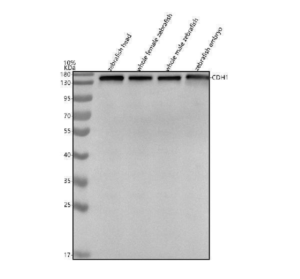
Western blot analysis of E-cadherin/CDH1 using anti-E-cadherin/CDH1 antibody (AZQ90Z37).
Electrophoresis was performed on a 10% SDS-PAGE gel at 80V (Stacking gel) / 120V (Resolving gel) for 2 hours. The sample well of each lane was loaded with 30 ug of sample under reducing conditions.
Lane 1: zebrafish head tissue lysates.
Lane 2: whole female zebrafish tissue lysates.
Lane 3: whole male zebrafish tissue lysates.
Lane 4: zebrafish embryo tissue lysates.
After electrophoresis, proteins were transferred to a nitrocellulose membrane at 150 mA for 50-90 minutes. Blocked the membrane with 5% non-fat milk/TBS for 1.5 hour at RT. The membrane was incubated with rabbit anti-E-cadherin/CDH1 antigen affinity purified polyclonal antibody (AZQ90Z37) at 0.5 μg/mL overnight at 4°C, then washed with TBS-0.1%Tween 3 times with 5 minutes each and probed with a goat anti-rabbit IgG-HRP secondary antibody at a dilution of 1:5000 for 1.5 hour at RT. The signal is developed using an ECL Plus Western Blotting Substrate (Catalog # AR1196-200) with Tanon 5200 system. A specific band was detected for E-cadherin/CDH1 at approximately 130 kDa. The expected band size for E-cadherin/CDH1 is at 120 kDa.

Click image to see more details
IHC analysis of E-cadherin/CDH1 using anti-E-cadherin/CDH1 antibody (AZQ90Z37).
E-cadherin/CDH1 was detected in a paraffin-embedded section of zebrafish inner ear tissue. Heat mediated antigen retrieval was performed in EDTA buffer (pH 8.0, epitope retrieval solution). The tissue section was blocked with 10% goat serum. The tissue section was then incubated with 2 μg/ml rabbit anti-E-cadherin/CDH1 Antibody (AZQ90Z37) overnight at 4°C. Peroxidase Conjugated Goat Anti-rabbit IgG was used as secondary antibody and incubated for 30 minutes at 37°C. The tissue section was developed using HRP Conjugated Rabbit IgG Super Vision Assay Kit (Catalog # SV0002) with DAB as the chromogen.

Click image to see more details
IHC analysis of E-cadherin/CDH1 using anti-E-cadherin/CDH1 antibody (AZQ90Z37).
E-cadherin/CDH1 was detected in a paraffin-embedded section of zebrafish colon tissue. Heat mediated antigen retrieval was performed in EDTA buffer (pH 8.0, epitope retrieval solution). The tissue section was blocked with 10% goat serum. The tissue section was then incubated with 2 μg/ml rabbit anti-E-cadherin/CDH1 Antibody (AZQ90Z37) overnight at 4°C. Peroxidase Conjugated Goat Anti-rabbit IgG was used as secondary antibody and incubated for 30 minutes at 37°C. The tissue section was developed using HRP Conjugated Rabbit IgG Super Vision Assay Kit (Catalog # SV0002) with DAB as the chromogen.

Click image to see more details
IHC analysis of E-cadherin/CDH1 using anti-E-cadherin/CDH1 antibody (AZQ90Z37).
E-cadherin/CDH1 was detected in a paraffin-embedded section of zebrafish esophageal epithelium tissue. Heat mediated antigen retrieval was performed in EDTA buffer (pH 8.0, epitope retrieval solution). The tissue section was blocked with 10% goat serum. The tissue section was then incubated with 2 μg/ml rabbit anti-E-cadherin/CDH1 Antibody (AZQ90Z37) overnight at 4°C. Peroxidase Conjugated Goat Anti-rabbit IgG was used as secondary antibody and incubated for 30 minutes at 37°C. The tissue section was developed using HRP Conjugated Rabbit IgG Super Vision Assay Kit (Catalog # SV0002) with DAB as the chromogen.

Click image to see more details
IHC analysis of E-cadherin/CDH1 using anti-E-cadherin/CDH1 antibody (AZQ90Z37).
E-cadherin/CDH1 was detected in a paraffin-embedded section of zebrafish heart tissue. Heat mediated antigen retrieval was performed in EDTA buffer (pH 8.0, epitope retrieval solution). The tissue section was blocked with 10% goat serum. The tissue section was then incubated with 2 μg/ml rabbit anti-E-cadherin/CDH1 Antibody (AZQ90Z37) overnight at 4°C. Peroxidase Conjugated Goat Anti-rabbit IgG was used as secondary antibody and incubated for 30 minutes at 37°C. The tissue section was developed using HRP Conjugated Rabbit IgG Super Vision Assay Kit (Catalog # SV0002) with DAB as the chromogen.

Click image to see more details
IHC analysis of E-cadherin/CDH1 using anti-E-cadherin/CDH1 antibody (AZQ90Z37).
E-cadherin/CDH1 was detected in a paraffin-embedded section of zebrafish kidney tissue. Heat mediated antigen retrieval was performed in EDTA buffer (pH 8.0, epitope retrieval solution). The tissue section was blocked with 10% goat serum. The tissue section was then incubated with 2 μg/ml rabbit anti-E-cadherin/CDH1 Antibody (AZQ90Z37) overnight at 4°C. Peroxidase Conjugated Goat Anti-rabbit IgG was used as secondary antibody and incubated for 30 minutes at 37°C. The tissue section was developed using HRP Conjugated Rabbit IgG Super Vision Assay Kit (Catalog # SV0002) with DAB as the chromogen.
Specific Publications For Anti-Zebrafish E-cadherin/CDH1 Antibody Picoband® (AZQ90Z37)
Loading publications
Recommended Resources
Here are featured tools and databases that you might find useful.
- Boster's Pathways Library
- Protein Databases
- Bioscience Research Protocol Resources
- Data Processing & Analysis Software
- Photo Editing Software
- Scientific Literature Resources
- Research Paper Management Tools
- Molecular Biology Software
- Primer Design Tools
- Bioinformatics Tools
- Phylogenetic Tree Analysis
Customer Reviews
Have you used Anti-Zebrafish E-cadherin/CDH1 Antibody Picoband®?
Share your experimental results or join a short interview to earn up to $1,000 in product credits or other rewards.
0 Reviews For Anti-Zebrafish E-cadherin/CDH1 Antibody Picoband®
Customer Q&As
Have a question?
Find answers in Q&As, reviews.
Can't find your answer?
Submit your question
