This website uses cookies to ensure you get the best experience on our website.


- Table of Contents
Plan ACTB chromogenic IHC in paraffin sections using the catalog antibody at 1:25 (datasheet: M01263-3). Assess myoepithelial staining (HPA tissue IHC) and account for possible detection of products from more than one gene (HPA tissue IHC).

Expected localisation, validated protocols, controls and antibodies — the at-a-glance facts below, then the full design guide.
| Expected localisation | Cytoskeletal and nuclear locations are annotated; the tissue compartment is unreported (UniProt; HPA tissue IHC) | |
| Staining pattern | High myoepithelial staining across tissues; compartment unreported (HPA tissue IHC) | |
| Antigen retrieval | Citrate pH 6 HIER, heat-mediated (datasheet M01263-3) | |
| Positive control | Appendix+4 more · see all | |
| Negative control | Adrenal gland+4 more · see all |
| Fixation | Formaldehyde-fixed paraffin sections are shown (selected-SKU IHC image M01263-3); Matched tissue-IHC evidence does not establish the fixation claim. Validate the specimen-specific method before use. (selected-SKU IHC image M01263-3) | |
| Caveat | The antibody may detect products of more than one gene (HPA tissue IHC) | |
| Regulation | Low tissue specificity; an intensity regulator is unreported (HPA tissue IHC) | |
| Isoform / epitope | No annotated isoforms; N-terminal processing may affect epitopes (UniProt) |
The catalog antibody protocol and the published ACTB IHC protocol below both use citrate pH 6 retrieval (datasheet: citrate pH 6; PMC12446288: citrate pH 6).
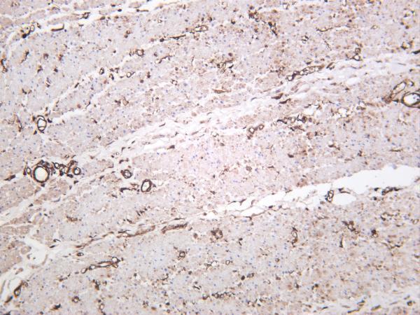
| Sample | Formaldehyde-fixed, paraffin-embedded human heart tissue (datasheet M01263-3) |
| Fixation | Image formalin-fixed; duration unreported (datasheet M01263-3); verify before use. |
| Sectioning | 4–5 µm sections on charged slides (standard) |
| Deparaffinisation | Xylene, graded ethanol series to water (standard) |
| Antigen retrieval | Heat retrieval: Citrate pH 6 (datasheet M01263-3); 20 min, 95–100 °C (standard) |
| Peroxidase block | 3% H2O2, 10 min, room temperature (standard) |
| Blocking | 10% normal serum of the secondary host, 30 min, room temperature (standard) |
| Primary antibody | Mouse monoclonal (clone 137CT26.1.1) anti-ACTB, 1:25 (datasheet M01263-3) |
| Primary incubation | 1 hours at 37°C (datasheet M01263-3) |
| Detection | HRP-polymer secondary, DAB chromogen 5–10 min (standard) |
| Counterstain | Hematoxylin, blue, dehydrate and mount (standard) |
| Expected result | ACTB-positive staining in enterocytes - Microvilli of appendix (HPA tissue IHC: High). HPA tissue profile: High myoepithelial expression in all tissues. No signal in the no-primary control. |
ACTB localises to the cytoplasmic cytoskeleton and can also occur in the nucleus; it has no transmembrane segment (UniProt P60709). Expect high myoepithelial staining across tissues and high enterocyte microvillar staining in appendix, colon and duodenum (HPA: tissue IHC). HPA rates the tissue pattern Supported, with medium consistency and a caution that staining may detect products of more than one gene (HPA: reliability).
| Strong breast myoepithelial staining (HPA: High). | Matches the reported ACTB tissue pattern (HPA: breast myoepithelial cells High). |
| Extracellular-only chromogen. | Inconsistent with intracellular ACTB; suspect artefact (UniProt P60709 topology). |
| Strong bronchial respiratory epithelial staining. | Unexpected (HPA: Not detected); consider cross-reactivity or endogenous enzyme activity (standard IHC practice). |
| Diffuse chromogen over cells and empty areas. | Suggests nonspecific background or detection artefact (standard IHC practice). |
| No signal in breast myoepithelial cells. | Unexpected in a reported positive cell type (HPA: breast myoepithelial cells High). |
| Antibody specificity (HPA: reliability) | IHC is Supported, but staining may detect products of more than one gene (HPA: caution). |
| Nuclear localisation (UniProt P60709) | Nuclear signal can be plausible alongside cytoskeletal staining (UniProt P60709). |
| N-terminal processing (UniProt P60709) | Residues 1–375 and 2–375 are annotated; check epitope position if known (UniProt P60709). |
| Cell-type variation (HPA: tissue IHC) | High breast myoepithelial and undetected bronchial epithelial staining are reported (HPA: tissue IHC). |
| Situation | Likely cause | Next action |
|---|---|---|
| No breast myoepithelial signal (HPA: High). | Assay failure is possible (standard IHC practice). | Check positive control, retrieval, antibody dilution and detection (standard IHC practice). |
| Diffuse slide background. | Inadequate blocking or washing is possible (standard IHC practice). | Review blocking, washes and detection controls (standard IHC practice). |
| Extracellular-only chromogen. | Precipitate or detection artefact is possible (standard IHC practice). | Inspect reagent cleanliness and a no-primary control (standard IHC practice). |
| Strong bronchial respiratory epithelial signal (HPA: Not detected). | Cross-reactivity or endogenous enzyme activity is possible (HPA: caution; standard IHC practice). | Compare an independent antibody and a no-primary control (standard IHC practice). |
| Nuclear-only breast myoepithelial signal. | The expected cytoskeletal component is absent (UniProt P60709). | Review controls and intracellular staining distribution (standard IHC practice). |
| IF/ICC: is a nuclear signal plausible? | ACTB can be nuclear; HPA provides no ICC-IF images (UniProt P60709; HPA: subcellular). | Interpret with IF controls and the separate IF/ICC guide (standard IF practice). |


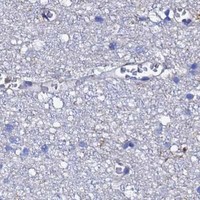


Comprehensive Human Protein Atlas IHC scoring per tissue (reliability: Supported — Medium consistency between antibody staining and RNA expression data. Caution, targets protein from more than one gene.). Rows are taken directly from the HPA tissue chart — click any row's HPA link to view the source.
| Tissue | Cell type | Level | Evidence | Source |
|---|---|---|---|---|
| Appendix | Enterocytes - Microvilli | High | Protein (IHC) | HPA → |
| Breast | Myoepithelial cells | High | Protein (IHC) | HPA → |
| Colon | Enterocytes - Microvilli | High | Protein (IHC) | HPA → |
| Duodenum | Enterocytes - Microvilli | High | Protein (IHC) | HPA → |
| Epididymis | Glandular cells | High | Protein (IHC) | HPA → |
| Tissue | Cell type | Level | Evidence | Source |
|---|---|---|---|---|
| Adrenal gland | Glandular cells | Not detected | Protein (IHC) | HPA → |
| Bronchus | Respiratory epithelial cells | Not detected | Protein (IHC) | HPA → |
| Caudate | Glial cells | Not detected | Protein (IHC) | HPA → |
| Cerebellum | Cells in granular layer | Not detected | Protein (IHC) | HPA → |
| Cerebral cortex | Glial cells | Not detected | Protein (IHC) | HPA → |
Use the catalog antibody’s tissue staining conditions as a starting point, then assess ACTB signal by compartment, cell type, and controls.
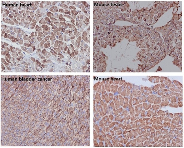
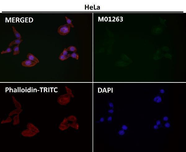
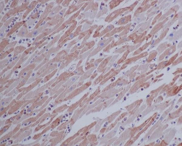
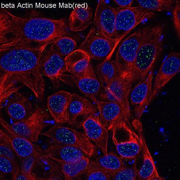
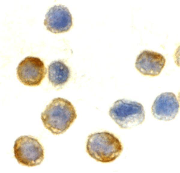
Anti-ACTB antibodies have IHC images from human and mouse tissues and IF/ICC images from HeLa cells; catalog reactivity spans human, mouse, rat, chicken, pig and rabbit (catalog image captions and reactivity).
M01263-3 shows human heart IHC-P (M01263-3 image caption); M01263 shows human heart, human bladder cancer, mouse testis and mouse heart IHC (M01263 image caption); M01263-4 shows human heart IHC (M01263-4 image caption); M01263-8 shows human colon staining (M01263-8 image caption). A01263-1 shows HeLa ICC and IF (A01263-1 image captions), while A01263-2 shows HeLa ICC and IF (A01263-2 image captions).
Which to pick: For paraffin-section IHC, choose M01263-3: its human heart image specifies formaldehyde fixation, citrate retrieval at pH 6 and a 1:25 primary dilution (M01263-3 image caption). For IF/ICC, A01263-1 lists both applications and has HeLa images for each (A01263-1 applications and image captions). For broader listed species reactivity, M01263-4 covers chicken, human, mouse, pig, rabbit and rat; its IHC image shows paraffin-embedded human heart, but the fixative is unreported (M01263-4 catalog reactivity and IHC image caption).