HSP90AA1 / Heat shock protein HSP 90-alpha · IHC design guide

Design Immunohistochemistry for HSP90AA1

Plan chromogenic HSP90AA1 IHC around the cytoplasmic staining reported in several tissues (HPA tissue IHC). High staining in ciliated cells of the fallopian tube and nasopharynx offers reference material, but low concordance between antibody staining and RNA data warrants cautious interpretation (HPA tissue IHC).

Evidence assembled Oct 2026 · For research use; verify linked source records and product datasheet before use
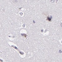
Immunohistochemistry protocol sheet for HSP90AA1 (IHC for HSP90AA1): expected localisation Cytoplasmic staining in several tissues (HPA tissue IHC), antibody M01103-2, validated IHC image, and IHC protocol steps
Printable HSP90AA1 IHC protocol sheet — expected localisation Cytoplasmic staining in several tissues (HPA tissue IHC), antibody M01103-2, controls and protocol steps. Open the full HSP90AA1 IHC guide →

HSP90AA1 Immunohistochemistry Experimental Design Guide

Expected localisation, validated protocols, controls and antibodies — the at-a-glance facts below, then the full design guide.

Must know before staining
Expected localisation Cytoplasmic staining in several tissues (HPA tissue IHC)
Staining pattern Ciliated cells show cytoplasmic staining (HPA tissue IHC)
Antigen retrieval EDTA pH 8.0 HIER, heat-mediated (datasheet M01103-2)
Positive control ⓘ Fallopian tube+4 more · see all
Negative control ⓘ Adipose tissue+4 more · see all
Important caveats
Reasons your staining may differ from the expected pattern.
Fixation Keep fixation consistent across sections (standard IHC practice; not target-specific). Selected-image fixative and duration unreported (datasheet M01103-2); verify before use.
Caveat Antibody staining has low concordance with RNA data (HPA tissue IHC)
Regulation Stress-response association (UniProt)
Isoform / epitope 2 isoforms; epitope differences unspecified (UniProt)
Section 1

Recommended HSP90AA1 IHC & IF Protocols

The catalog antibody protocol is followed by four published HSP90AA1 IHC protocols for paraffin sections (datasheet; PMC12783301; PMC13164253; PMC10398399; PMC9934426).

Recommended immunohistochemistry (IHC-P) protocol parameters
SampleParaffin-embedded human brest cancer tissue; fixative not specified (datasheet M01103-2)
FixationImage fixative and duration unreported (datasheet M01103-2); verify before use.
Sectioning4–5 µm sections on charged slides (standard)
DeparaffinisationXylene, graded ethanol series to water (standard)
Antigen retrievalHeat retrieval: EDTA pH 8.0 (datasheet M01103-2); 20 min, 95–100 °C (standard)
Peroxidase block3% H2O2, 10 min, room temperature (standard)
Blocking10% goat serum (datasheet M01103-2)
Primary antibodyRabbit monoclonal (clone EBD-8) anti-HSP90AA1, 1:50 (datasheet M01103-2)
Primary incubationOvernight at 4 °C (datasheet M01103-2)
DetectionHRP-conjugated secondary, DAB chromogen (datasheet M01103-2)
CounterstainHematoxylin, blue, dehydrate and mount (standard)
Expected resultHSP90AA1-positive staining in ciliated cells (cell body) of fallopian tube (HPA tissue IHC: High). HPA tissue profile: Cytoplasmic expression in several tissues. No signal in the no-primary control.
💡Decision noteStart with heat-mediated EDTA pH 8.0 retrieval (datasheet); citrate at 100°C for 30 min is a published alternative (PMC10398399).
Section 2

What Is the Expected HSP90AA1 Staining Pattern?

HSP90AA1 staining should be predominantly cytoplasmic in several tissues, with high staining reported in ciliated cell bodies of the fallopian tube and nasopharynx and in cells of testicular seminiferous ducts (HPA: tissue IHC). UniProt also lists nuclear, membrane, mitochondrial and melanosomal locations, but no transmembrane segment (UniProt P07900: subcellular location and topology). Treat tissue predictions cautiously: HPA rates the IHC pattern Approved while reporting low consistency with RNA expression (HPA: tissue IHC reliability).

What am I looking at on my slide?
Clear cytoplasmic staining in fallopian tube or nasopharyngeal ciliated cell bodies, or in cells of testicular seminiferous ducts (HPA: High in these cells).This matches the strongest supplied tissue IHC examples and the reported cytoplasmic profile (HPA: tissue IHC). Compare the identified cells with adjacent structures, and score intensity in those cells; staining elsewhere in the section does not replace a cell-specific check (general IHC practice).
Predominantly nuclear, sharply membranous or exclusively punctate staining, with little cytoplasmic signal (HPA: cytoplasmic tissue IHC profile).Investigate localisation before accepting it: HPA supports cytosol in ICC-IF and calls nucleoplasm uncertain, while UniProt lists several additional locations (HPA: subcellular ICC-IF; UniProt P07900: subcellular location). An unusual compartment alone cannot prove artefact, but a pattern lacking the expected cytoplasmic component needs independent assessment.
Strong staining in a cell population listed as undetected, such as adipocytes or glomerular cells (HPA: Not detected in these cells).Check cell identity and staining controls before assigning HSP90AA1 expression; cross-reactivity or endogenous chromogenic activity may explain discordant staining (general IHC practice). HPA's entries describe particular cell populations, so an undetected entry must not be read as a claim that every cell in that tissue is negative (HPA: tissue IHC).
Diffuse colour across tissue, extracellular space or the negative-control section, without a cell-shaped cytoplasmic pattern (HPA: cytoplasmic tissue IHC profile).This is less convincing than staining confined to the expected cell bodies (HPA: High in fallopian tube and nasopharyngeal ciliated cells). Consider nonspecific antibody binding, residual endogenous detection activity or chromogen deposition; compare a matched negative control and section morphology (general IHC practice).
No cytoplasmic signal in correctly identified ciliated cell bodies from fallopian tube or nasopharynx (HPA: High in these cells).A failed positive reference raises a technical or reagent question before it supports a biological negative call (general IHC practice). Confirm that the expected cells are present, then review the catalog antibody's IHC-P instructions and detection controls; HPA's Approved rating and reported low RNA–staining consistency warrant cautious interpretation (HPA: tissue IHC reliability).
💡Expected HSP90AA1 appearanceCall a result consistent when identifiable ciliated cell bodies in fallopian tube or nasopharynx show strong, predominantly cytoplasmic staining (HPA: High; cytoplasmic tissue IHC); colour spread through unrelated structures or a negative control suggests background rather than a cell-specific positive (general IHC practice).
How each factor affects the staining
Cell population and reference tissue (HPA: tissue IHC).HPA reports High staining in fallopian tube and nasopharyngeal ciliated cell bodies and seminiferous duct cells, Medium in bronchial ciliated and several glandular cell populations, and Low in selected neuronal and epithelial populations (HPA: tissue IHC). Choose a reported High cell population for the positive reference and identify it morphologically before scoring.
Compartment evidence (HPA: subcellular ICC-IF; UniProt P07900: subcellular location).Cytosol is supported in ICC-IF; nucleoplasm is uncertain (HPA: subcellular ICC-IF). UniProt also lists nucleus, melanosome, cell membrane and mitochondrion, with no transmembrane segment (UniProt P07900: location and topology). These annotations permit context-dependent localisation but do not make isolated membrane or nuclear staining the expected tissue IHC pattern.
Antibody evidence and scope (HPA: antibody validation).CAB002058 is IHC Approved and ICC Uncertain; HPA047290 is ICC Enhanced with no IHC status supplied (HPA: antibody validation). Do not transfer an ICC validation grade to paraffin-section IHC. HPA also reports low agreement between tissue staining and RNA data, pending external verification, so corroborate a consequential discordant result (HPA: tissue IHC reliability).
Chromogenic detection background (general IHC practice).Endogenous enzyme activity, incomplete blocking or nonspecific antibody binding can create colour unrelated to target localisation (general IHC practice). Interpret the pattern against a matched negative control and the expected cell bodies. The supplied HPA and UniProt records provide no target-specific fixation-sensitivity or antigen-retrieval effect, so do not predict either from staining levels or topology.
Why is my staining missing, weak or wrong?
SituationLikely causeNext action
Expected ciliated cells are present, but the positive reference has no signal (HPA: High in fallopian tube and nasopharyngeal ciliated cells).A failed staining or detection step is possible; absence alone does not identify its cause (general IHC practice).Check the catalog antibody's IHC-P instructions, primary-antibody and detection setup, and a known working control; repeat before calling the sample negative (general IHC practice).
Colour appears throughout the section, including areas outside identifiable cells (HPA: cytoplasmic tissue IHC profile).Nonspecific binding, endogenous enzyme activity or chromogen deposition may obscure a cell-specific result (general IHC practice).Inspect a matched negative control, review blocking and detection steps, and score only interpretable cell-shaped staining after background is resolved (general IHC practice).
Adipocytes, glomerular cells or other HPA-undetected cell populations stain strongly (HPA: Not detected in those cells).Misidentified cells, cross-reactivity or endogenous chromogenic activity are possibilities; an HPA cell-level entry does not define every cell in the tissue (HPA: tissue IHC; general IHC practice).Recheck morphology and negative controls, then verify the unexpected cell-specific pattern with an independent antibody or orthogonal evidence if it changes the conclusion (general IHC practice).
Staining is mainly nuclear or membrane-like, with little cytoplasmic signal (HPA: cytoplasmic tissue IHC profile).The pattern differs from the principal tissue profile; nuclear localisation is listed by UniProt, while HPA calls nucleoplasm uncertain in ICC-IF (UniProt P07900: location; HPA: subcellular ICC-IF).Confirm morphology and controls, then seek independent localisation evidence before accepting or rejecting the atypical pattern; do not treat an unusual compartment by itself as proof of artefact.
Two tissue samples show different intensity in nominally similar cells (HPA: low tissue RNA specificity and low RNA–staining consistency).Biological or technical variation is possible; HPA's Approved tissue profile is pending external verification and includes a reported consistency caveat (HPA: tissue IHC reliability).Compare the same cell population under matched staining and scoring conditions, retain a positive reference, and avoid interpreting intensity differences as expression changes without corroboration (general IHC practice).
IF/ICC question: should nucleoplasmic fluorescence count as the expected HSP90AA1 pattern?HPA supports cytosol and marks nucleoplasm uncertain in ICC-IF; UniProt lists nucleus among several locations (HPA: subcellular ICC-IF; UniProt P07900: location).Use cytosolic fluorescence as the primary localisation reference and assess a nuclear claim separately with controls and independent evidence (HPA: subcellular ICC-IF; general IF practice).

Sample controls for HSP90AA1 IHC & IF

🧪Run fallopian tube first: ciliated cell bodies should stain strongly (HPA: High in fallopian tube ciliated cells). Use adipose tissue as the negative tissue, where adipocytes are not detected (HPA: Not detected in adipocytes); on the positive slide, score only morphologically identifiable cells lacking specific staining as internal negatives, with background-level signal (standard IHC practice).
Positive control tissue: Fallopian tube (Ciliated cells (cell body), HPA High)
Negative control tissue: Adipose tissue (HPA Not detected)
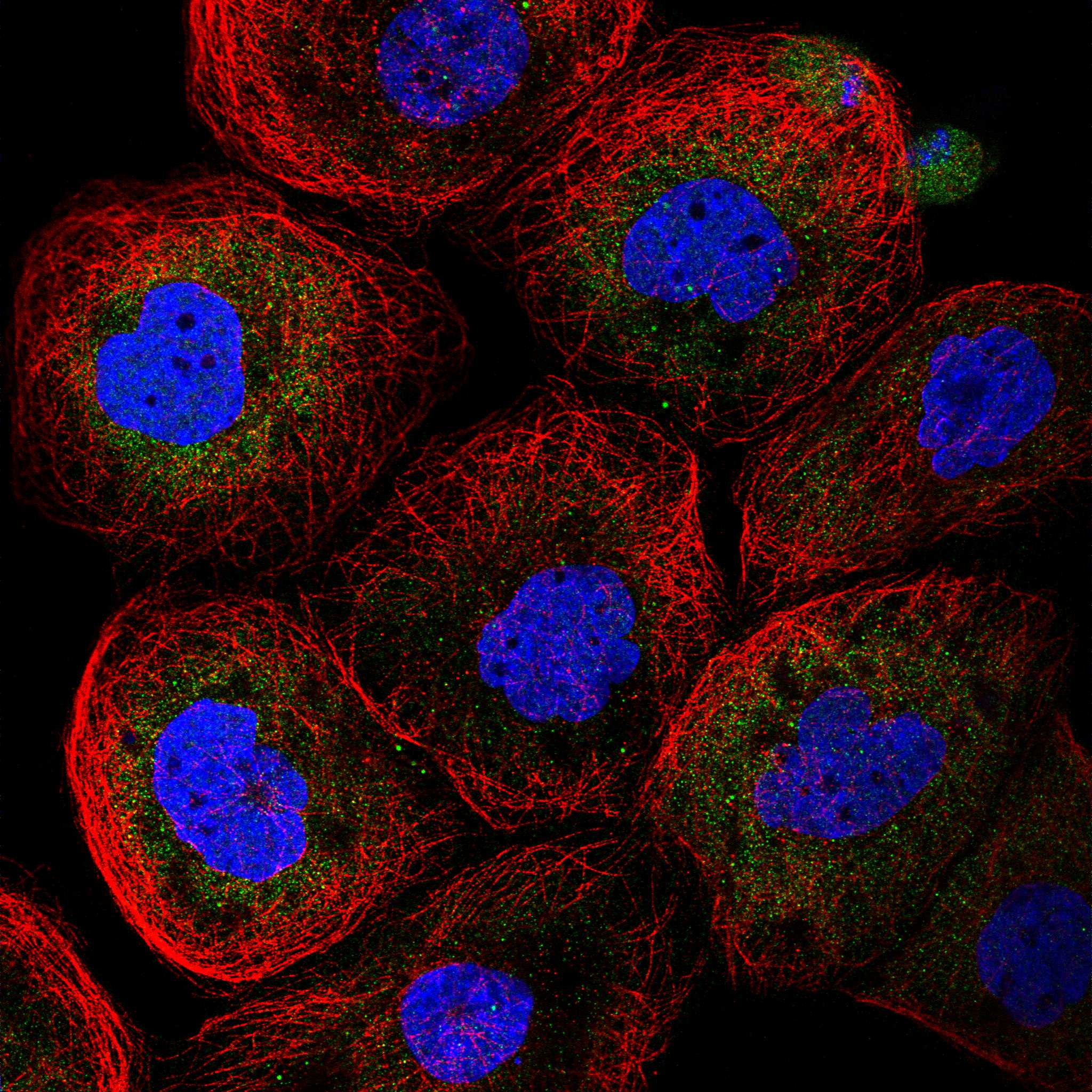
ICC-IF cell lines (HPA subcellular resource): HPA ICC-IF images show HSP90AA1 in A-431, U-251MG, U2OS, MCF-7, PC-3, RPTEC/TERT1, hTERT-RPE1 (serum starved), KOLF2.1J, HeLa BAC 6644, HeLa , with annotated localisation: Cytosol (supported) (HPA subcellular).
Technical controls: Include a no-primary, secondary-only control and an isotype control matched to the rabbit primary antibody’s host and clonality; use HSP90AA1 knockout material as a biological specificity control if available (caption: rabbit primary; standard IHC practice). For DAB detection, quench endogenous peroxidase and inspect background in the fallopian tube section (caption: peroxidase secondary and DAB; standard IHC practice).
⚠️Feasibility: A target-specific fixation window or fixation effect is unreported, and the selected M01103-2 tissue-IHC caption does not state a fixative (caption: fixative unreported). The reported paraffin-section procedure uses heat-mediated EDTA retrieval at pH 8.0, but its necessity for other specimens is unreported (caption: EDTA retrieval, pH 8.0). The supplied evidence does not establish whether frozen sections or IF are easier; ciliated cell bodies provide the positive readout, so assess epithelial background when scoring (HPA: High in fallopian tube ciliated cells; standard IHC practice).

HPA tissue IHC evidence for HSP90AA1

Comprehensive Human Protein Atlas IHC scoring per tissue (reliability: Approved — Low consistency between antibody staining and RNA expression data. Pending external verification. Significant inter-individual differences was observed between vagina RNA expression values.). Rows are taken directly from the HPA tissue chart — click any row's HPA link to view the source.

Positive expression · recommended positive controls

TissueCell typeLevelEvidenceSource
Fallopian tube Ciliated cells (cell body) High Protein (IHC) HPA →
Nasopharynx Ciliated cells (cell body) High Protein (IHC) HPA →
Testis Cells in seminiferous ducts High Protein (IHC) HPA →
Bronchus Ciliated cells (cell body) Medium Protein (IHC) HPA →
Duodenum Glandular cells Medium Protein (IHC) HPA →

Undetected expression · recommended negative controls

TissueCell typeLevelEvidenceSource
Adipose tissue Adipocytes Not detected Protein (IHC) HPA →
Bone marrow Hematopoietic cells Not detected Protein (IHC) HPA →
Caudate Glial cells Not detected Protein (IHC) HPA →
Esophagus Squamous epithelial cells Not detected Protein (IHC) HPA →
Heart muscle Cardiomyocytes Not detected Protein (IHC) HPA →
Section 3

Advanced HSP90AA1 IHC Tips

Troubleshoot HSP90AA1 staining in paraffin sections by checking retrieval, cell compartment, antibody specificity, and how staining is scored.

What retrieval should I use when HSP90AA1 staining is weak?
Start with heat-mediated retrieval in EDTA at pH 8.0 (datasheet M01103-2). The selected paraffin-section example used this retrieval before overnight incubation at 4°C with the catalog antibody at 1:50 (datasheet M01103-2). If signal remains weak, compare retrieval durations on adjacent sections while keeping antibody dilution, detection, and development conditions constant; record any tissue damage alongside signal. Include a known positive section, such as fallopian tube ciliated cells, and assess cell-body staining rather than treating stronger background as improved retrieval (HPA: High in fallopian tube ciliated cells; HPA: cytoplasmic expression in several tissues).
Could fixation explain weak or uneven HSP90AA1 staining?
Target-specific fixation sensitivity is unknown: the selected paraffin-section caption does not state a fixative (datasheet M01103-2). Record the actual fixative and fixation duration for each specimen, then compare sections processed and stained together before attributing a weak result to fixation. Keep retrieval at EDTA pH 8.0 and antibody incubation conditions consistent during that comparison (datasheet M01103-2). Check tissue preservation and whether staining follows recognizable cell bodies; the expected tissue-IHC pattern is cytoplasmic in several tissues, but that pattern does not establish how HSP90AA1 responds to any particular fixative (HPA: cytoplasmic expression in several tissues).
How should I assess nuclear or membrane-like HSP90AA1 staining?
Use cytoplasmic cell-body staining as the main tissue-IHC reference pattern (HPA: cytoplasmic expression in several tissues). HSP90AA1 is also annotated in the nucleus, cytoplasm, cell membrane, and mitochondrion, while supported subcellular imaging places it mainly in the cytosol and rates nucleoplasmic localisation uncertain (UniProt P07900 subcellular location; HPA: cytosol supported, nucleoplasm uncertain). Review nuclear staining against a counterstain and neighboring cytoplasm on the same section, then confirm a reproducible pattern across specimens before assigning a compartment. A membrane outline alone needs particular scrutiny because HSP90AA1 has no annotated transmembrane segment (UniProt P07900 topology).
Can the antibody distinguish HSP90AA1 isoforms or modified epitopes?
Do not assign staining to a particular HSP90AA1 isoform without an antibody epitope and isoform-specific validation; the record lists 2 isoforms (UniProt P07900 isoforms). HSP90AA1 has annotated phosphorylation and acetylation sites, including phosphoserine 231, but those annotations alone do not show that this antibody detects a modification (UniProt P07900 modified residues). Check the catalog antibody's stated immunogen or epitope, if available, before interpreting differential staining as a molecular change. Compare the staining pattern with an independently validated antibody or orthogonal measurement when isoform identity or modification state is central to the conclusion.
How can I compare HSP90AA1 IF with the chromogenic IHC pattern?
Treat IF as a separate assay and compare its cell-body pattern with the cytoplasmic pattern reported for tissue IHC (HPA: cytoplasmic expression in several tissues). Multiplex HSP90AA1 with a marker for the cell population being assessed; for example, identify ciliated cells when examining fallopian tube, where their cell bodies show high tissue-IHC staining (HPA: High in fallopian tube ciliated cells). Select a fluorophore channel after checking the specimen's autofluorescence, and include single-stain controls when assessing overlap. For intracellular epitopes, assess permeabilisation because HSP90AA1 is mainly cytosolic in subcellular imaging; the antibody's epitope location is not supplied here (HPA: cytosol supported).
How do I separate diffuse HSP90AA1 signal from chromogenic background?
First inspect whether DAB follows intact cell bodies; the reported tissue-IHC pattern is cytoplasmic across several tissues (HPA: cytoplasmic expression in several tissues). The selected example used 10% goat serum blocking, a 1:50 rabbit primary overnight at 4°C, and peroxidase-based DAB detection (datasheet M01103-2). Compare a primary-omission control and a matched positive section, and check whether color develops in tissue edges, damaged regions, or cells expected to lack signal. If background dominates, optimize blocking, washes, antibody concentration, and DAB development one variable at a time; use a peroxidase block as a general chromogenic-IHC control.
What is a defensible way to score HSP90AA1 IHC? ⚠ ANSWER MARKED FOR VERIFICATION
Define the cell population and compartment before scoring; tissue IHC describes cytoplasmic expression in several tissues (HPA: cytoplasmic expression in several tissues). For cellular staining, record the percentage of positive cells and an intensity category, then calculate an H-score only when the same thresholds and scoring rules apply across sections. Normalize counts to the number of eligible intact cells, or report positive-cell density per mm² of viable tissue when cell counts cannot be compared directly. Keep exposure-independent chromogenic development, counterstaining, and section selection consistent, and report excluded necrotic or damaged areas separately from negative cells.
What makes an HSP90AA1 positive result convincing?
Look for reproducible cell-body staining in morphologically intact cells, with cytoplasm as the principal tissue-IHC reference compartment (HPA: cytoplasmic expression in several tissues). Fallopian tube and nasopharynx ciliated cells are high-staining examples, whereas adipocytes are listed as not detected; use these observations as context, not absolute specimen-level controls (HPA: High in fallopian tube and nasopharynx ciliated cells; HPA: Not detected in adipocytes). Recheck isolated nuclear or sharp membrane-only signal against the reported compartment evidence (HPA: cytosol supported, nucleoplasm uncertain; UniProt P07900 topology). Edge staining, necrotic regions, or color persisting after primary omission warrant an artefact check, including endogenous peroxidase in this DAB workflow.
Boster reagents

Best HSP90AA1 / Heat shock protein HSP 90-alpha IHC Antibodies

Catalog images show HSP90AA1 staining in paraffin-embedded human and mouse tissues and IF in HeLa cells; listed reactivity spans human, mouse, rat and monkey (catalog image captions; catalog reactivity).

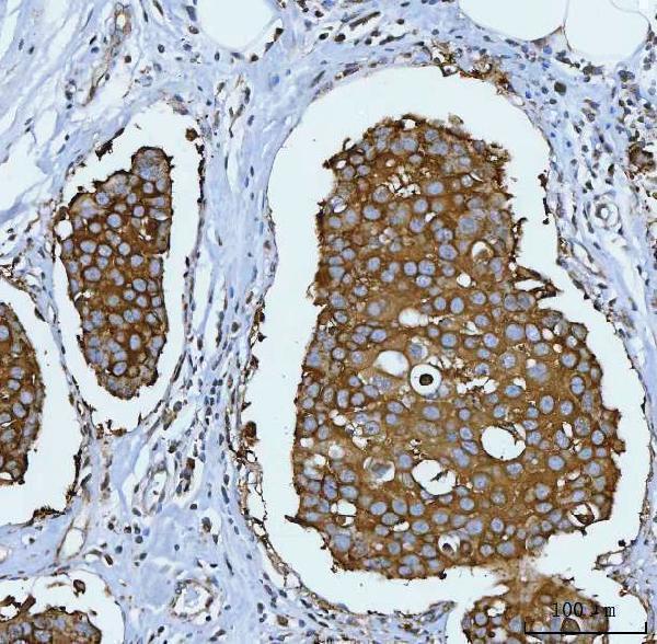
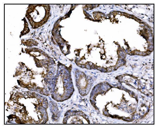
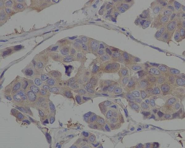
Real IHC data IHC analysis of Hsp90 using anti-Hsp90 antibody (M01103-2). Hsp90 was detected in a paraffin-embedded section of human brest cancer tissue. Heat mediated antigen retrieval was performed in EDTA buffer (pH 8.0, epitope retrieval solution). The tissue section was blocked with 10% goat serum. The tissue section was then incubated with 1:50 rabbit anti-Hsp90 Antibody (M01103-2) overnight at 4°C. Peroxidase Conjugated Goat Anti-rabbit IgG was used as secondary antibody and incubated for 30 minutes at 37°C. The tissue section was developed using HRP Conjugated Rabbit IgG Super Vision Assay Kit (Catalog # SV0002) with DAB as the chromogen.
Anti-Hsp90 alpha + beta HSP90AA1 Rabbit Monoclonal Antibody
Cat # M01103-2
Real IHC data IHC analysis of Hsp90 alpha using anti-Hsp90 alpha antibody (M01103-4). Hsp90 alpha was detected in paraffin-embedded section of human cervical cancer tissue. Heat mediated antigen retrieval was performed in EDTA buffer (pH8.0, epitope retrieval solution). The tissue section was blocked with 10% goat serum. The tissue section was then incubated with 2μg/ml mouse anti-Hsp90 alpha Antibody (M01103-4) overnight at 4°C. Biotinylated goat anti-mouse IgG was used as secondary antibody and incubated for 30 minutes at 37°C. The tissue section was developed using Strepavidin-Biotin-Complex (SABC) (Catalog # SA1021) with DAB as the chromogen.
Anti-Hsp90 alpha Antibody ® (monoclonal, 6B5)
Cat # M01103-4
Real IHC data IHC analysis of Hsp90 Alpha using anti-Hsp90 Alpha antibody (PB9089). Hsp90 Alpha was detected in a paraffin-embedded section of human intestinal cancer tissue. Heat mediated antigen retrieval was performed in EDTA buffer (pH 8.0, epitope retrieval solution). The tissue section was blocked with 10% goat serum. The tissue section was then incubated with 1 μg/ml rabbit anti-Hsp90 Alpha Antibody (PB9089) overnight at 4°C. Biotinylated goat anti-rabbit IgG was used as secondary antibody and incubated for 30 minutes at 37°C. The tissue section was developed using Strepavidin-Biotin-Complex (SABC) (Catalog # SA1022) with DAB as the chromogen.
Anti-Hsp90 alpha/HSP90AA1 Antibody ®
Cat # PB9089
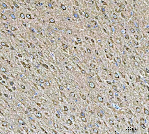
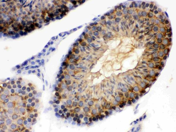
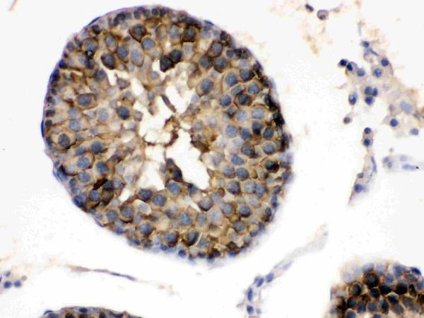
Real IHC data IHC analysis of Hsp90 alpha using anti-Hsp90 alpha antibody (PB9635). Hsp90 alpha was detected in a paraffin-embedded section of mouse testis tissue. Heat mediated antigen retrieval was performed in EDTA buffer (pH 8.0, epitope retrieval solution). The tissue section was blocked with 10% goat serum. The tissue section was then incubated with 1 μg/ml rabbit anti-Hsp90 alpha Antibody (PB9635) overnight at 4°C. Biotinylated goat anti-rabbit IgG was used as secondary antibody and incubated for 30 minutes at 37°C. The tissue section was developed using Strepavidin-Biotin-Complex (SABC) (Catalog # SA1022) with DAB as the chromogen.
Anti-Hsp90 alpha/HSP90AA1 Antibody ®
Cat # PB9635
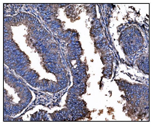
Real IHC data Immunohistochemical analysis of paraffin-embedded human breast cancer, using Hsp90 alpha antibody.
Anti-Hsp90 alpha HSP90AA1 Rabbit Monoclonal Antibody
Cat # M01103-1
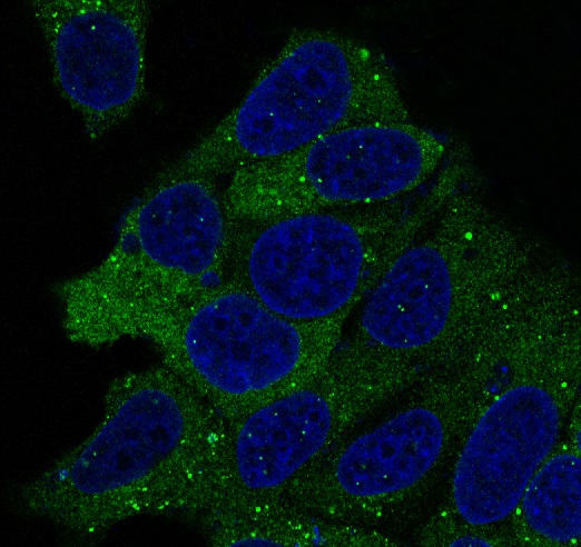
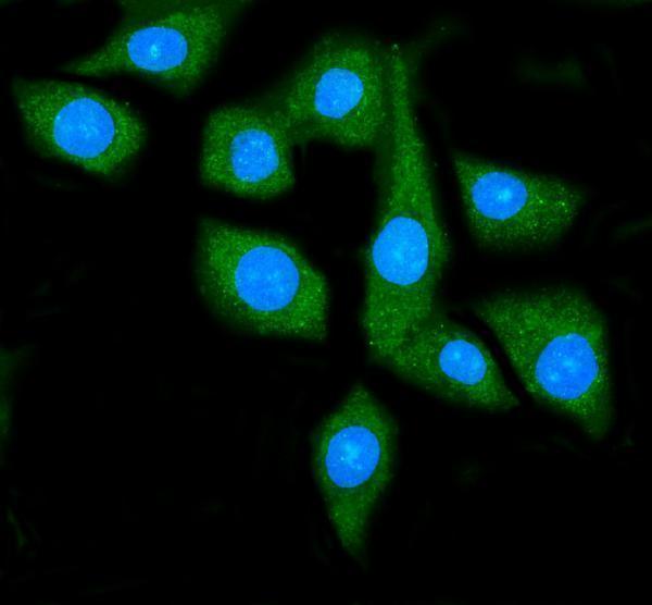
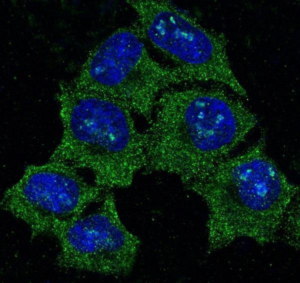
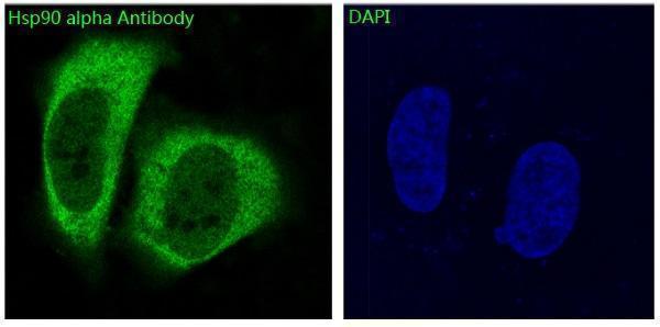
Real IF data Immunofluorescent analysis of Hela cells, using Hsp90 alpha Antibody .
Anti-Hsp90 alpha HSP90AA1 Rabbit Monoclonal Antibody
Cat # M01103-3

M01103-2 shows IHC in human breast cancer, M01103-4 in human cervical cancer, and PB9089 in human intestinal cancer (respective IHC captions). PB9635 shows IHC in mouse testis, M01103-1 in paraffin-embedded human breast cancer, and M01103-3 shows IF in HeLa cells (respective image captions).

Which to pick: For human tissue IHC, M01103-4 is an alpha-directed mouse monoclonal with a paraffin-section cervical cancer image; its IHC caption does not report the fixative (catalog title; M01103-4 IHC caption). For IF/ICC, M01103-3 is an alpha-directed rabbit monoclonal with a HeLa IF image and IF/ICC listed among its applications (catalog title; M01103-3 IF caption and applications). For IHC across species, PB9635 has paraffin-section images from human, mouse and rat testis; monkey is listed in its reactivity, but has no IHC image in the payload (PB9635 IHC captions; catalog reactivity).

Each figure is that product's own IHC / IF validation image from its datasheet.

References

  1. UniProt Consortium. UniProt entry P07900 (HS90A_HUMAN, Heat shock protein HSP 90-alpha).
  2. Human Protein Atlas. HSP90AA1 tissue IHC expression (reliability: Approved).
  3. Human Protein Atlas. HSP90AA1 subcellular location (ICC-IF): Mainly localized to the cytosol. In addition localized to the nucleoplasm..
  4. Human Protein Atlas. HSP90AA1 antibody validation summary (2 antibodies).
  5. HSP90AA1 restrains clear cell renal cell carcinoma progression by promoting CADM1 expression and suppressing the PI3K-AKT pathway through interaction with FBXO7. Cell death discovery 2026 — PMC12783301.
  6. Nephrotoxicity of Evodiamine in Mice: Mechanistic Insights from Integrated Network Toxicology and Transcriptomic Profiling. International journal of molecular sciences 2026 — PMC13164253.
  7. HSP90AA1 promotes lymphatic metastasis of hypopharyngeal squamous cell carcinoma by regulating epithelial-mesenchymal transition. Oncology research 2023 — PMC10398399.
  8. Construction and validation of a prognostic signature based on necroptosis-related genes in hepatocellular carcinoma. PloS one 2023 — PMC9934426.
  9. PubMed PMID:2780322 — UniProt-cited evidence.
  10. PubMed PMID:1368637 — UniProt-cited evidence.
  11. PubMed PMID:2527334 — UniProt-cited evidence.