This website uses cookies to ensure you get the best experience on our website.


- Table of Contents
Plan MAPK14 staining in paraffin sections using the cytoplasmic and nuclear tissue pattern reported by HPA (HPA tissue IHC). The guide covers antibody selection, staining controls and interpretation of total versus phospho-specific signal (UniProt; datasheet: A00176-2, A00176T180Y182).

Expected localisation, validated protocols, controls and antibodies — the at-a-glance facts below, then the full design guide.
| Expected localisation | General cytoplasmic and nuclear staining (HPA tissue IHC) | |
| Staining pattern | Cytoplasmic and nuclear staining in multiple cell types (HPA tissue IHC) | |
| Antigen retrieval | Citrate pH 6 HIER, heat-mediated (datasheet M00176-1) | |
| Positive control | Adrenal gland+4 more · see all | |
| Negative control | Adipose tissue+4 more · see all |
| Fixation | Matched tissue-IHC evidence does not establish the fixation claim. Validate the specimen-specific method before use. (selected-SKU IHC image M00176-1) | |
| Caveat | Total MAPK14 staining does not establish activation (UniProt) | |
| Regulation | Cytokines and stress activate MAPK14 (UniProt) | |
| Isoform / epitope | 5 isoforms; check epitope coverage for the chosen antibody (UniProt) |
Start with the catalog antibody’s citrate pH 6 IHC-P protocol (datasheet: M00176-1). The published methods below cover colorectal, liver, patient and mouse, and mouse carotid samples (PMC8818961; PMC7967205; PMC7519103; PMC6377391).
| Sample | Paraffin-embedded tissue sections; fixative not specified (datasheet M00176-1; sample unspecified) |
| Fixation | Image fixative and duration unreported (datasheet M00176-1); verify before use. |
| Sectioning | 4–5 µm sections on charged slides (standard) |
| Deparaffinisation | Xylene, graded ethanol series to water (standard) |
| Antigen retrieval | Heat retrieval: Citrate pH 6, 20 min (datasheet M00176-1) |
| Peroxidase block | 3% H2O2, 10 min, room temperature (standard) |
| Blocking | 10% goat serum (datasheet M00176-1) |
| Primary antibody | Rabbit monoclonal (clone EGC-13) anti-MAPK14, 1:50 recommended; image 1ug/ml (datasheet M00176-1) |
| Primary incubation | Overnight at 4 °C (datasheet M00176-1) |
| Detection | Streptavidin-biotin complex (SABC), DAB chromogen (datasheet M00176-1) |
| Counterstain | Hematoxylin, blue, dehydrate and mount (standard) |
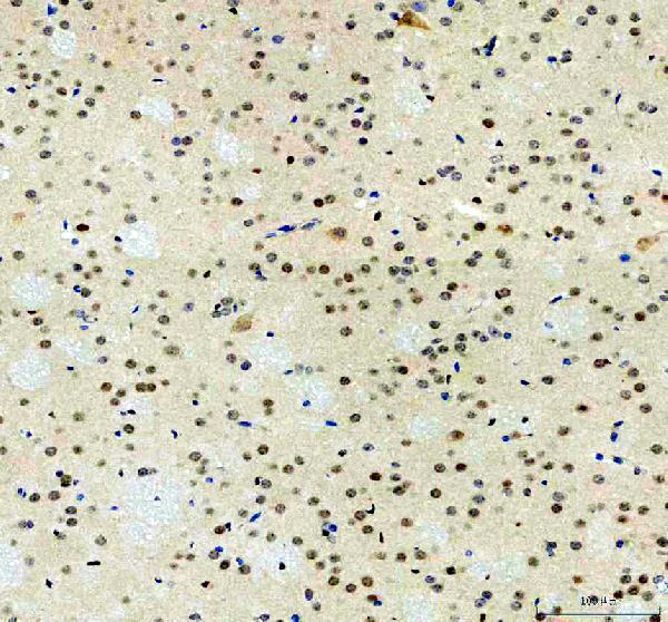
| Expected result | MAPK14-positive staining in glandular cells of adrenal gland (HPA tissue IHC: Medium). HPA tissue profile: General cytoplasmic and nuclear expression. No signal in the no-primary control. |
MAPK14 is expected in cytoplasm and nuclei, with tissue dependent staining in defined cell populations (UniProt Q16539 subcellular location; HPA tissue IHC: general cytoplasmic and nuclear expression). Its lack of a transmembrane segment makes a membrane restricted pattern unexpected (UniProt Q16539 topology). HPA rates the tissue IHC profile Approved, with medium consistency between antibody staining and RNA expression; interpret individual cells and controls accordingly (HPA tissue IHC: reliability).
| Cytoplasmic and nuclear signal in cerebral cortical neurons or bone marrow hematopoietic cells. | This fits the reported compartments and medium staining in those cell populations (UniProt Q16539 subcellular location; HPA tissue IHC: cerebral cortex and bone marrow). Compare cells within the section; the HPA rating describes observed staining, not a guaranteed intensity for every specimen (HPA tissue IHC: Approved, medium consistency). |
| Signal appears exclusively along cell borders, with little cytoplasmic or nuclear staining. | A membrane restricted pattern conflicts with the reported intracellular locations and absence of a transmembrane segment (UniProt Q16539 subcellular location and topology). Check the antibody and detection controls before assigning it to MAPK14; a cell border signal alone does not establish target localization (general IHC practice). |
| Strong signal appears in adipocytes or smooth muscle cells. | HPA reports MAPK14 as not detected in those cell populations (HPA tissue IHC: adipose tissue and smooth muscle). Investigate antibody cross reactivity or endogenous detection activity with appropriate controls (general IHC practice). A discordant result needs validation because HPA's Approved profile has only medium staining–RNA consistency (HPA tissue IHC: reliability). |
| Color spreads across tissue or appears similarly in cells and surrounding spaces. | That distribution is difficult to assign to the reported cytoplasmic and nuclear compartments (UniProt Q16539 subcellular location; HPA tissue IHC: profile). Review the no primary control, blocking, washes and chromogen development to distinguish background from cell associated staining (general IHC practice). Do not score a diffuse deposit as a positive cell (general IHC practice). |
| No signal is seen in cerebral cortical neurons on a test section. | HPA reports medium neuronal staining in cerebral cortex, so absence warrants a technical check (HPA tissue IHC: cerebral cortex). Confirm that the expected cells are present, then review the antibody's IHC-P instructions and detection controls (general IHC practice). An HPA positive example is a reference pattern, not proof that every specimen must stain (HPA tissue IHC: Approved, medium consistency). |
| Compartment and topology | MAPK14 is reported in cytoplasm and nucleus and has no transmembrane segment (UniProt Q16539 subcellular location and topology). Assess staining inside identifiable cells; a membrane restricted result requires scrutiny rather than automatic acceptance as the expected pattern (general IHC practice). |
| Tissue and cell context | HPA lists medium staining in cortical neurons, marrow hematopoietic cells and several epithelial or glandular populations, but low staining in lung alveolar cells and no detection in adipocytes (HPA tissue IHC: positive, low and negative lists). Select comparison regions by cell type, not tissue name alone (general IHC practice). |
| Evidence strength and antibody choice | The tissue profile is Approved with medium staining–RNA consistency; CAB010285 and CAB040578 are IHC Approved, while HPA051825 is ICC Supported with no IHC status supplied (HPA tissue IHC: reliability; HPA antibodies). These labels do not establish that every antibody will reproduce every listed tissue pattern (HPA antibody validation summary). |
| Isoforms and modifications | UniProt lists 5 isoforms and multiple modified residues, including phosphorylation and acetylation (UniProt Q16539 isoforms and modified residues). The payload gives no epitope for the IHC antibodies, so neither isoform coverage nor modification dependent staining can be inferred from these entries (HPA antibodies; UniProt Q16539). |
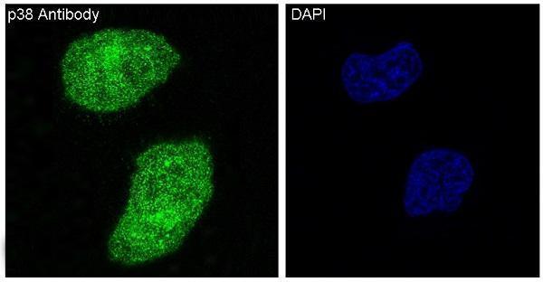
| IF localization, separate application | HPA ICC-IF places MAPK14 mainly in supported nuclear speckles, with supported cytosolic localization (HPA subcellular ICC-IF). This higher resolution IF observation can inform interpretation, but an IHC paraffin section need not resolve speckles (general microscopy practice); the supplied IHC evidence describes general nuclear and cytoplasmic staining (HPA tissue IHC: profile). |
| Situation | Likely cause | Next action |
|---|---|---|
| No staining in an HPA positive cell population. | The expected cells may be absent, or the IHC workflow or detection may have failed (general IHC practice); HPA reports medium staining in cortical neurons and marrow hematopoietic cells (HPA tissue IHC: positive list). | Verify the cell population, run a known positive section, and review the IHC-validated antibody's instructions plus detection controls (general IHC practice). Avoid declaring a specimen negative from a failed control (general IHC practice). |
| Diffuse color obscures cell boundaries. | Background from blocking, washing or chromogen development can obscure intracellular signal (general IHC practice). | Compare a no primary control and adjust the general IHC blocking, washing or development workflow as indicated (general IHC practice). Score only identifiable cellular staining in the reported compartments (HPA tissue IHC: profile; general IHC practice). |
| Adipocytes or smooth muscle cells stain prominently. | These populations are listed as not detected, so cross reactivity or endogenous detection activity is plausible (HPA tissue IHC: adipose tissue and smooth muscle; general IHC practice). | Check no primary and detection controls, then verify the antibody's IHC validation before interpreting the signal as MAPK14 (general IHC practice; HPA antibodies: IHC status). |
| Only cell borders stain. | The pattern conflicts with MAPK14's reported cytoplasmic and nuclear location and lack of a transmembrane segment (UniProt Q16539 subcellular location and topology). | Inspect control sections and detection artifacts, and seek reproducible intracellular staining in an HPA listed cell population before calling the result positive (general IHC practice; HPA tissue IHC: positive list). |
| Staining varies between cell types within one section. | HPA reports different levels by cell population, including medium cortical neuronal staining, low lung alveolar staining and no detection in adipocytes (HPA tissue IHC: positive, low and negative lists). | Record compartment and intensity separately for each identifiable cell type, using the corresponding HPA cell population as the comparison (HPA tissue IHC: tissue entries; general IHC practice). |
| IF/ICC Q: Must a paraffin IHC section show nuclear speckles? | HPA's supported nuclear speckle observation comes from ICC-IF, whereas its tissue IHC profile reports general nuclear and cytoplasmic expression (HPA subcellular ICC-IF; HPA tissue IHC: profile). | A: Judge IHC by recognizable intracellular staining and its cell type; consult the separate IF/ICC guide for that application (HPA tissue IHC: profile; general microscopy practice). |


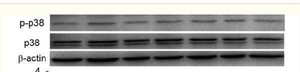
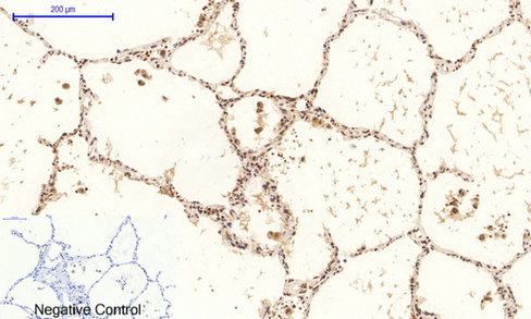
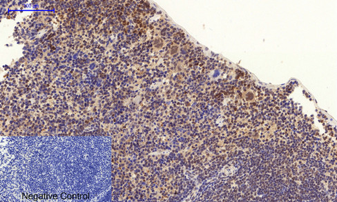
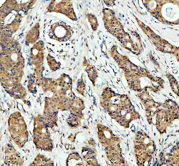


Comprehensive Human Protein Atlas IHC scoring per tissue (reliability: Approved — Medium consistency between antibody staining and RNA expression data.). Rows are taken directly from the HPA tissue chart — click any row's HPA link to view the source.
| Tissue | Cell type | Level | Evidence | Source |
|---|---|---|---|---|
| Adrenal gland | Glandular cells | Medium | Protein (IHC) | HPA → |
| Bone marrow | Hematopoietic cells | Medium | Protein (IHC) | HPA → |
| Cerebral cortex | Neuronal cells | Medium | Protein (IHC) | HPA → |
| Cervix | Squamous epithelial cells | Medium | Protein (IHC) | HPA → |
| Duodenum | Glandular cells | Medium | Protein (IHC) | HPA → |
| Tissue | Cell type | Level | Evidence | Source |
|---|---|---|---|---|
| Adipose tissue | Adipocytes | Not detected | Protein (IHC) | HPA → |
| Caudate | Glial cells | Not detected | Protein (IHC) | HPA → |
| Parathyroid gland | Glandular cells | Not detected | Protein (IHC) | HPA → |
| Prostate | Glandular cells | Not detected | Protein (IHC) | HPA → |
| Smooth muscle | Smooth muscle cells | Not detected | Protein (IHC) | HPA → |
Troubleshoot MAPK14 staining in paraffin sections by checking retrieval, controls, compartment assignment and cell-level scoring before interpreting signal as pathway activity.
IHC images show human brain, lung, and breast cancer paraffin sections and mouse and rat brain paraffin sections (catalog IHC captions); IF images show HeLa cells (catalog IF captions).
M00176-1 has human brain paraffin IHC and HeLa IF images (catalog image captions); A00176T180Y182 has human lung paraffin IHC and HeLa IF images (catalog image captions). A00176-2 has human breast cancer, mouse brain, and rat brain paraffin IHC images (catalog IHC captions); M00176 has a HeLa IF image and lists ICC as an application (catalog IF caption; catalog applications).
Which to pick: For tissue IHC, choose A00176-2 when its documented EDTA pH 8.0 retrieval and 2 μg/ml primary incubation suit the experiment (A00176-2 IHC caption). For IF/ICC, M00176 is a rabbit monoclonal with a HeLa IF image and listed ICC use (catalog clone and applications; M00176 IF caption). For IHC across human, mouse, and rat samples, A00176-2 has paraffin-section images in all three species; the captions do not report the fixative (A00176-2 IHC captions).