This website uses cookies to ensure you get the best experience on our website.
- Table of Contents
131 Citations 17 Q&As
72 Citations 14 Q&As
63 Citations 16 Q&As
60 Citations 5 Q&As
24 Citations
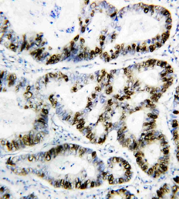
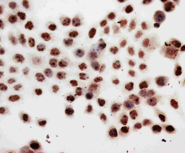
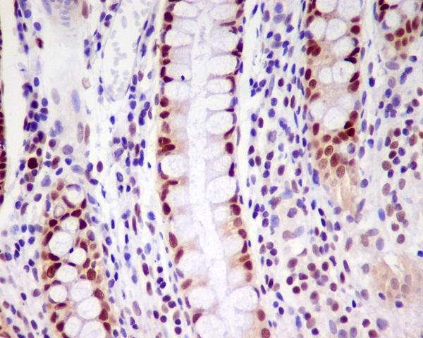
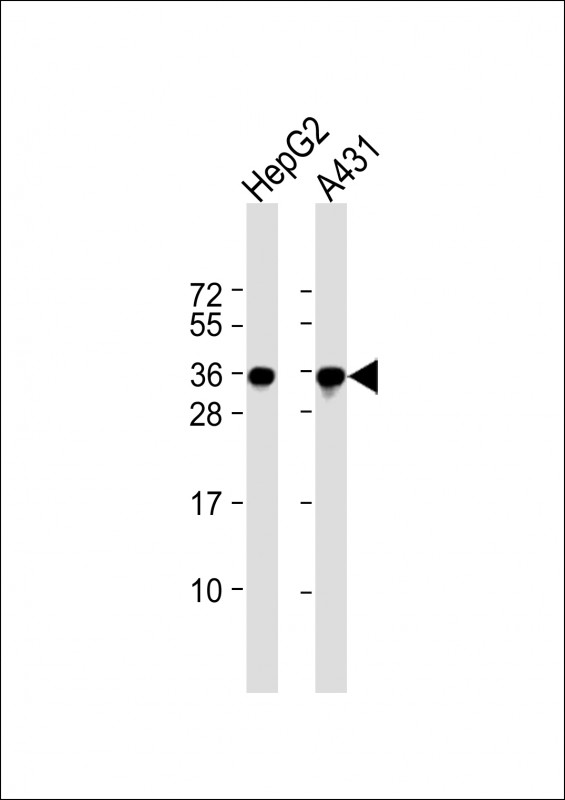
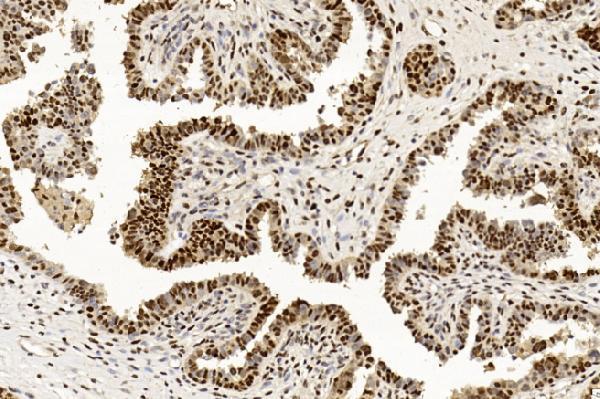
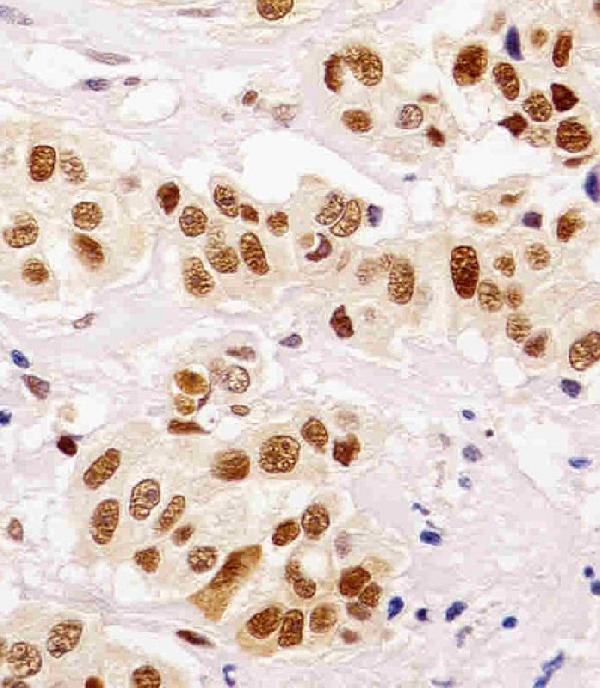
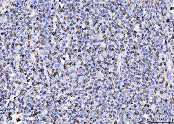
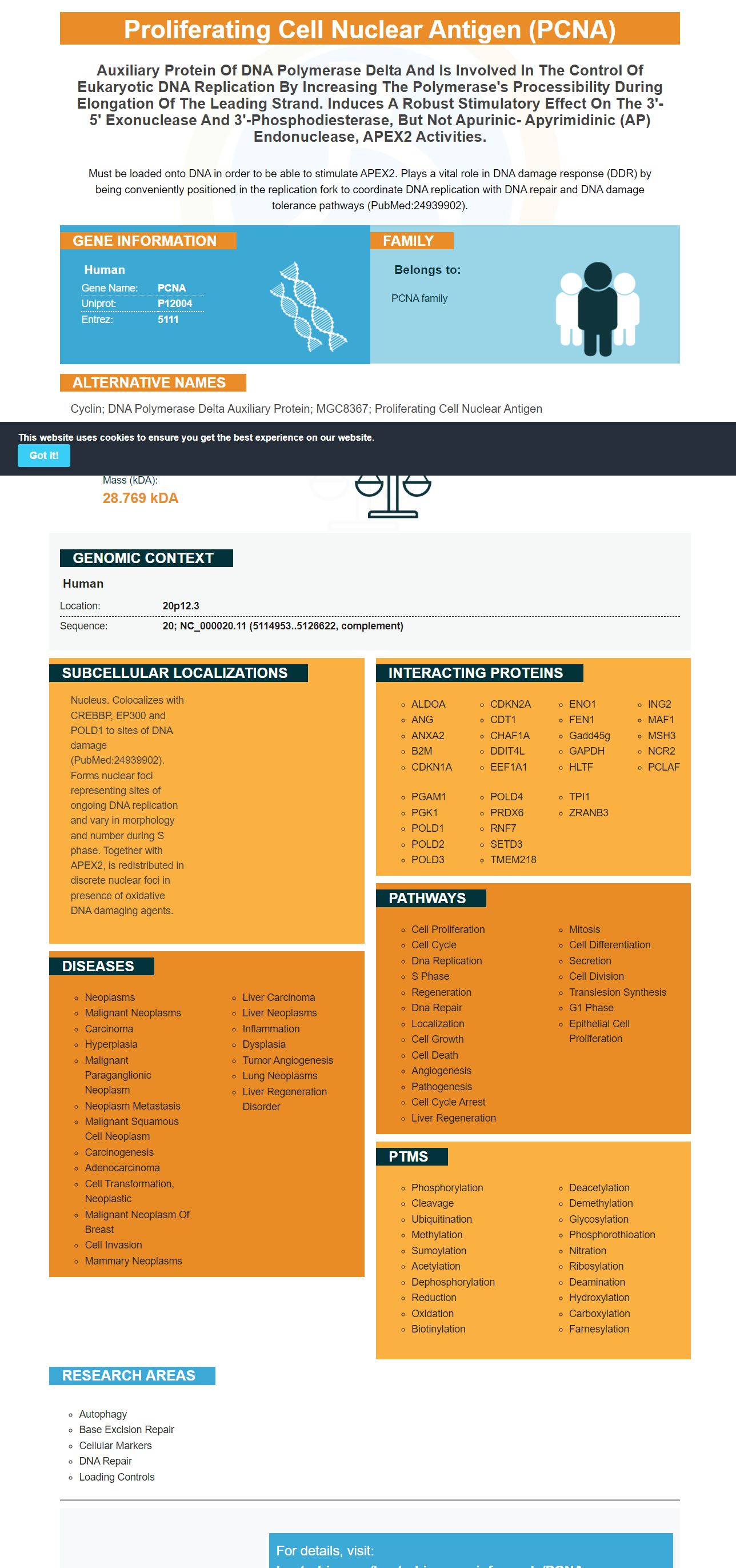
Facts about Proliferating cell nuclear antigen.
Must be loaded onto DNA in order to be able to stimulate APEX2. Plays a vital role in DNA damage response (DDR) by being conveniently positioned in the replication fork to coordinate DNA replication with DNA repair and DNA damage tolerance pathways (PubMed:24939902).
Human | |
---|---|
Gene Name: | PCNA |
Uniprot: | P12004 |
Entrez: | 5111 |
Belongs to: |
---|
PCNA family |
cyclin; DNA polymerase delta auxiliary protein; MGC8367; proliferating cell nuclear antigen
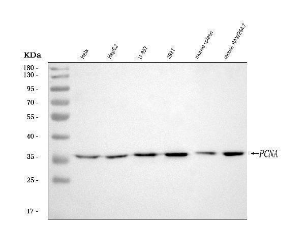
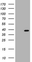
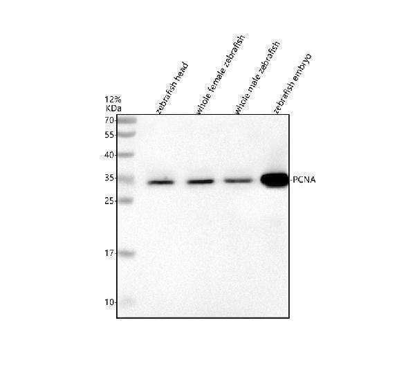
Mass (kDA):
28.769 kDA
Human | |
---|---|
Location: | 20p12.3 |
Sequence: | 20; NC_000020.11 (5114953..5126622, complement) |
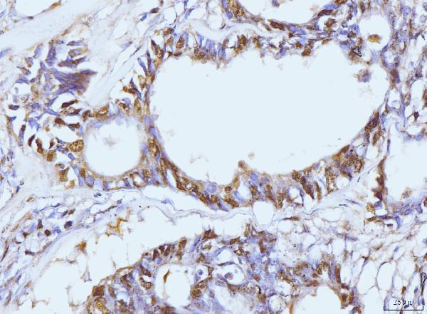
Nucleus. Colocalizes with CREBBP, EP300 and POLD1 to sites of DNA damage (PubMed:24939902). Forms nuclear foci representing sites of ongoing DNA replication and vary in morphology and number during S phase. Together with APEX2, is redistributed in discrete nuclear foci in presence of oxidative DNA damaging agents.
In addition to PCNA as an primary antibody researchers may also submit results on specific species or applications that use this marker. Boster scientists could also be qualified to receive credits for their research. The PCNA Marker can be used to all researchers across the globe. Boster Bio has more information. We are available to answer any questions you may have. For more information, please visit our FAQ section. In addition to providing answers to your questions, we also provide a product credit application form.
Anti-PCNA antibodies are immunofluorescent proteins. They have been used to detect lymphocytes that are malignant and activated. Boster Bio's anti PCNA Marker is monoclonal and reacts to human mouse, rat, and PCNA. It is stable and is able to store at temperatures ranging from -20 to 4 degrees Celsius. The reagent is made up of mouse ascites fluid, BSA and a preservative (0.01mg N3). Additionally, you can purchase a blocking peptide that will enhance the sensitivity your anti-PCNA antibody.
The amount of K164 is the main factor that determines PCNA ubiquitination. It can also occur in cells that aren't capable of repair of photoproducts. The mechanism that triggers ubiquitination of PCNA may involve a DNA structure that is caused by lesions. Ubiquitination of PCNA is not dependent on DNA replication, but instead, it may be triggered by transcription complexes that build up at the location of damage.
LAM cells can have different shapes. The majority of cells that were strongly positive had a nucleus with a small size that was spindle-shaped. However, there was no evidence of a significant connection between PCNA and HMB45 antibody reactivity. The relationship between the two markers could suggest that they may have different mechanisms for recognizing antigens. If your patient is positive for PCNA the Anti-PCNA Marker in Boster Bio can be used to detect LAM cells.
In contrast the cytoplasmic extract from adult cardiomyocytes can also influence the levels of PCNA protein in S phase nuclei of mammalian cells. In these cells the inhibition of cdk2 function inhibits the cytoplasmic PCNA protein expression, whereas the reduction of Rb did not alter the levels of PCNA. This result was in line with the Boster Bio study.
We recently isolated primary antibodies against PCNA from Citrus depressa. The antibody was then diluted to 1:50 or 1:100 , and incubated for 12 hours at 4degC. The HRP-conjugated goat monoclonal antibodies were then used as an after-care. The images were captured using a microscope and an electronic camera. In the next section we will describe the techniques we employed to create the antibodies.
We first showed that DNCREB exerts an influence on PCNA promoter activity. In addition, we utilized cells transfected with pCMV PCNA-CR133 as the vector for transfection. The cells were then subjected to Western analysis of the total protein in the cell. We also found that the over-expression of PCNA influences CREB effects, and pCMV-PCNA transfection resulted in twofold increase in PCNA protein levels.
Surprisingly, PCNA overexpression reverses the effects of DNCREB upon cell survival. In addition, co-transfected cells with constitutively-expressed PCNA protein exhibited restored radiosensitivity to wild-type levels. In this way, primary antibodies against PCNA have a role in determining the longevity of cells. The relationship between PCNA levels, radiosensitivity and PCNA is not yet understood. The current understanding of PCNA's role in cancer repair can help us better understand this disease.
The protein is also involved in TLS as well, and PCNA is essential for DNA replication. These TLS polymerases interact with PCNA and alter their specificity and coordination. With the help of an REV3L APIM construct, we have found that the full-length protein is colocalized with PCNA in replication foci. In contrast the colocalization between REV3L-YFP with PCNA is significantly diminished when using an APIM CFP construct.
When conserving PCNA, it is essential to store the antibody at the temperature that doesn't exceed 4deg C. The antibody is stable at this temperature for a few weeks. To use it immediately, it should be diluted, and then stored at the temperature of -20deg C. After the antibody is diluted it should be placed in the freezer. The storage procedure may vary and the antibody might need to be stored in a refrigerator for a few weeks.
The nucleus is the most common storage area for this protein. A PCNA marker can be found in DNA samples from a range of ages. This marker is vital for the synthesis of DNA, and plays a variety of roles in cell division and the progression of the cell cycle. It is highly conserved across species and is a crucial part of DNA replication. Its storage in the nucleus can be an effective tool for studying the progress of cell growth.
PCNA immunoreactivity in paraffin-embedded tissues is affected by the nature of cell, duration, temperature and fixative. Different types of cells have different requirements for packaging. This means that antibodies might not recognize PCNA within the nucleus. Finally, dilution of antibodies could affect PCNA expression in the nucleus. All of these elements can cause PCNA immunereactivity difficult or even impossible.
The PCNA Marker is an essential nuclear protein used in prognostication, immunohistochemistry, and other diagnostic procedures. Its function is crucial for DNA replication, mismatch repair, and even DNA excision. It also connects to CDK inhibitor p21 and to structure-specific endonucleases, such as Fen1 and XPG. It is a homotrimer that is located in the nucleus. It is a crucial element in DNA replication and plays an important role in the regulation of cell cycle and leading the synthesis of strands.
This protein is vital for cell growth. It is expressed at lower levels than active cells in cells that are quiescent. It is present in the G1/S cycle of cells and functions as an accessory protein to DNA polymerase alpha. PCNA also regulates cell growth by creating a ring around part of the DNA. This is why PCNA expression is a biomarker that can be used to determine cell expansion, and its evaluation can have a significant impact on the prognosis of cancer patients. Anti-PCNA antibodies can be used to identify subpopulations which are in S-phase or cycling.
The PCNA Marker may be utilized in retrospective as well in prospective studies. Researchers may be able to make use of its capability to be preserved in paraffin-embedded and fixed tissues to test its potential as a marker for human tumors. Once we know its function, we will understand the benefits and limitations of PCNA's use as a proliferation marker. Further the PCNA marker could be used in clinical trials to screen patients for a variety cancers, ranging from a single tumor to an entire tumor.
PCNA is an essential element of the cell cycle. It must be properly defined molecularly and biochemically in order to ensure the accuracy of cell expansion as an indicator. It is essential to distinguish it from other proteins that impact DNA synthesis such as cyclins. To better determine its role in cell expansion, PCNA must be identified as an important target for various cell cycle regulators. Many of these inhibitors inhibit the interaction between PCNA and DNA synthesizing.
Although it is not known what role PCNA plays, it is a hub protein which is involved in a variety of cellular interactions. Pep8, for example, targets intracellular PCNA and triggers cell death. The R11-NLSCPP is not designed to kill cancer cells and may also harm non-cancer proliferating tissue. The applications of PCNA Marker are numerous, and are growing rapidly.
PMID: 2882507 by Almendral J.M., et al. Cloning and sequence of the human nuclear protein cyclin: homology with DNA-binding proteins.
PMID: 2565339 by Travali S., et al. Structure of the human gene for the proliferating cell nuclear antigen.
*Showing only the more recent 20. More publications can be found for each product on its corresponding product page