
Click image to see more details
-

-
-
-
-
+25
Product Info Summary
| SKU: | MA1083 |
|---|---|
| Size: | 100 μg/vial |
| Reactive Species: | Human, Mouse, Rat |
| Host: | Mouse |
| Application: | IHC, ICC, WB |
Customers Who Bought This Also Bought

Product info
Product Name
Anti-PCNA Antibody (Monoclonal, PC 10)
SKU/Catalog Number
MA1083
BM0104 is an alternative SKU for this antibody, used in previous lots.
Size
100 μg/vial
Form
Lyophilized
Description
Boster Bio Anti-PCNA Antibody (Monoclonal, PC 10) catalog # MA1083. Tested in IHC, ICC, WB applications. This antibody reacts with Human, Mouse, Rat.
Storage & Handling
Store at -20˚C for one year from date of receipt. After reconstitution, at 4˚C for one month. It can also be aliquotted and stored frozen at -20˚C for six months. Avoid repeated freeze-thaw cycles.
Cite This Product
Anti-PCNA Antibody (Monoclonal, PC 10) (Boster Biological Technology, Pleasanton CA, USA, Catalog # MA1083)
Host
Mouse
Contents
Mouse IgG in stabilizing components, 1.2% sodium acetate and 0.01mg NaN3.
Clonality
Monoclonal
Clone Number
PC 10
Isotype
Mouse IgG2a
Immunogen
Protein A fusion protein.
Cross-reactivity
No cross-reactivity with other proteins
Reactive Species
MA1083 is reactive to Pcna in Human, Mouse, Rat
Observed Molecular Weight
35 kDa
Calculated molecular weight
28.7 kDa
Background of Pcna
Proliferating cell nuclear antigen (PCNA) was originally identified by immunofluorescence as a nuclear protein whose appearance correlated with the proliferative state of the cell. PCNA/cyclin has been localized by in situ hybridization to the short arm of human chromosome 20 with a peak of grains over band 20p13. PCNA gene is present in single copy and has 6 exons. It spans 4,961 bp. Synthesis of the nuclear protein cyclin and DNA in quiescent mouse fibroblasts is coordinately induced by serum and purified growth factors. PCNA controls establishment of sister chromatid cohesion during S phase.
Antibody Validation
Boster validates all antibodies on WB, IHC, ICC, Immunofluorescence, and ELISA with known positive control and negative samples to ensure specificity and high affinity, including thorough antibody incubations.
Application & Images
Applications
MA1083 is guaranteed for IHC, ICC, WB Boster Guarantee
Recommend Dilution
| Application | Dilution | Species |
|---|---|---|
| Immunohistochemistry (Paraffin-embedded Section) | 0.4-1μg/ml | Human, mouse, rat |
| Immunocytochemistry | 1μg/ml | Human, mouse, rat, - |
| Immunohistochemistry (Frozen Section) | 0.4-1μg/ml | Human, mouse, rat, - |
| Western blot | 2μg/ml | Human, mouse, rat |
Tested application
Suggested blocking solution with 5% non-fat milk or BSA; (*)Recommended protein loading: 20-40 µg per lane
Use TE buffer pH 9.0 for antigen retrieval; (*) citrate buffer pH 6.0 is an alternative.
Validation Images & Assay Conditions
Click image to see more details
Western blot analysis of PCNA using anti-PCNA antibody (MA1083).
Electrophoresis was performed on a 5-20% SDS-PAGE gel at 70V (Stacking gel) / 90V (Resolving gel) for 2-3 hours. The sample well of each lane was loaded with 50ug of sample under reducing conditions.
Lane 1: human Caco-2 whole cell lysates,
Lane 2: human MDA-MB-231 whole cell lysates,
Lane 3: human Jurkat whole cell lysates,
Lane 4: human HT1080 whole cell lysates.
After Electrophoresis, proteins were transferred to a Nitrocellulose membrane at 150mA for 50-90 minutes. Blocked the membrane with 5% Non-fat Milk/ TBS for 1.5 hour at RT. The membrane was incubated with mouse anti-PCNA antigen affinity purified monoclonal antibody (Catalog # MA1083) at 0.5 μg/mL overnight at 4°C, then washed with TBS-0.1%Tween 3 times with 5 minutes each and probed with a goat anti-mouse IgG-HRP secondary antibody at a dilution of 1:10000 for 1.5 hour at RT. The signal is developed using an Enhanced Chemiluminescent detection (ECL) kit (Catalog # EK1001) with Tanon 5200 system. A specific band was detected for PCNA at approximately 35KD. The expected band size for PCNA is at 29KD.

Click image to see more details
IHC analysis of PCNA using anti-PCNA antibody (MA1083).
PCNA was detected in paraffin-embedded section of human Rectal cancer tissues. Heat mediated antigen retrieval was performed in citrate buffer (pH6, epitope retrieval solution) for 20 mins. The tissue section was blocked with 10% goat serum. The tissue section was then incubated with 1μg/ml mouse anti-PCNA Antibody (MA1083) overnight at 4°C. Biotinylated goat anti-mouse IgG was used as secondary antibody and incubated for 30 minutes at 37°C. The tissue section was developed using Strepavidin-Biotin-Complex (SABC)(Catalog # SA1021) with DAB as the chromogen.

Click image to see more details
IHC analysis of PCNA using anti-PCNA antibody (MA1083).
PCNA was detected in immunocytochemical section of human HELA Cell. Enzyme antigen retrieval was performed using IHC enzyme antigen retrieval reagent (AR0022) for 15 mins. The cells were blocked with 10% goat serum. And then incubated with 1μg/ml mouse anti-PCNA Antibody (MA1083) overnight at 4°C. Biotinylated goat anti-mouse IgG was used as secondary antibody and incubated for 30 minutes at 37°C. The section was developed using Strepavidin-Biotin-Complex (SABC)(Catalog # SA1021) with DAB as the chromogen.

Click image to see more details
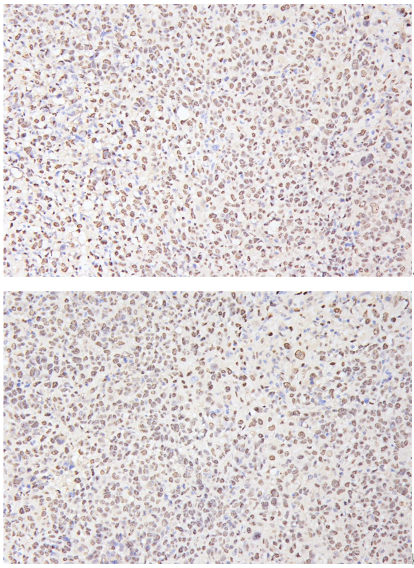
IHC analysis of PCNA using anti-PCNA antibody (MA1083).
PCNA was detected in paraffin-embedded section of HepG2 subcutaneous xenograft in nude mice tissues. Heat mediated antigen retrieval was performed in citrate buffer (pH6, epitope retrieval solution) for 20 mins. The tissue section was blocked with 10% goat serum. The tissue section was then incubated with 1:500 mouse anti-PCNA Antibody (MA1083) overnight at 4°C. Two-step IHC detection kit was used as secondary antibody and incubated for 30 minutes at 37°C. The tissue section was developed using Strepavidin-Biotin-Complex (SABC)(Catalog # SA1021) with DAB as the chromogen.

Click image to see more details
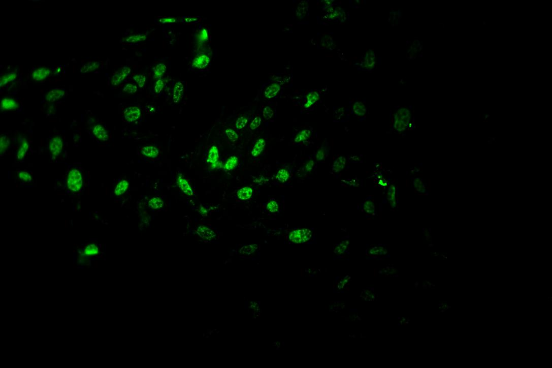
IF analysis of PCNA using anti-PCNA antibody (MA1083).
PCNA was detected in an immunocytochemical section of HepG2 cells. Enzyme antigen retrieval was performed using IHC enzyme antigen retrieval reagent (AR0022) for 15 mins. The cells were blocked with 10% goat serum. And then incubated with 1:1000 mouse anti-PCNA Antibody (MA1083) overnight at 4°C. DyLight®488 Conjugated Goat Anti-Rabbit IgG (BA1127) was used as secondary antibody at 1:500 dilution and incubated for 30 minutes at 37°C. The section was counterstained with DAPI. Visualize using a laser confocol.

Click image to see more details
KAZN overexpression inhibits cell proliferation and induces apoptosis.
(A and B) Western blot analysis (upper) and quantification (lower) of cleaved-caspase 3 (A) and PCNA (B) levels in A549 cells overexpressing KAZN.
(C and D) Western blot analysis (upper) and quantification (lower) of cleaved-caspase 3 (C) and PCNA (D) levels in NCI-H1299 cells overexpressing KAZN.
(E and F) EdU staining of A549 cells overexpressing KAZN and quantification of EdU-positive cells.
(G and H) EdU staining of NCI-H1299 cells overexpressing KAZN and quantification of EdU-positive cells. Scale bar: 200 μm.
Data were presented as mean ± SD from at least 3 independent experiments. p values were calculated using the unpaired Student’s t test. ∗p < 0.05.
Index in PubMed under a CC BY license. PMID: 41126879

Click image to see more details
Combination of PO and PD-1 blockade inhibited proliferation and induced apoptosis of tumor in vivo . (A) Paraffin sections of CT26 tumor tissues were analyzed by H&E staining (n = 3). (B) Expression and quantification of PCNA-positive staining in CT26 tumor tissues was examined by IHC using Image-Pro Plus 6.0 and in three random fields (n = 3). Scale bar, 50 μm. (C) TUNEL staining and the quantification of TUNEL-positive cells in CT26 tumor tissues (n = 3). Scale bar, 20 μm *p < 0.05, **p < 0.01, versus as indicated. ns, not significant.
Index in PubMed under a CC BY license. PMID: 39990679

Click image to see more details
knockdown of RIPK2 suppressed GC cell proliferation and apoptosis in vivo. (A-D) IHC was used to detect the expression of RIPK2 and PCNA in tumor section, and quantification. (E-F) TUNEL was performed to detect the apoptosis cells in tumor section, and the quantification of fluorescent intensity. * P < 0.05, ** P < 0.01.
Index in PubMed under a CC BY license. PMID: 38164277

Click image to see more details
FGF-2 impedes the AAD-induced anti-EC effect via FGFR1-ERK-MYC signaling. A Cell growth of human ECs receiving the conditioned medium of scrambled- or FGF2 shRNA-transfected NPC tumor cells ( n = 5 samples per group). B Representative micrographs of PCNA + proliferative cells and DAPI signals in ECs treated with vehicle or recombinant human FGF-2. Scale bar = 50 μm. Quantification of PCNA + signals in and human ECs ( n = 8 random fields per group). C Vehicle- or VEGF-treated ECs were challenged with or without sunitinib or FGF-2. Phosphorylation of AKT and ERK in ECs was detected. β-actin marks the loading level in each lane ( n = 3 samples per group). D QPCR quantification of Fgfr1 , Fgfr2 , Fgfr3 , and Fgfr4 mRNA levels in ECs ( n = 3 samples per group). E Vehicle- or FGF-2-treated ECs were challenged with or without various FGFR inhibitors. Phosphorylation of ERK in ECs was detected. β-actin marks the loading level in each lane ( n = 3 samples per group). F Downstream of VEGF signaling transcription factors were selected and detected in vehicle- or FGF-2-treated ECs. Heatmap of qPCR array screened out Myc as the highest upregulated transcription factor. G Correlation of FGF2 and MYC transcriptomic expression levels of human NPCs (NPC, n = 113 samples). Data was extracted from dataset GSE102349. H QPCR quantification of Myc mRNA levels in isolated CD31 + ECs from scrambled- or FGF2 shRNA-transfected NPC tumor tissues ( n = 3 samples per group). I QPCR quantification of Myc mRNA levels in various groups of ECs ( n = 3 samples per group). J Vehicle- or VEGF-treated ECs were treated with or without AAD or FGF-2. MYC expression in ECs was detected. β-actin marks the loading level in each lane ( n = 3 samples per group). K QPCR quantification of Myc mRNA levels in vehicle- or FGF-2-treated ECs, with or without various inhibitors ( n = 3 samples per group). L Diagram of ETS-binding site prediction. M ChIP detection of ETS binding to the Myc gene promoter. Nonimmune IgG and Myc exon 2 regions served as controls ( n = 3 samples per group). N QPCR quantification of EC proliferative marker Kdr , Plxnd1 , Ptgs2 , Robo4 in scrambled- or Myc siRNA-transfected ECs administrated with vehicle or FGF-2 ( n = 3 samples per group). * P < 0.05; ** P < 0.01; *** P < 0.001. NS not significant. Data presented as mean ± SD.
Index in PubMed under a CC BY license. PMID: 35985991

Click image to see more details
Immunohistochemistry staining of wound tissues on days 7 and 14. (A) Representative images for CD31, PCNA, α-SMA, and CD68 staining (scale bar = 100 μm). (B–E) Quantification of CD31, PCNA, α-SMA, and CD68 protein expressions, respectively. * p < 0.05, ** p < 0.01, *** p < 0.001 vs . control group; # p < 0.05, ## p < 0.01, ### p < 0.001 vs . CAH group as statistically significant.
Index in PubMed under a CC BY license. PMID: 35784748

Click image to see more details
Effect of asiaticoside on the transformation of fibroblasts into myofibroblasts, collagen deposition and fibroblast proliferation in vivo . Immunohistochemical staining of tissue around silicone rubber (SR) and carbon silicone rubber (C-SR) with α-SMA, vimentin, PCNA and COL-1A1 (100X).
Index in PubMed under a CC BY license. PMID: 35646845

Click image to see more details
Increase proliferation of muscle cells in tibialis anterior in DMD mice. (A) Immunohistochemical staining of muscle cells was performed with the PCNA antibody. PCNA positive cells are shown as brown (red arrow). The PCNA positive cells were counted under microscope. Numbers of PCNA positive cells were demonstrated in the visual field Data were presented at means ± SEM ( n = 6) with independent sample t -test. ** p < 0.01. (B) Gene expression of tibialis anterior cells in WT and DMD mice was detected by RT-PCR. Data are expressed as the mean ± SEM ( n = 6). ** p < 0.01.
Index in PubMed under a CC BY license. PMID: 35372342

Click image to see more details
Effects of GT3 on proliferation of muscle cells. (A) The tibialis anterior was extracted 48 h after GT3 treatment in 40-week-old WT and DMD mice. PCNA positive cells were stained with brown (red arrow, left panel). Numbers of PCNA positive cells were counted in each field. Data were presented at means ± SEM ( n = 6) with two-way ANOVA analysis. ** p < 0.01, *** p < 0.001. (B) Gene expression of tibialis anterior muscle cells was detected by RT-PCR after GT3 treatment. Data were presented at means ± SEM ( n = 6) with two-way ANOVA analysis. * p < 0.05, ** p < 0.01.
Index in PubMed under a CC BY license. PMID: 35372342

Click image to see more details
AIF blocks hypoxia-induced progression of PH in vitro and in vivo. A Hypoxia increased the viability of PASMCs after growth arrest for 24 h, and this effect was decreased by AIF (n = 4). B Pretreatment with an AIF overexpression plasmid blocked the effects of hypoxia on EdU incorporation in cells (n = 6). Scale bars: 50 μm. C Cell cycle analysis by flow cytometry indicated that hypoxia stimulated cell progression into G 2 /M + S phase, and this effect was inhibited by AIF overexpression (n = 3). D Effects of AIF on the expression of PCNA, Cyclin A and Cyclin D under hypoxia (n = 4–5). E Represents weight ratio of the right ventricular (RV)/left ventricular (LV) + Septum (n = 6); F Represents the right ventricular systolic pressure (RVSP) from mice (n = 5); G pulmonary artery velocity time integral (PAVTI), pulmonary artery acceleration time (PAAT) and left ventricular ejection fraction (LVEF) of the hypoxic mouse model infected with AAV5-NC and AAV5-AIF (n = 6). All data are presented as the means ± standard deviation. *p < 0.05; **p < 0.01; ***p < 0.001; Nor normoxia, Hyp hypoxia, NC negative control
Index in PubMed under a CC BY license. PMID: 35090552

Click image to see more details
AIF blocks hypoxia-induced pulmonary vascular remodeling in vivo. A Morphological analysis of the pulmonary artery was performed using HE staining and Masson staining, and the thickness of pulmonary vascular medium was measured by α-SMA staining (n = 5). B Increased proliferation of the pulmonary vascular cells was visualized by PCNA-positive staining per vascular area under hypoxia compared with exposure to normal conditions at the same time, these effects were reversed by the administration of AAV5-AIF (n = 3). C Homology analysis of the AIF gene among humans, mice and rats. Nor normoxia, Hyp hypoxia, NC negative control
Index in PubMed under a CC BY license. PMID: 35090552

Click image to see more details
SOX1 inhibits CCA cell proliferation in vitro and suppresses tumor growth in vivo. A TFK-1 and HUCCT-1 cells were transfected with NC-SOX1, LV-SOX1 and SOX1-KD for 72 h. SOX1 protein level was assessed by Western blotting. B Representative images of colony formation assay (left panel) and analysis of colony numbers (right panel). *p < 0.05, **p < 0.01, ****p < 0.0001. C Cell proliferation was assessed by CCK-8 assay. ***p < 0.001, ****p < 0.0001. D Above panel: Xenograft tumors at day 18 after implantation of NC-SOX1 or LV-SOX1 cells into the right flank of nude mice. Below panel: comparison of tumor volumes between NC-SOX1 and LV-SOX1 xenograft mice. **p < 0.01, ***p < 0.001. E Protein of xenograft tumors was extract and PCNA, BCL2, SOX1 was assessed by Western blotting. F Flow cytometry detected apoptotic cells after cells were transfected with LV-SOX1 and NC-SOX1
Index in PubMed under a CC BY license. PMID: 34876142

Click image to see more details
Mir-155-5p inhibits SOX1 leading to activation of the Raf/MEK/ERK pathway. A Cells were transfected with lentiviral negative control vector (NC-SOX1) or lentiviral SOX1 (LV-SOX1) for 72 h. Protein expressions of SOX1, HES1, PROX1, p-AKT, p-JNK, and p-P38 were examined by Western blot. B Above panel: protein levels of ERK and p-ERK in HUCCT-1 and TFK-1 cells transfected with miR-negative control (miR-NC), miR-155-5p-mimic (miR-155-5p), and miR-155-5p-inhibitor (miR-155-5pI). Below panel: TFK-1 and HUCCT-1 cells was treated with different concentrations of miR-155-5pI. The protein level of ERK and p-ERK were detected by Western blot. C Protein levels of central members of MAPK/ERK signaling (RAF, p-RAF, MEK, p-MEK, ERK and p-ERK) and downstream of ERK (PCNA) in TFK-1 and HUCCT-1 cells were detected by Western blot. D TFK-1 and HUCCT-1 cells was transfected with miR-negative control (miR-NC) and miR-15-5p inhibitor (miR-155-5pI), then protein levels of central members of MAPK/ERK signaling (RAF, p-RAF, MEK, p-MEK, ERK and p-ERK) and PCNA were detected by Western blot. E TFK-1 and HUCCT-1 cells was transfected with lenvisual carrying SOX1-RNAi (SOX1-KD), then protein levels of central members of MAPK/ERK signaling (RAF, p-RAF, MEK, p-MEK, ERK and p-ERK) and PCNA were detected by Western blot. F Cells were infected with NC-SOX1, LV-SOX1, LV-SOX1 + NC-ERK and LV-SOX1 + LV-ERK, and then detect changes in ERK and PCNA protein levels by western blot. G Protein levels of central members of MAPK/ERK signaling (RAF, p-RAF, MEK, p-MEK, ERK and p-ERK) was detected in xenograft tumor samples which had been transfected with NC-SOX1 and LV-SOX1
Index in PubMed under a CC BY license. PMID: 34876142

Click image to see more details
Immunohistochemical staining to evaluate the activity of carcinoma-associated fibroblast, the status of mediated epithelial-mesenchymal transition of cancer cells, as well as the proliferation and apoptosis of cancer cells. a H&E staining. b Anti-α-SMA staining. c Picrosirius red staining. d Anti-E-cadherin staining. e Anti-Vimentin staining. f Anti-Ki-67 staining. g Anti-PCNA staining. h Anti-cleaved-Caspase 3. For quantitative analysis, the integral optical density (IOD) of values the IHC stainings were calculated. * p < 0.05, ** p < 0.01, *** p < 0.001, and **** p < 0.0001 denote the significant difference relative to YM101 treatment. Scale bars, 100 μm. α-TGF-β: anti-TGF-β, α-PD-L1: anti-PD-L1
Index in PubMed under a CC BY license. PMID: 33593403

Click image to see more details
Representative images of different LAIR-1 immunohistochemistry staining intensities in OS tissues. The proportion of positively stained cells for LAIR-1 was calculated by assessing the entire image. Based on the LAIR-1 staining intensities in OS tumor samples, the staining patterns were categorized as follows: weak (+), moderate (++), and intense (+++). Upper panel, original magnification × 200; lower panel, original magnification × 400. b Kaplan–Meier plot of survival rates of patients with tumors exhibiting high (blue line) or low (red line) LAIR-1 expression; data were obtained using the R2 platform. c Western blotting for determining LAIR-1 expression and PCNA proliferation marker levels in HOS cells following LV-NC or LV-LAIR-1 lentivirus infection or without treatment (blank). β-actin was used as a loading control
Index in PubMed under a CC BY license. PMID: 32563267

Click image to see more details
Anti-TNBC efficacy of Nano-DOX in comparison with DOX. a Effects of Nano-DOX and DOX on the viability of 4T1 cells in vitro assayed by the CCK-8 test. b Effects of Nano-DOX and DOX on the proliferation of 4T1 cells in vitro assayed by CFSE staining. c , d Apoptosis of 4T1 cells after 24-h treatment of Nano-DOX or DOX assayed by annexin V immunofluorescent staining and FACS. e , f Size and weight of orthotopic 4T1 tumor xenografts in mice at the end of 3-week treatment of Nano-DOX or DOX. g Immunohistochemical staining of Ki67, PCNA (markers of cancer cell proliferation), and caspase 3 (marker of cancer cell apoptosis) in mouse orthotopic 4T1 tumor xenografts at the end of 3-week treatment of Nano-DOX or DOX. (Duration of Nano-DOX or DOX treatment was 24 h for the in vitro cell experiments.) In FACS analysis, geometric means were used to quantify fluorescence intensity. Values were mean ± SD (n = 3 for in vitro experiments and n = 8 for in vivo experiments, *p < 0.05, **p < 0.01)
Index in PubMed under a CC BY license. PMID: 31623629

Click image to see more details
hAECs facilitated endometrial recovery in the IUA mouse model. A IHC staining of vWF reflected the MVD of the endometrium. The microvessels, which were vWF-positive, are indicated by arrows in the figure. MVD was reduced in the IUA group and increased in the hAEC-treated group. B IHC staining showed that the expression of VEGF was higher in the hAEC-treated group than in the IUA group. C The expression of PCNA decreased in the IUA group and reached almost normal levels in the hAEC-treated group. a–c, scale bar = 100 μm; d–f, scale bar = 50 μm
Index in PubMed under a CC BY license. PMID: 31412924

Click image to see more details
hAECs facilitated endometrial recovery in the IUA mouse model. A According to IHC staining, the number of ER-positive cells was higher in the hAEC-treated group than in the IUA group. B There was no difference in PR expression among these three groups. C VEGF expression was semi-quantified, and the number of positive cells per field was calculated. D MVD was valued by counting microvascular vessels, which were vWF-positive. E – G . PCNA, ER, and PR expression levels were semi-quantified by calculating the percentage of positive cells per field (* p < 0.05; ** p < 0.01; *** p < 0.001; NS, p ≥ 0.05). a–c, scale bar = 100 μm; d–f, scale bar = 50 μm
Index in PubMed under a CC BY license. PMID: 31412924

Click image to see more details
hAECs promoted autophagy in hEnSCs in vitro. A The cell viability of H 2 O 2 -treated hEnSCs significantly decreased. B hEnSCs were cocultured with hAECs in a Transwell system. C After 2.5 h of H 2 O 2 treatment and another 24 h of culture, hEnSCs shrank severely, but hAEC coculture repaired the cell morphology of hEnSCs damaged by H 2 O 2 . D Western blot analysis showed that p62 expression increased significantly in H 2 O 2 -treated hEnSCs and decreased in hEnSCs cocultured with hAECs. The relative expression of LC3-II/LC3-I was decreased in H 2 O 2 -treated hEnSCs and increased in hEnSCs cocultured with hAECs. The expression level of ER was downregulated in H 2 O 2 -treated hEnSCs but upregulated in hEnSCs cocultured with hAECs. The expression of VEGF, PCNA, and PR did not change prominently. E The grayscale values of the western blots were evaluated. The ratios of LC3-II/LC3-I were standardized to those of the control group. The protein levels of p62, PCNA, VEGF, ER, and PR were normalized to that of β-tubulin ( n = 3; * p < 0.05; ** p < 0.01; *** p < 0.001; NS, p ≥ 0.05)
Index in PubMed under a CC BY license. PMID: 31412924

Click image to see more details
The effect of TEAS on irradiation-induced expression of Bax, Bcl-2 and PCNA proteins in ovary. a The protein expression levels of Bax, Bcl-2, PCNA were determined by western blot analysis. b – d Quantitative analysis of total proteins was represented using a bar graph. The data represent the mean ± SEM. * P < 0.05, ** P < 0.01 vs. control- group; # P < 0.05, ## P < 0.01 vs. IR 2D- group
Index in PubMed under a CC BY license. PMID: 31324205

Click image to see more details
PNU-282987 treatment prevents and reveres pulmonary vascular remodeling. (A) Representative images of hematoxylin and eosin (HE), immunostaining of alpha-smooth muscle actin (α-SMA) and proliferating cell nuclear antigen (PCNA) from the lung in the sham, MCT, prevention and treatment groups. HE shows the thickness of the pulmonary artery. Brown staining with α-SMA indicates pulmonary artery smooth muscle, whereas brown-stained cells with PCNA represent proliferating pulmonary artery smooth muscle. The arrow indicates PCNA positive cells of the pulmonary artery. Occlusion (%), %vessels and PCNA positive cell (%) were calculated in a millimeter from 10 separate images of different fields. Original magnification: x400. (B) Graph showing the percentage of the median thickness of the arteriole. (C) Graph showing percentage of muscularization after α-SMA immunostaining. (D) Graph showing the percentage of PCNA positive cells. The data are summarized as means ± SD. ∗ P < 0.05 versus the sham group and # P < 0.05 versus the MCT group. Results shown are from one experiment (sham group, n = 15; MCT group, n = 8; MCT+protective group, n = 13; MCT+treatment group, n = 11).
Index in PubMed under a CC BY license. PMID: 30863307

Click image to see more details
Pathway of phycocyanobilin-accelerated liver regeneration. A-E: Results of real-time quantitative PCR detection at 1, 2, 3 and 5 d after CCl 4 treament. A: The expression of HGF; B: The expression of TGF-α; C: The expression of TGF-β; D: The expression of TNF-α; E: The expression of IL-6; F: The western blotting result of PCNA, TNF-α, and cytochrome C in liver tissue. C1-C5 indicates the results of the control group from day 1 to day 5, D1-D5 indicates the results of the PCB group from day 1 to day 5. a P < 0.05, b P < 0.01 and d P < 0.001 vs control.
Index in PubMed under a CC BY license. PMID: 25987768

Click image to see more details
Tumorigenicity assay in nude mice. A : The growth rates of tumors formed from untransfected MFE-296 cells (MFE-296) and MFE-296 cells transfected with NC (MFE-296/NC) or shFOXA1 (MFE-296/shFOXA1). After injection, tumor volumes were calculated every seven days. B and C : Six weeks after injection of MFE-296, MFE-296-NC, and MFE-296-shFOXA1 cells, tumors were removed, and the tumor weights and volumes were determined. Arithmetic means and SD are shown. D : Staining with hematoxylin and eosin (H&E) or immunohistochemical staining for FOXA1, AR, Notch1, Hes1, Ki67, and PCNA in mouse tumor tissues (immunohistochemical staining, 200×). *p < 0.05 compared with the NC group.
Index in PubMed under a CC BY license. PMID: 24512546

Click image to see more details
Localization of transplanted cells and in situ proliferation at the infarct border in experimental model . (A-C) are images of coronal cerebral slices showing the impact of occlusion 6 hours post-occlusion, with the infarct appearing as pale area marked with broken line. Hematoxylin and eosin (H.E.) stain illustrates loss of relatively large cells and infiltration of small cells at the border of the infarct (D). Paired cellular profiles are seen peripheral to the infarct border or the infarct penumbra (E). At low magnification, a large number of transplanted bone marrow cells pre-labeled by PKH26 (red fluorescence) are present in the infarct penumbra 7 days post lesion (F). The labeled cells are small and may occur in cluster (G). No fluorescent cells exist in the cerebral cortex of mice received vehicle infusion (H). In situ cell division reflected by immunoreactivity of proliferating cell nuclear antigen (PCNA) occurs predominantly at the infarct border (I) and penumbra (J), appearing as brown immunoreactive nuclei in hematoxylin counter-stained section.
Index in PubMed under a CC BY license. PMID: 20973978

Click image to see more details
Identification of PKH26-labeled cells with proliferating cell nuclear antigen and doublecortin after transplantation . Colocalization of PKH26-labeled bone marrow cells with proliferating cell nuclear antigen (PCNA) (A-D) and doublecortin (DCX) (E-G) around the infarct penumbra 7 and 14 days following transplantation. PKH26 and PCNA double-labeled cells are small and occur in pair or small cluster (C, D). PKH26 and DCX double-labeled cells have round or oval somata with visible neuronal processes. PKH26 fluorescence appears weaker in these double-labeled cells relative to those seen at 7 days post cell transplantation. Scale bar (in B) = 200 μm for A, B and 50 μm for C-G.
Index in PubMed under a CC BY license. PMID: 20973978
Specific Publications For Anti-PCNA Antibody (Monoclonal, PC 10) (MA1083)
Loading publications
Recommended Resources
Here are featured tools and databases that you might find useful.
- Boster's Pathways Library
- Protein Databases
- Bioscience Research Protocol Resources
- Data Processing & Analysis Software
- Photo Editing Software
- Scientific Literature Resources
- Research Paper Management Tools
- Molecular Biology Software
- Primer Design Tools
- Bioinformatics Tools
- Phylogenetic Tree Analysis
Customer Reviews
Have you used Anti-PCNA Antibody (Monoclonal, PC 10)?
Share your experimental results or join a short interview to earn up to $1,000 in product credits or other rewards.
2 Reviews For Anti-PCNA Antibody (Monoclonal, PC 10)
The PCNA antibody (Cat# MA1083) showed clear and distinct nuclear positivity in HepG2-derived subcutaneous tumors, indicating reliable detection.
Excellent
| SKU | MA1083 |
|---|---|
| Application | Immunohistochemistry |
| Sample | HepG2 subcutaneous xenograft in nude mice |
| Sample Processing Description | HepG2 cells were expanded and then implanted subcutaneously into nude mice. After 2 weeks, tumors formed, which were excised, fixed in formalin for 48 hours, and processed for paraffin embedding and sectioning. |
| Other Reagents | Goat serum, DAB chromogen solution |
| Primary Antibody | PCNA Antibody (Monoclonal, PC 10) |
| Primary Incubation | 1:500, overnight at 4 ℃ |
| Secondary Antibody | Two-step IHC detection kit |
| Secondary Incubation | 30 min in 37℃ |
| Detection | Image system: Leica DM2500 |
| Results Summary | PCNA is a key marker for cell proliferation studies; however, besides being a proliferation marker, it is a core component of the DNA replication and repair complex. Its long half-life and involvement in DNA damage repair mean that PCNA positivity does not solely indicate cell proliferation but may also reflect cells responding to DNA damage. Combined with Ki67 staining, it can accurately indicate proliferative status. In our experiment, the staining was clear, with distinct nuclear positivity. |
Fengtong Wang, The First Affiliated Hospital of Xinjiang Medical University
Verified customer
Submitted 2026-03-31
Highly prone to positive signals, with precise localization of expression
Excellent
| SKU | MA1083 |
|---|---|
| Application | Immunofluorenscence |
| Sample | Mouse MC-8 cells |
| Sample Processing Description | 4% paraformaldehyde for 15 minutes |
| Primary Antibody | Anti-PCNA Antibody (Monoclonal, PC 10) |
| Primary Incubation | 1:1000, overnight at 4 ℃ |
| Blocking Agent | Goat serum |
| Secondary Antibody | DyLight 550-conjugated goat anti-rabbit antibody. |
| Secondary Incubation | Incubate at room temperature for 1 hour |
| Detection | Laser confocal microscopy |
| Results Summary | I will purchase Boster products again and recommend them to my classmates and colleagues. |
Guangyu Mei, Tongji University
Verified customer
Submitted 2025-10-17
Customer Q&As
Have a question?
Find answers in Q&As, reviews.
Can't find your answer?
Submit your question
17 Customer Q&As for Anti-PCNA Antibody (Monoclonal, PC 10)
Question
Is this MA1083 anti-PCNA antibody (Monoclonal, PC 10) reactive to the isotypes of PCNA?
Verified Customer
Verified customer
Asked: 2020-04-03
Answer
The immunogen of MA1083 anti-PCNA antibody (Monoclonal, PC 10) is Protein A fusion protein. Could you tell me which isotype you are interested in so I can help see if the immunogen is part of this isotype?
Boster Scientific Support
Answered: 2020-04-03
Question
We have seen staining in rat bone marrow lung. Are there any suggestions? Is anti-PCNA antibody (Monoclonal, PC 10) supposed to stain bone marrow lung positively?
Verified Customer
Verified customer
Asked: 2020-01-16
Answer
From literature bone marrow lung does express PCNA. From Uniprot.org, PCNA is expressed in oocyte, skeletal muscle, bone marrow lung, fetal brain cortex, placenta, liver, among other tissues. Regarding which tissues have PCNA expression, here are a few articles citing expression in various tissues:
Bone marrow, and Lung, Pubmed ID: 15489334
Fetal brain cortex, Pubmed ID: 9305916
Liver, Pubmed ID: 24275569
Placenta, Pubmed ID: 12522211
Skeletal muscle, Pubmed ID: 14702039
Boster Scientific Support
Answered: 2020-01-16
Question
Does anti-PCNA antibody (Monoclonal, PC 10) MA1083 work for WB with oocyte?
Verified Customer
Verified customer
Asked: 2019-12-11
Answer
According to the expression profile of oocyte, PCNA is highly expressed in oocyte. So, it is likely that anti-PCNA antibody (Monoclonal, PC 10) MA1083 will work for WB with oocyte.
Boster Scientific Support
Answered: 2019-12-11
Question
Please see the WB image, lot number and protocol we used for oocyte using anti-PCNA antibody (Monoclonal, PC 10) MA1083. Please let me know if you require anything else.
Verified Customer
Verified customer
Asked: 2019-10-16
Answer
Thank you very much for the data. Our lab team are working to resolve this as quickly as possible, and we appreciate your patience and understanding! You have provided everything we needed. Please let me know if there is anything you need in the meantime.
Boster Scientific Support
Answered: 2019-10-16
Question
I am looking for to test anti-PCNA antibody (Monoclonal, PC 10) MA1083 on human oocyte for research purposes, then I may be interested in using anti-PCNA antibody (Monoclonal, PC 10) MA1083 for diagnostic purposes as well. Is the antibody suitable for diagnostic purposes?
Verified Customer
Verified customer
Asked: 2019-09-16
Answer
The products we sell, including anti-PCNA antibody (Monoclonal, PC 10) MA1083, are only intended for research use. They would not be suitable for use in diagnostic work. If you have the means to develop a product into diagnostic use, and are interested in collaborating with us and develop our product into an IVD product, please contact us for more discussions.
Boster Scientific Support
Answered: 2019-09-16
Question
I was wanting to use your anti-PCNA antibody (Monoclonal, PC 10) for WB for human oocyte on frozen tissues, but I want to know if it has been tested for this particular application. Has this antibody been tested and is this antibody a good choice for human oocyte identification?
Verified Customer
Verified customer
Asked: 2019-08-06
Answer
As indicated on the product datasheet, MA1083 anti-PCNA antibody (Monoclonal, PC 10) has been validated for IHC, ICC, WB on human, mouse, rat tissues. We have an innovator award program that if you test this antibody and show it works in human oocyte in IHC-frozen, you can get your next antibody for free.
Boster Scientific Support
Answered: 2019-08-06
Question
Do you have a BSA free version of anti-PCNA antibody (Monoclonal, PC 10) MA1083 available?
Verified Customer
Verified customer
Asked: 2019-07-12
Answer
Thank you for your recent telephone inquiry. I can confirm that some lots of this anti-PCNA antibody (Monoclonal, PC 10) MA1083 are BSA free. For now, these lots are available and we can make a BSA free formula for you free of charge. It will take 3 extra days to prepare. If you require this antibody BSA free again in future, please do not hesitate to contact me and I will be pleased to check which lots we have in stock that are BSA free.
Boster Scientific Support
Answered: 2019-07-12
Question
I was wanting to use using your anti-PCNA antibody (Monoclonal, PC 10) for liver regeneration studies. Has this antibody been tested with western blotting on human hela? We would like to see some validation images before ordering.
Verified Customer
Verified customer
Asked: 2019-06-21
Answer
Thank you for your inquiry. This MA1083 anti-PCNA antibody (Monoclonal, PC 10) is validated on human hela, jurkat whole cell lysates. It is guaranteed to work for IHC, ICC, WB in human, mouse, rat. Our Boster guarantee will cover your intended experiment even if the sample type has not been be directly tested.
Boster Scientific Support
Answered: 2019-06-21
Question
Is a blocking peptide available for product anti-PCNA antibody (Monoclonal, PC 10) (MA1083)?
Verified Customer
Verified customer
Asked: 2019-06-21
Answer
We do provide the blocking peptide for product anti-PCNA antibody (Monoclonal, PC 10) (MA1083). If you would like to place an order for it please contact support@bosterbio.com and make a special request.
Boster Scientific Support
Answered: 2019-06-21
Question
Would anti-PCNA antibody (Monoclonal, PC 10) MA1083 work on primate IHC with skeletal muscle?
Verified Customer
Verified customer
Asked: 2018-03-19
Answer
Our lab technicians have not tested anti-PCNA antibody (Monoclonal, PC 10) MA1083 on primate. You can run a BLAST between primate and the immunogen sequence of anti-PCNA antibody (Monoclonal, PC 10) MA1083 to see if they may cross-react. If the sequence homology is close, then you can perform a pilot test. Keep in mind that since we have not validated primate samples, this use of the antibody is not covered by our guarantee. However we have an innovator award program that if you test this antibody and show it works in primate skeletal muscle in IHC, you can get your next antibody for free.
Boster Scientific Support
Answered: 2018-03-19
Question
I see that the anti-PCNA antibody (Monoclonal, PC 10) MA1083 works with WB, what is the protocol used to produce the result images on the product page?
Verified Customer
Verified customer
Asked: 2017-07-10
Answer
You can find protocols for WB on the "support/technical resources" section of our navigation menu. If you have any further questions, please send an email to support@bosterbio.com
Boster Scientific Support
Answered: 2017-07-10
Question
We are currently using anti-PCNA antibody (Monoclonal, PC 10) MA1083 for rat tissue, and we are happy with the ICC results. The species of reactivity given in the datasheet says human, mouse, rat. Is it true that the antibody can work on canine tissues as well?
Verified Customer
Verified customer
Asked: 2017-05-26
Answer
The anti-PCNA antibody (Monoclonal, PC 10) (MA1083) has not been validated for cross reactivity specifically with canine tissues, though there is a good chance of cross reactivity. We have an innovator award program that if you test this antibody and show it works in canine you can get your next antibody for free. Please contact me if I can help you with anything.
Boster Scientific Support
Answered: 2017-05-26
Question
My team were satisfied with the WB result of your anti-PCNA antibody (Monoclonal, PC 10). However we have been able to see positive staining in skeletal muscle nucleus using this antibody. Is that expected? Could you tell me where is PCNA supposed to be expressed?
Verified Customer
Verified customer
Asked: 2017-05-12
Answer
According to literature, skeletal muscle does express PCNA. Generally PCNA expresses in nucleus. Regarding which tissues have PCNA expression, here are a few articles citing expression in various tissues:
Bone marrow, and Lung, Pubmed ID: 15489334
Fetal brain cortex, Pubmed ID: 9305916
Liver, Pubmed ID: 24275569
Placenta, Pubmed ID: 12522211
Skeletal muscle, Pubmed ID: 14702039
Boster Scientific Support
Answered: 2017-05-12
Question
My question regarding product MA1083, anti-PCNA antibody (Monoclonal, PC 10). I was wondering if it would be possible to conjugate this antibody with biotin. I would need it to be without BSA or sodium azide. I am planning on using a buffer exchange of sodium azide with PBS only. Would there be problems for me to conjugate the antibody and store it in -20 degrees in small aliquots?
N. Banerjee
Verified customer
Asked: 2017-01-04
Answer
We do not advise storing this antibody with PBS buffer only in -20 degrees. If you want to store it in -20 degrees it is best to add some cryoprotectant like glycerol. If you want carrier free MA1083 anti-PCNA antibody (Monoclonal, PC 10), we can provide it to you in a special formula with trehalose and/or glycerol. These molecules will not interfere with conjugation chemistry and provide a good level of protection for the antibody from degradation. Please be sure to specify this in your purchase order.
Boster Scientific Support
Answered: 2017-01-04
Question
We appreciate helping with my inquiry over the phone. Here are the WB image, lot number and protocol we used for oocyte using anti-PCNA antibody (Monoclonal, PC 10) MA1083. Let me know if you need anything else.
K. Moore
Verified customer
Asked: 2016-05-09
Answer
We appreciate the data. You have provided everything we needed. Our lab team are working to resolve your inquiry as quickly as possible, and we appreciate your patience and understanding! Please let me know if there is anything you need in the meantime.
Boster Scientific Support
Answered: 2016-05-09
Question
We purchased anti-PCNA antibody (Monoclonal, PC 10) for IHC on skeletal muscle a few years ago. I am using mouse, and We intend to use the antibody for ICC next. My question regards examining skeletal muscle as well as oocyte in our next experiment. Could you please give me some suggestion on which antibody would work the best for ICC?
G. Huang
Verified customer
Asked: 2014-08-29
Answer
I looked at the website and datasheets of our anti-PCNA antibody (Monoclonal, PC 10) and I see that MA1083 has been tested on mouse in both IHC and ICC. Thus MA1083 should work for your application. Our Boster satisfaction guarantee will cover this product for ICC in mouse even if the specific tissue type has not been validated. We do have a comprehensive range of products for ICC detection and you can check out our website bosterbio.com to find out more information about them.
Boster Scientific Support
Answered: 2014-08-29
Question
Will MA1083 anti-PCNA antibody (Monoclonal, PC 10) work on parafin embedded sections? If so, which fixation method do you recommend we use (PFA, paraformaldehyde, other)?
T. Singh
Verified customer
Asked: 2013-03-04
Answer
It shows on the product datasheet, MA1083 anti-PCNA antibody (Monoclonal, PC 10) as been validated on WB. It is best to use PFA for fixation because it has better tissue penetration ability. PFA needs to be prepared fresh before use. Long term stored PFA turns into formalin, as the PFA molecules congregate and become formalin.
Boster Scientific Support
Answered: 2013-03-04
