Click image to see more details
-

-
-
-
-
+9
Product Info Summary
| SKU: | A00125 |
|---|---|
| Size: | 100 μg/vial |
| Reactive Species: | Human, Mouse, Rat |
| Host: | Rabbit |
| Application: | ELISA, Flow Cytometry, IF, IHC, ICC, WB |
Customers Who Bought This Also Bought

Product info
Product Name
Anti-PCNA Antibody Picoband®
SKU/Catalog Number
A00125
Size
100 μg/vial
Form
Lyophilized
Description
Boster Bio Anti-PCNA Antibody Picoband® catalog # A00125. Tested in ELISA, Flow Cytometry, IF, IHC, ICC, WB applications. This antibody reacts with Human, Mouse, Rat. The brand Picoband indicates this is a premium antibody that guarantees superior quality, high affinity, and strong signals with minimal background in Western blot applications. Only our best-performing antibodies are designated as Picoband, ensuring unmatched performance.
Storage & Handling
Store at -20˚C for one year from date of receipt. After reconstitution, at 4˚C for one month. It can also be aliquotted and stored frozen at -20˚C for six months. Avoid repeated freeze-thaw cycles.
Cite This Product
Anti-PCNA Antibody Picoband® (Boster Biological Technology, Pleasanton CA, USA, Catalog # A00125)
Host
Rabbit
Contents
Each vial contains 4 mg Trehalose, 0.9 mg NaCl and 0.2 mg Na2HPO4.
Clonality
Polyclonal
Isotype
Rabbit IgG
Immunogen
E.coli-derived human PCNA recombinant protein (Position: M1-S261).
Cross-reactivity
No cross-reactivity with other proteins.
Reactive Species
A00125 is reactive to PCNA in Human, Mouse, Rat
Observed Molecular Weight
36 kDa
Calculated molecular weight
28.8 kDa
Background of PCNA
Proliferating cell nuclear antigen (PCNA) is a DNA clamp that acts as a processivity factor for DNA polymerase δ in eukaryotic cells and is essential for replication. It is mapped to 20p12.3. The protein encoded by this gene is found in the nucleus and is a cofactor of DNA polymerase delta. The encoded protein acts as a homotrimer and helps increase the processivity of leading strand synthesis during DNA replication. In response to DNA damage, this protein is ubiquitinated and is involved in the RAD6-dependent DNA repair pathway. Two transcript variants encoding the same protein have been found for this gene. Pseudogenes of this gene have been described on chromosome 4 and on the X chromosome.
Antibody Validation
Boster validates all antibodies on WB, IHC, ICC, Immunofluorescence, and ELISA with known positive control and negative samples to ensure specificity and high affinity, including thorough antibody incubations.
Application & Images
Applications
A00125 is guaranteed for ELISA, Flow Cytometry, IF, IHC, ICC, WB Boster Guarantee
Recommend Dilution
| Application | Dilution | Species |
|---|---|---|
| Western blot | 0.25-0.5μg/ml | Human, Mouse, Rat |
| Immunohistochemistry (Paraffin-embedded Section) | 2-5μg/ml | Human, Rat |
| Immunocytochemistry/Immunofluorescence | 5 μg/ml | Human |
| Flow Cytometry (Fixed) | 1-3μg/1x106 cells | Human |
| ELISA | 0.1-0.5μg/ml | - |
Tested application
Suggested blocking solution with 5% non-fat milk or BSA; (*)Recommended protein loading: 20-40 µg per lane
Use TE buffer pH 9.0 for antigen retrieval; (*) citrate buffer pH 6.0 is an alternative.
Validation Images & Assay Conditions
Click image to see more details
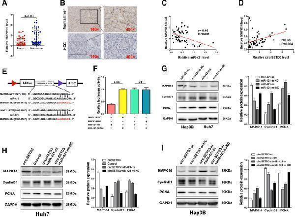
CircSETD3 inhibits the growth of HCC through thecircSETD3/miR-421/MAPK14 pathway. a and b Both qRT-PCR and IHC showed MAPK14 was significantly downregulated in HCC tissues compared with matched non-tumorous tissues. c and d MAPK14 negatively correlated with miR-421 whereas positively correlated with circSETD3 in HCC tissues. e Schematic of MAPK14 wild-type (WT) and mutant (Mut) luciferase reporter vectors. f The relative luciferase activities were analyzed in 293 T cells co-transfected with miR-421 mimics or miR-mimics-NC and WT or Mut luciferase reporter vectors. g MiR-421 inhibitor up-regulated MAPK14 and down-regulated cyclinD1 and PCNA in Hep3B cells. MiR-421 mimics down-regulated MAPK14 and up-regulated cyclinD1 and PCNA in Huh7 cells. h CircSETD3 letivirus up-regulated MAPK14 and down-regulated cyclinD1 and PCNA in Huh7 cells, this effect could be reversed by co-transfected with miR-421 mimics. i circSETD3 siRNA down-regulated MAPK14 and up-regulated cyclinD1 and PCNA in Hep3B cells, this effect can be reversed by co-transfected with miR-421 inhibitors. HCC, hepatocellular carcinoma; qRT-PCR, quantitative reverse transcription polymerase chain reaction; in, inhibitors; mi, mimics; IHC, immunohistochemistry. ***P < 0.001. Error bars indicate SD
Index in PubMed under a CC BY license. PMID: 30795787

Click image to see more details
CircSETD3 stably maintained in xenograft tumor models and inhibit tumor growth by targeting MAPK14. a and b Smaller tumor size and lower tumor weight were observed in circSETD3-overexpressing group. c Over-expressed circSETD3 could stably maintained in xenograft tumor models. d The expression of MAPK14 was increased, Ki-67, PCNA and Cyclin D1 were decreased in circSETD3-overexpressing group when compared with control group. **P < 0.01, ***P < 0.001. Error bars indicate SD
Index in PubMed under a CC BY license. PMID: 30795787

Click image to see more details
Effects of each treatment on the growth of human OS xenograft tumors. Nude mice bearing 143 human OS xenograft tumors were treated with CAP (20 mg/kg) and DDP (4 mg/kg) alone or in combination. The volumes of the xenograft tumors were measured at the indicated time points ( a ). The weight of each nude was measured at the indicated time points ( b ). After the last treatment, the mice were sacrificed, and representative images of the subcutaneous tumor xenografts in the nude mice and the morphology of the tumors are presented ( c ). Tumors were collected, and the tumor weights were measured and compared ( d ). Xenograft tumors were sectioned and stained with PCNA and Ki67 via IHC ( e ). Statistical analyses of the expression of PCNA and Ki67 in different groups ( f ). Tumors were sectioned and stained with H&E, and representative histopathological images are presented ( g ). Effects of the different treatments on renal histology. Representative histological profiles of kidneys after the different treatments were detected by H&E staining ( h ). Effects of the different treatments on BUN and creatinine levels in mice ( i ). The quantitative data are shown as the mean ± SD of 5 independent experiments; * p < 0.05, ** p < 0.01 and *** p < 0.001 vs. the control (CAP-, DDP-)
Index in PubMed under a CC BY license. PMID: 30326933

Click image to see more details
Western blot analysis of PCNA using anti-PCNA antibody (A00125).
Electrophoresis was performed on a 5-20% SDS-PAGE gel at 70V (Stacking gel) / 90V (Resolving gel) for 2-3 hours. The sample well of each lane was loaded with 30 ug of sample under reducing conditions.
Lane 1: human Hela whole cell lysates,
Lane 2: human MCF-7 whole cell lysates,
Lane 3: human 293T whole cell lysates,
Lane 4: human HepG2 whole cell lysates,
Lane 5: rat PC-12 whole cell lysates,
Lane 6: mouse NIH/3T3 whole cell lysates.
After electrophoresis, proteins were transferred to a nitrocellulose membrane at 150 mA for 50-90 minutes. Blocked the membrane with 5% non-fat milk/TBS for 1.5 hour at RT. The membrane was incubated with rabbit anti-PCNA antigen affinity purified polyclonal antibody (Catalog # A00125) at 0.5 μg/mL overnight at 4°C, then washed with TBS-0.1%Tween 3 times with 5 minutes each and probed with a goat anti-rabbit IgG-HRP secondary antibody at a dilution of 1:5000 for 1.5 hour at RT. The signal is developed using an Enhanced Chemiluminescent detection (ECL) kit (Catalog # EK1002) with Tanon 5200 system. A specific band was detected for PCNA at approximately 36 kDa. The expected band size for PCNA is at 29 kDa.

Click image to see more details
IHC analysis of PCNA using anti-PCNA antibody (A00125).
PCNA was detected in a paraffin-embedded section of human breast cancer tissue. Heat mediated antigen retrieval was performed in EDTA buffer (pH 8.0, epitope retrieval solution). The tissue section was blocked with 10% goat serum. The tissue section was then incubated with 2 μg/ml rabbit anti-PCNA Antibody (A00125) overnight at 4°C. Peroxidase Conjugated Goat Anti-rabbit IgG was used as secondary antibody and incubated for 30 minutes at 37°C. The tissue section was developed using HRP Conjugated Rabbit IgG Super Vision Assay Kit (Catalog # SV0002) with DAB as the chromogen.

Click image to see more details
IHC analysis of PCNA using anti-PCNA antibody (A00125).
PCNA was detected in a paraffin-embedded section of human colorectal adenocarcinoma tissue. Heat mediated antigen retrieval was performed in EDTA buffer (pH 8.0, epitope retrieval solution). The tissue section was blocked with 10% goat serum. The tissue section was then incubated with 2 μg/ml rabbit anti-PCNA Antibody (A00125) overnight at 4°C. Peroxidase Conjugated Goat Anti-rabbit IgG was used as secondary antibody and incubated for 30 minutes at 37°C. The tissue section was developed using HRP Conjugated Rabbit IgG Super Vision Assay Kit (Catalog # SV0002) with DAB as the chromogen.

Click image to see more details
IHC analysis of PCNA using anti-PCNA antibody (A00125).
PCNA was detected in a paraffin-embedded section of human tonsil tissue. Heat mediated antigen retrieval was performed in EDTA buffer (pH 8.0, epitope retrieval solution). The tissue section was blocked with 10% goat serum. The tissue section was then incubated with 2 μg/ml rabbit anti-PCNA Antibody (A00125) overnight at 4°C. Peroxidase Conjugated Goat Anti-rabbit IgG was used as secondary antibody and incubated for 30 minutes at 37°C. The tissue section was developed using HRP Conjugated Rabbit IgG Super Vision Assay Kit (Catalog # SV0002) with DAB as the chromogen.

Click image to see more details
IHC analysis of PCNA using anti-PCNA antibody (A00125).
PCNA was detected in a paraffin-embedded section of human papillary thyroid carcinoma tissue. Heat mediated antigen retrieval was performed in EDTA buffer (pH 8.0, epitope retrieval solution). The tissue section was blocked with 10% goat serum. The tissue section was then incubated with 2 μg/ml rabbit anti-PCNA Antibody (A00125) overnight at 4°C. Peroxidase Conjugated Goat Anti-rabbit IgG was used as secondary antibody and incubated for 30 minutes at 37°C. The tissue section was developed using HRP Conjugated Rabbit IgG Super Vision Assay Kit (Catalog # SV0002) with DAB as the chromogen.

Click image to see more details
IHC analysis of PCNA using anti-PCNA antibody (A00125).
PCNA was detected in a paraffin-embedded section of human glioblastoma tissue. Heat mediated antigen retrieval was performed in EDTA buffer (pH 8.0, epitope retrieval solution). The tissue section was blocked with 10% goat serum. The tissue section was then incubated with 2 μg/ml rabbit anti-PCNA Antibody (A00125) overnight at 4°C. Peroxidase Conjugated Goat Anti-rabbit IgG was used as secondary antibody and incubated for 30 minutes at 37°C. The tissue section was developed using HRP Conjugated Rabbit IgG Super Vision Assay Kit (Catalog # SV0002) with DAB as the chromogen.

Click image to see more details
IHC analysis of PCNA using anti-PCNA antibody (A00125).
PCNA was detected in a paraffin-embedded section of human liver cancer tissue. Heat mediated antigen retrieval was performed in EDTA buffer (pH 8.0, epitope retrieval solution). The tissue section was blocked with 10% goat serum. The tissue section was then incubated with 2 μg/ml rabbit anti-PCNA Antibody (A00125) overnight at 4°C. Peroxidase Conjugated Goat Anti-rabbit IgG was used as secondary antibody and incubated for 30 minutes at 37°C. The tissue section was developed using HRP Conjugated Rabbit IgG Super Vision Assay Kit (Catalog # SV0002) with DAB as the chromogen.

Click image to see more details
IHC analysis of PCNA using anti-PCNA antibody (A00125).
PCNA was detected in a paraffin-embedded section of rat colon tissue. Heat mediated antigen retrieval was performed in EDTA buffer (pH 8.0, epitope retrieval solution). The tissue section was blocked with 10% goat serum. The tissue section was then incubated with 2 μg/ml rabbit anti-PCNA Antibody (A00125) overnight at 4°C. Peroxidase Conjugated Goat Anti-rabbit IgG was used as secondary antibody and incubated for 30 minutes at 37°C. The tissue section was developed using HRP Conjugated Rabbit IgG Super Vision Assay Kit (Catalog # SV0002) with DAB as the chromogen.

Click image to see more details
IF analysis of PCNA using anti-PCNA antibody (A00125) and anti-Beta Tubulin antibody (M01857-3).
PCNA was detected in immunocytochemical section of A431 cell. Enzyme antigen retrieval was performed using IHC enzyme antigen retrieval reagent (AR0022) for 15 mins. The cells were blocked with 10% goat serum. And then incubated with 5 μg/mL rabbit anti-PCNA Antibody (A00125) and mouse anti-Beta Tubulin antibody (M01857-3) overnight at 4°C. DyLight®488 Conjugated Goat Anti-Rabbit IgG (BA1127) and Cy3 Conjugated Goat Anti-Mouse IgG (BA1031) were used as secondary antibody at 1:500 dilution and incubated for 30 minutes at 37°C. Visualize using a fluorescence microscope and filter sets appropriate for the label used.

Click image to see more details
Flow Cytometry analysis of 293T cells using anti-PCNA antibody (A00125).
Overlay histogram showing 293T cells stained with A00125 (Blue line). To facilitate intracellular staining, cells were fixed with 4% paraformaldehyde and permeabilized with permeabilization buffer. The cells were blocked with 10% normal goat serum. And then incubated with rabbit anti-PCNA Antibody (A00125, 1 μg/1x106 cells) for 30 min at 20°C. DyLight®488 conjugated goat anti-rabbit IgG (BA1127, 5-10 μg/1x106 cells) was used as secondary antibody for 30 minutes at 20°C. Isotype control antibody (Green line) was rabbit IgG (1 μg/1x106) used under the same conditions. Unlabelled sample (Red line) was also used as a control.
Specific Publications For Anti-PCNA Antibody Picoband® (A00125)
Loading publications
Recommended Resources
Here are featured tools and databases that you might find useful.
- Boster's Pathways Library
- Protein Databases
- Bioscience Research Protocol Resources
- Data Processing & Analysis Software
- Photo Editing Software
- Scientific Literature Resources
- Research Paper Management Tools
- Molecular Biology Software
- Primer Design Tools
- Bioinformatics Tools
- Phylogenetic Tree Analysis
Customer Reviews
Have you used Anti-PCNA Antibody Picoband®?
Share your experimental results or join a short interview to earn up to $1,000 in product credits or other rewards.
0 Reviews For Anti-PCNA Antibody Picoband®
Customer Q&As
Have a question?
Find answers in Q&As, reviews.
Can't find your answer?
Submit your question
5 Customer Q&As for Anti-PCNA Antibody Picoband®
Question
We have been able to see staining in rat liver. Do you have any suggestions? Is anti-PCNA antibody supposed to stain liver positively?
Verified Customer
Verified customer
Asked: 2020-03-23
Answer
According to literature liver does express PCNA. According to Uniprot.org, PCNA is expressed in oocyte, skeletal muscle, bone marrow lung, fetal brain cortex, placenta, liver, among other tissues. Regarding which tissues have PCNA expression, here are a few articles citing expression in various tissues:
Bone marrow, and Lung, Pubmed ID: 15489334
Fetal brain cortex, Pubmed ID: 9305916
Liver, Pubmed ID: 24275569
Placenta, Pubmed ID: 12522211
Skeletal muscle, Pubmed ID: 14702039
Boster Scientific Support
Answered: 2020-03-23
Question
We are currently using anti-PCNA antibody A00125 for mouse tissue, and we are content with the Flow Cytometry results. The species of reactivity given in the datasheet says human, mouse, rat. Is it true that the antibody can work on feline tissues as well?
C. Mangal
Verified customer
Asked: 2019-12-17
Answer
The anti-PCNA antibody (A00125) has not been tested for cross reactivity specifically with feline tissues, but there is a good chance of cross reactivity. We have an innovator award program that if you test this antibody and show it works in feline you can get your next antibody for free. Please contact me if I can help you with anything.
Boster Scientific Support
Answered: 2019-12-17
Question
We ordered your anti-PCNA antibody for IHC-P on bone marrow lung in a previous project. I am using human, and We intend to use the antibody for Flow Cytometry next. I am looking for examining bone marrow lung as well as oocyte in our next experiment. Could you please give me some suggestion on which antibody would work the best for Flow Cytometry?
Verified Customer
Verified customer
Asked: 2019-09-24
Answer
I looked at the website and datasheets of our anti-PCNA antibody and it seems that A00125 has been validated on human in both IHC-P and Flow Cytometry. Thus A00125 should work for your application. Our Boster satisfaction guarantee will cover this product for Flow Cytometry in human even if the specific tissue type has not been validated. We do have a comprehensive range of products for Flow Cytometry detection and you can check out our website bosterbio.com to find out more information about them.
Boster Scientific Support
Answered: 2019-09-24
Question
My lab would like using your anti-PCNA antibody for removal of the flap intermediate studies. Has this antibody been tested with western blotting on 293t cells? We would like to see some validation images before ordering.
Verified Customer
Verified customer
Asked: 2019-06-14
Answer
I appreciate your inquiry. This A00125 anti-PCNA antibody is tested on rat c6 whole cell lysates, mouse nih3t3 whole cell lysates, 293t cells. It is guaranteed to work for ELISA, Flow Cytometry, IHC-P, WB in human, mouse, rat. Our Boster guarantee will cover your intended experiment even if the sample type has not been be directly tested.
Boster Scientific Support
Answered: 2019-06-14
Question
Our team were satisfied with the WB result of your anti-PCNA antibody. However we have seen positive staining in skeletal muscle nucleus using this antibody. Is that expected? Could you tell me where is PCNA supposed to be expressed?
Verified Customer
Verified customer
Asked: 2017-06-02
Answer
According to literature, skeletal muscle does express PCNA. Generally PCNA expresses in nucleus. Regarding which tissues have PCNA expression, here are a few articles citing expression in various tissues:
Bone marrow, and Lung, Pubmed ID: 15489334
Fetal brain cortex, Pubmed ID: 9305916
Liver, Pubmed ID: 24275569
Placenta, Pubmed ID: 12522211
Skeletal muscle, Pubmed ID: 14702039
Boster Scientific Support
Answered: 2017-06-02
