This website uses cookies to ensure you get the best experience on our website.
- Table of Contents
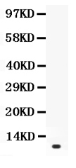
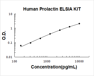
2 Citations 6 Q&As
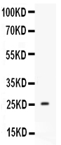
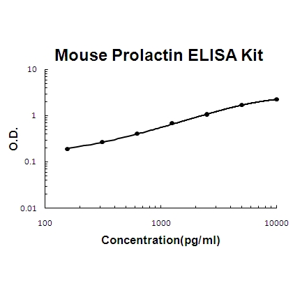
4 Citations 13 Q&As

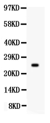
1 Citations
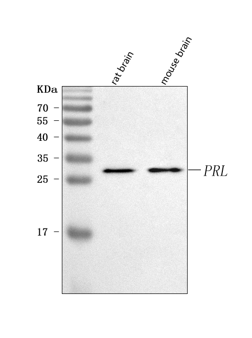
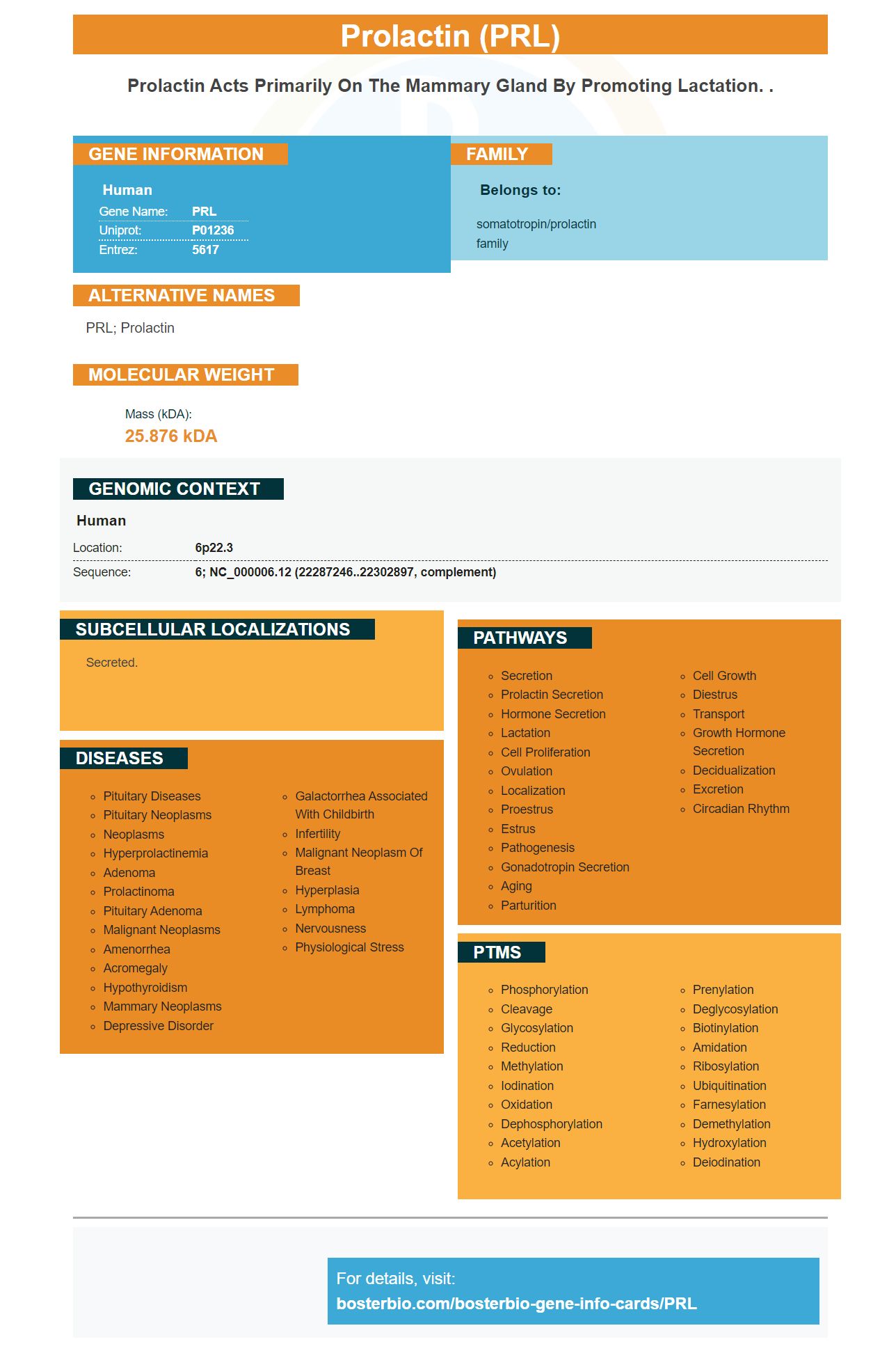
Facts about Prolactin.

| Human | |
|---|---|
| Gene Name: | PRL |
| Uniprot: | P01236 |
| Entrez: | 5617 |

| Belongs to: |
|---|
| somatotropin/prolactin family |

PRL; Prolactin
Mass (kDA):
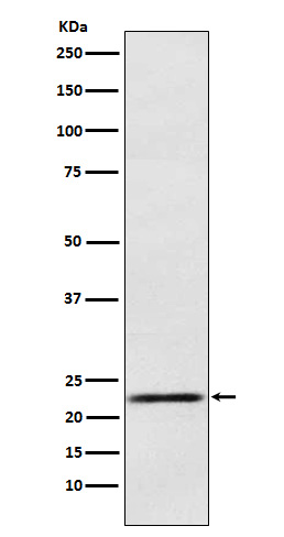
25.876 kDA

| Human | |
|---|---|
| Location: | 6p22.3 |
| Sequence: | 6; NC_000006.12 (22287246..22302897, complement) |
Secreted.


PMID: 6260780 by Cooke N.E., et al. Human prolactin. cDNA structural analysis and evolutionary comparisons.
PMID: 6325171 by Truong A.T., et al. Isolation and characterization of the human prolactin gene.
*More publications can be found for each product on its corresponding product page