This website uses cookies to ensure you get the best experience on our website.
- Table of Contents
6 Citations 15 Q&As
4 Citations 5 Q&As
1 Citations 17 Q&As
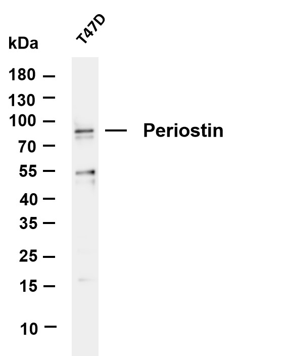
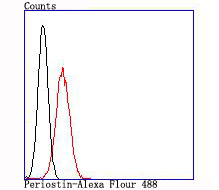
Facts about Periostin.
.
Human | |
---|---|
Gene Name: | POSTN |
Uniprot: | Q15063 |
Entrez: | 10631 |
Belongs to: |
---|
No superfamily |
Fasciclin I-like; MGC119510; MGC119511; OSF2; OSF-2; OSF-2osteoblast specific factor 2 (fasciclin I-like); OSF2periodontal ligament-specific periostin; Osteoblast-specific factor 2; PDLPOSTN; periostin isoform thy2; periostin isoform thy4; periostin isoform thy6; periostin isoform thy8; Periostin; periostin, osteoblast specific factor; PNRP11-412K4.1; POSTN; TRIF52
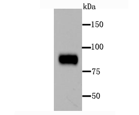
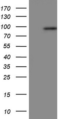
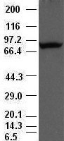
Mass (kDA):
93.314 kDA
Human | |
---|---|
Location: | 13q13.3 |
Sequence: | 13; NC_000013.11 (37562582..37598844, complement) |
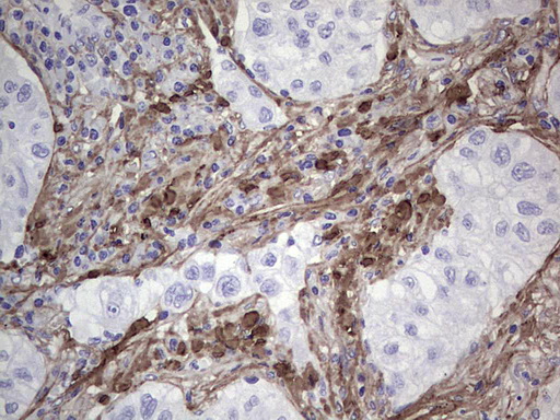
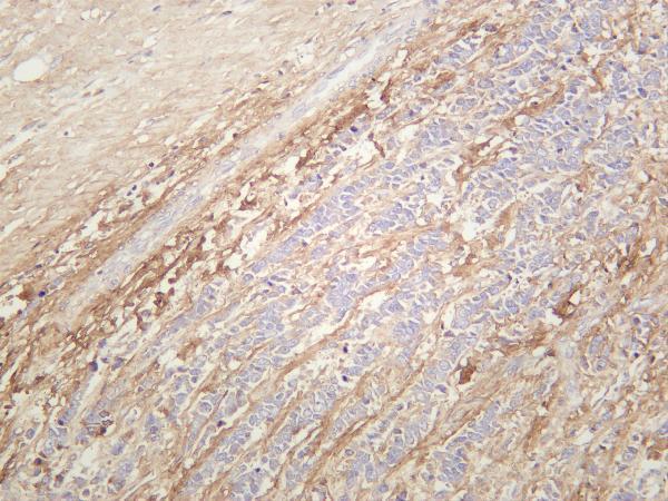
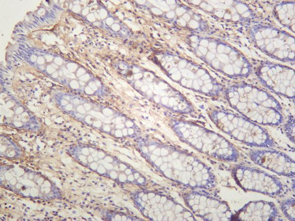
Widely expressed with highest levels in aorta, stomach, lower gastrointestinal tract, placenta, uterus, thyroid tissue and breast. Up-regulated in epithelial ovarian tumors. Not expressed in normal ovaries. Also highly expressed at the tumor periphery of lung carcinoma tissue but not within the tumor. Overexpressed in breast cancers.
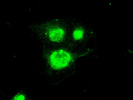
Golgi apparatus. Secreted. Secreted, extracellular space, extracellular matrix. Colocalizes with BMP1 in the Golgi.
The POSTN Marker is a protein in the body and plays numerous important roles. It is responsible for the maintenance of cancer stem cells, tissue growth and metastasis. This article will discuss the ways this protein is utilized in various ways. It will also provide information on its role in the development of tissues. Boster Bio The Best uses for the POSTN Marker
The POSTN marker performs a variety of functions, including regulation of cell adhesion and movement and angiogenesis-related genes expression. It also plays a part in EMT by regulating VEGF C and EMT-related genes. It also regulates the interaction between GSCs, their environment, and the treatment of anti-VEGFA. By targeting POSTN and the agents that cause disease, they could be able to defeat the highly infiltrative manifestation of the glioblastoma.
The expression of POSTN and VEGF-1 was measured by using human ELISA kits. The results were expressed as the mean concentration in picograms per liter (pg/mL). Additionally, POSTN and pSTAT3 were detected using reverse-phase protein array analysis (RPPA) in GSCs, gliomas and tissues of mice. RPPA was carried out according to guidelines for RPPA. The samples were analyzed using 218 validated primary antibodies. The RPPA core facility at MD Anderson was used for the analysis.
In addition, knockdown of POSTN decreased resistance of human GSCs to anti-VEGF-A therapy. Additionally, POSTN expression was inversely associated with survival in adults with glioma. Elimination of POSTN resulted in a reduction in tumor volume and an increase in survival in mice with GSC cells. And, in a recent study, POSTN knockdown increased survival and decreased the size of tumors in mice implanted with GSC cells. Researchers found that POSTN knockdown inhibited tumor growth and improved survival.
The POSTN marker is essential to cardiac fibroblast function. Myofibroblasts release this protein in adult tissues. It is involved in the collagen organization as well as cellular adhesion and cell division. A mouse lacking Postn has diminished cardiac fibroblast function. Normal is a homozygous variation of the gene. Further the POSTN gene is essential to produce a protective scar.
To investigate the role of POSTN Marker in the development of fibroblasts Researchers have employed mice that have PostnMCM knockout. Mice were crossed with Rosa26 reporter mice that have the loxP site that is flanked by the stop cassette. This allowed for the tracing of lineages dependent on Cre. Mice were harvested at 16 weeks. A representative histological slice from the heart of PostnMCM mouse reveals rare interstitial cells. Inset: A-actin-stained cardiomyocytes are located around an interstitial cell.
The POSTN Marker is a constituent of the extracellular matrix (ECM), and its significant expression is associated with metastasis in a variety of cancer. POSTN is expressed by tumour stroma, and it blocks colonisation. It also facilitates the growth of mutant cancer stem cells and activates the NF-kB pathway, which is a key regulator of tumour growth and development.
This discovery provides new insights into cancer stem cells maintenance. In the last few years scientists haven't been able to pinpoint exactly how the POSTN Marker works. However, recent studies have discovered its role in the circulating tumor stem cells and are trying to determine if it plays a role in this process. One of these studies has been published in the journal Proc Natl Acad Sci.
A subset of tumour cells stem cells that are cancerous have similar characteristics to normal stem cells. They are self-renewing and undergo asymmetric division, and differentiate into a variety of types of cells. Furthermore, they are capable of initiating tumours, and they can even survive the presence of chemotherapy. Additionally, CSCs have been implicated in the progression of different kinds of cancers as well as in their resistance to conventional treatments.
Mesenchymal stem cells are one of the most vital components of the tumor microenvironment. They assist in the progression of tumors through the stimulation of angiogenesis and metastasis and also create an immune-suppressive microenvironment. Cytokines, as well as other factors, are released by cancer cells, which stimulate tumor stemness, undifferentiated state, and tumour stemness. The cancer cells also produce cytokines which draw mesenchymal and stem cells.
A new candidate marker for cancer stem cells, Tenascin-C, was recently discovered by tissue microarrays. The study found that the protein stimulates the growth of cancerous melanoma cell lines. It also maintains a population of quiescent stem cells for cancer and protects them from immune surveillance. This discovery could be a key factor in the creation of new treatments and prevention of cancer.
POSTN markers play an important role in the metabolism of cancerous cells. This is essential for cancer stem cells, as they have a greater capacity to regulate oxygen and other nutrient levels. Hypoxia also promotes the growth of cancer cells in an environment with low oxygen levels, and is also linked to poor treatment outcomes. The tumor's microenvironment may increase the aggression of cancer cells and can even lead to metastasis.
POSTN Marker regulates the growth and development of tumor-associated fibroblasts in addition to its function in maintaining cancer stem cells. It regulates the microenvironment as well as microRNA-29b. For the maintenance of cancer stem cells, osteopontin and bone-morphogenetic proteins are vital. Cancer can alter the balance between pro-growth and anti-growth signals. This leads to a tumor-related microenvironment that is characterized by an abnormal Wnt signalling pathway.
The POSTN Marker is expressed extensively in breast cancer and is associated with pathological TNM staging and lymph node metastasis. Its target miR-876 inhibits epithelial-to-mesenchymal transition (EMT) and fibrosis in hepatocellular carcinoma. These results suggest that POSTN could be a therapeutic target for hepatocellular carcinoma. POSTN also regulates EMT through the ERK signaling pathway. This can promote cancer cell migration, invasion, and metastasis.
This marker, also referred to as human osteoblast-specific factors (POSTN), was first discovered in mouse osteoblast cell cells by Takcshita. It has an N-terminal signaling peptide and the C-terminal Hydrophilic Domain. It also has a cysteine rich region and four internal homologous domains. POSTN is a key factor in osteogenesis aswell as the aggregation and maintenance of precursor cells. This marker is expressed predominantly in connective tissues, and its expression in the periodontal ligaments is high. In addition to its role in osteoblast migration, POSTN regulates cell adhesion and cell recruitment as well as growth.
Furthermore, the POSTN-shRNA-treated OS cells showed significantly reduced proliferation when transfected using the POSTN-shRNA. The decrease of POSTN MRNA was confirmed through western blot analysis. This revealed an important inhibition in cell invasion and migration. The shRNA-treated OS cells also showed lower levels proliferative nuclear antigen (PCNA) which is a marker for cell proliferative.
In the same way, CEA also plays a role in the process of liver metastasis in CRC. CEA-expressed CRC cell lines interact with Kupffer cells in livers and alter the microenvironment, favoring the formation of tumors. CRC cells can interact with CEA to trigger and stimulate signal transduction. Incredibly, the N domain is ineffective for colocalization with a5b1integrin.
PMID: 8363580 by Takeshita S., et al. Osteoblast-specific factor 2: cloning of a putative bone adhesion protein with homology with the insect protein fasciclin I.
PMID: 12235007 by Gillan L., et al. Periostin secreted by epithelial ovarian carcinoma is a ligand for alpha(V)beta(3) and alpha(V)beta(5) integrins and promotes cell motility.
*More publications can be found for each product on its corresponding product page