This website uses cookies to ensure you get the best experience on our website.
- Table of Contents


Facts about Mothers against decapentaplegic homolog 1.

SMAD1/OAZ1/PSMB4 complex mediates the degradation of this CREBBP/EP300 repressor SNIP1. May act synergistically with SMAD4 and YY1 in bone morphogenetic protein (BMP)-mediated cardiac-specific gene expression.
| Human | |
|---|---|
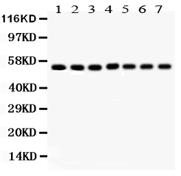
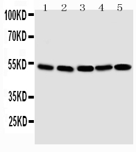
| Gene Name: | SMAD1 |
| Uniprot: | Q15797 |
| Entrez: | 4086 |

| Belongs to: |
|---|
| dwarfin/SMAD family |

BSP1; BSP-1; hSMAD1; JV4-1SMAD, mothers against DPP homolog 1 (Drosophila); MAD homolog 1; MADH1JV41; MADR1MAD, mothers against decapentaplegic homolog 1 (Drosophila); Mad-related protein 1; mothers against decapentaplegic homolog 1; Mothers against DPP homolog 1; SMAD family member 1SMAD 1; SMAD, mothers against DPP homolog 1; Smad1; TGF-beta signaling protein 1; transforming growth factor-beta signaling protein 1
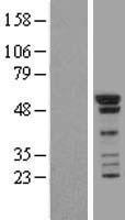
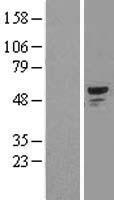
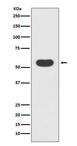
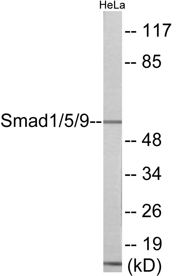
Mass (kDA):
52.26 kDA

| Human | |
|---|---|
| Location: | 4q31.21 |
| Sequence: | 4; NC_000004.12 (145481306..145559176) |
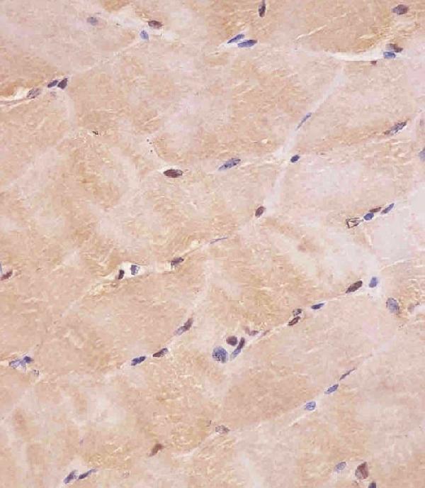
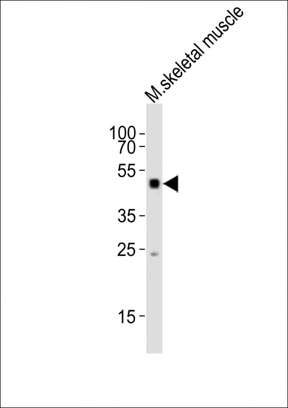
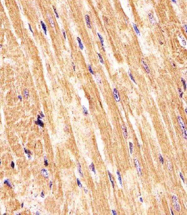
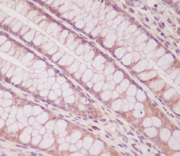
Ubiquitous. Highest expression seen in the heart and skeletal muscle.
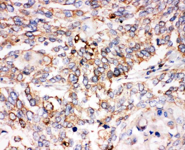
Cytoplasm. Nucleus. Cytoplasmic in the absence of ligand. Migrates to the nucleus when complexed with SMAD4 (PubMed:15647271). Co-localizes with LEMD3 at the nucleus inner membrane (PubMed:15647271). Exported from the nucleus to the cytoplasm when dephosphorylated (By similarity).


PMID: 8673135 by Riggins G.J., et al. Mad-related genes in the human.
PMID: 8637600 by Liu F., et al. A human Mad protein acting as a BMP-regulated transcriptional activator.
*More publications can be found for each product on its corresponding product page