This website uses cookies to ensure you get the best experience on our website.
- Table of Contents
2 Citations 3 Q&As
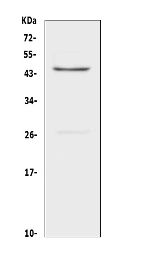
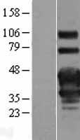
Facts about Tumor necrosis factor receptor superfamily member 4.
Receptor for TNFSF4/OX40L/GP34.
Is a costimulatory molecule implicated in long-term T-cell immunity..
Human | |
---|---|
Gene Name: | TNFRSF4 |
Uniprot: | P43489 |
Entrez: | 7293 |
Belongs to: |
---|
No superfamily |
ACT-135; ACT35 antigen; ACT35ATC35 antigen; CD134 antigen; CD134; Ly-70; OX40 cell surface antigen; OX40 homologue; OX40; OX40L receptor; OX40lymphoid activation antigene ACT35; TAX transcriptionally-activated glycoprotein 1 receptor; tax-transcriptionally activated glycoprotein 1 receptor; TNFRSF4; tumor necrosis factor receptor superfamily member 4; tumor necrosis factor receptor superfamily, member 4; Txgp1; TXGP1L; TXGP1LOX40 antigen
Mass (kDA):
29.341 kDA
Human | |
---|---|
Location: | 1p36.33 |
Sequence: | 1; NC_000001.11 (1211326..1216812, complement) |
Membrane; Single-pass type I membrane protein.
If you're looking for an exciting test to determine the expression of genes, TNFRSF4 is an excellent candidate. This gene shows increased expression in tumor cells. It's also present in tumors that have the TP53, FLT3 or NPM1 mutations. It's not found in the majority of cancers and isn't well-studied. If you're interested about the best uses of TNFRSF4 take a look!
The TNFRSF4 gene encodes one of the members of the superfamily of tumor necrosis receptors. It is involved in maintaining the dominating self-tolerance that is a result of the immune system of CD25+CD regulatory T cells. Knockout studies have also indicated its role in the activation of T cells by CD3 and programmed cell death. The exact role of TNFRSF4 is still unclear.
The TNFRSF4 gene is highly expressed on T-cells. OX40L which stimulates naive naive T cells, was also increased in asthmatic PBMCs that extracted the RNA. Therefore, this gene could play a role in asthmatic inflammation. The gene also regulates naive T-cell polarization. The gene that encodes OX40L is found on T cells, but it is mostly expressed on macrophages. However the OX40L gene has also been found in NK cells, mast cells, basophils, and dendritic cells.
Mutations in TP53 is a risk factor for poor survival in medulloblastoma cells. More than 50% of cancers in humans are the result of mutations in this gene. The effectiveness of radiation therapy is greater in the absence of normal p53 function. This means that knowing the patient's TP53 mutation status can help predict their response to radiation therapy. This mutation was identified in Boster Bio cells.
The zebrafish system is useful to study TP53 functions in living. This model system can be used for high-throughput screening of small molecules. In addition, a temperature-sensitive mutant line offers a vertebrate tp53-inducible system, allowing researchers to identify modifiers that act in downstream and parallel pathways. This is especially beneficial in studies of the role TP53 is involved in tumorigenesis.
In the study of an unmodified TP53 cell line, a TP53 mutation causing an inability to regulate the process of apoptosis was transiently transformed with an expression vector that contained a dominant-negative TP53 mutation, tp53M214K. The mutant embryos were irradiated and showed an increase in mid-S-phase cells, while wild-type embryos had an increased percentage of cells in G1.
The TP53 mutation was classified into four classes based on the location, nature, and suspected influence on the function of the protein p53. Apart from TP53 mutations and other genetic variants, other genetic variants were classified using the same classification system. Each mutation was classified according to its location, whereas the three others were classified according to their impact on the p53 protein function. These four classifications are in line with three different classification systems. The transcription class is related to the scale-invariant feature transformation algorithm.
TP53 mutations are associated with lower results in tests related to cancer. Certain kinds of mutations affect p53's ability to protect cells from DNA damage and increase the risk of cancer. Particularly amino acid substitutions within the hydrophobic core area of p53 as well as mutations on the non-DNA-binding surface of P53 are the most harmful. These mutations cause the DNA-binding surface to be damaged by the hydrophobic core.
Patients with TP53 mutations have a lower level of expression for the TNFRSF4 gene. The higher levels are associated with poor clinical outcomes. Patients with non-M3 AML and MDS (EB-1) were found to have higher levels of expression of TNFRSF4. Future functional studies are needed to understand the function of TNFRSF4 in these patient subtypes.
In a recent study researchers identified three subtypes of patients suffering from non-M3 AML. The presence of TNFRSF4 were associated with a shorter OS and DFS as compared to patients with low expression. The difference was not statistically significant. Patients with TP53-related mutations had significantly lower median survival. For patients with TP53 mutations and high levels of TNFRSF4 expression were associated with significantly shorter DFS.
Researchers found that TNFRSF4 expression was positively associated with bone blasts in the bone marrow. The Spearman's Rho was 0.372 when compared with 0.002 which suggests that the TNFRSF4 gene expression is linked with bone blasts in the bone marrow. Researchers believe the TNFRSF4 gene can be used to determine the risk of developing a blood disorder in patients with FLT3.
The TNFRSF4 gene has been identified as a biomarker for a variety of cancers. Researchers also discovered one gene that could be linked to tumor's expression of TNFRSF4. Although there is no clinical utility, a TNFRSF4 gene has shown a significant positive predictive value. A positive test can assist doctors decide on the best treatment.
The cut-off for acute myeloid leukemia (AML) is the percentage of bone marrow reblasts of 20% or higher. However this percentage is a topic of debate. Pathologists in the 1970s developed the French-American-British (FAB) classification system, which included refractory anemia with excess blasts (RAEB) and acute myeloid leukemia with more than 30% blasts. This classification system is the basis for the Revised International Scoring System (RIPS) and International Prognostic Scoring System (IPSS).
In each case, the B-cell count was recorded as well as the erythroid precursor count. The blast count was determined by counting 200 peripheral blood cells as well as 500 bone marrow cells. The percentages were then interpreted as percentages of total cells and erythroid precursor cells, and ring sideroblasts and nucleated erythroid cells respectively. Although B-cells constituted the majority of patients suffering from A-type erythroleukemia type had a higher number of B-cells than a healthy normal person.
R-IPSS scores patients who have marrowblasts of 10-20%, and younger patients with blasts that range from 11-29 percent. MDS-type chemotherapy should be given to patients over the age of 50 with blasts that exceed 20-30%. Patients with these blasts should also undergo the procedure of a stem cell transplant when their B-cell count falls below 20 percent. The treatment options are contingent on the stage and age of the disease.
B-cell-mediated lymphoma is a rare. However, AML is more common in patients suffering from de novo and has more bone blasts rate than those with acute myeloidleukemia. This kind of leukemia has a similar B-cell count to AML patients. The rarity of the disease prevents prospective randomized trials from being conducted.
PMID: 7510240 by Latza U., et al. The human OX40 homolog: cDNA structure, expression and chromosomal assignment of the ACT35 antigen.
PMID: 7704935 by Baum P.R., et al. Identification of OX40 ligand and preliminary characterization of its activities on OX40 receptor.
*More publications can be found for each product on its corresponding product page