This website uses cookies to ensure you get the best experience on our website.
- Table of Contents
Boster Bio Reporter-Labeled Cell Lines
Ready-to-use Renilla luciferase reporter cell lines for measuring pathway activation, promoter response, receptor signaling, and compound effects in functional cell-based assays.
Browse catalog reporter cell lines by pathway and cell background, or request a custom model for your assay.
Begin Inquiry
Boster Bio reporter cell lines support pathway activity assays, compound-response studies, transcription factor activity analysis, and custom functional cell-based assay development.
Monitor signaling responses through reporter systems such as NF-kB, ISRE, STAT, TLR/NF-kB, SBE, CRE, NFAT, and AP-1.
Use Renilla luciferase luminescence to quantify promoter activation, receptor signaling, and compound effects.
Choose from common research cell backgrounds including HEK293, Jurkat, RAW264.7, HeLa, Ba/F3, MCF7, and NIH 3T3.
Request custom reporter cell line generation when your assay requires a specific promoter, response element, receptor, or host cell.
Ready-to-use stable reporter cell lines give your team a faster starting point for functional assays, pathway activity studies, and compound-response screening.
Reporter cell lines help researchers convert pathway activation, promoter response, or receptor-mediated signaling into a quantitative luminescent readout. Instead of transfecting and validating a reporter construct for every experiment, ready-to-use stable reporter cell lines help reduce setup time and improve assay consistency.
Use this page to select a catalog reporter cell line by pathway and cell background, or request a custom reporter cell line when your assay requires a specific promoter, response element, receptor, or host cell model.

Start with the biological question first, then narrow by pathway, response element, and cell background.
Consider NF-kB, TLR/NF-kB, ISRE, iNOS, TNF-α, or IL-8 reporter cell lines.
Consider STAT1, STAT3, STAT4, STAT5, NFAT, AP-1, or cytokine-responsive reporter models.
Use stable reporter cell lines to compare activators, inhibitors, biologics, and small molecules.
Request a custom reporter cell line for a target pathway, promoter, receptor, or cell background not listed.
Select from validated reporter cell lines across common pathway readouts and cell backgrounds. Each product page includes catalog details, application information, and available validation figures.
Ba/F3 reporter model for STAT4 pathway activity studies.

1 image
Jurkat reporter model for FOXP3 transcriptional response studies.

1 image
HEK293 reporter model for interferon-stimulated response element activity.

1 image
Jurkat NF-kB reporter cell line for inflammation and immune signaling assays.

1 image
Macrophage-like reporter model for ISRE and immune signaling studies.

1 image
HEK293 reporter model for STAT4 cytokine signaling response.

1 image
HEK293 reporter model for TLR5-mediated NF-kB pathway activation.

1 image
HeLa reporter model for ATF6 and cellular stress response studies.

1 image
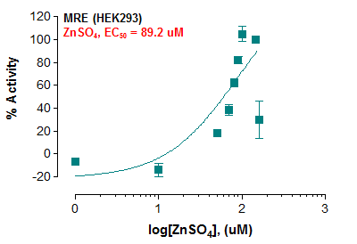
HEK293 reporter model for metal response element activity.
1 image
HEK293 reporter model for SBE-mediated pathway analysis.

1 image
Displaying Items 1-10 of
Need a pathway, promoter, receptor, or cell background not listed?
Boster Bio can support custom reporter cell line generation for specialized assay needs, including custom response elements, receptor systems, and host cell backgrounds.
Use the tabs below to review reporter-labeled pathway options across commonly used research cell backgrounds. Linked pathway names lead to Boster Bio pathway maps where available.
| ISRE | STAT4 | TLR/NF-kB | MRE |
| SBE | CRE | SRF-RE | SRE |
| TLR9/NF-kB | TLR8/NF-kB | TLR7/NF-kB | TLR3/NF-kB |
| TLR2/NF-kB | NF-kB | TLR3/ISRE | TLR3/IFNB |
| TCF/LEF | STAT3 | NFAT | GATA3 |
| TNF-β | TNF-α | AP-1 |
| ATF6 | P53 | TLR-4/IL-8 | HRE |
| STAT1 |
| STAT4 | STAT5 |
| FOXP3 | NF-kB |
| Nrf2 |
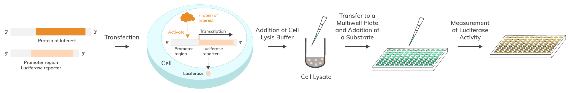
A luciferase reporter cell line is a stable cell model engineered to express a luciferase reporter gene under the control of a selected promoter or response element. When the target signaling pathway is activated, the reporter is expressed and produces a luminescent signal that can be measured as a quantitative readout.
Renilla luciferase reporter cell lines are commonly used to study transcriptional regulation, receptor-mediated signaling, promoter activity, and compound effects. Compared with transient transfection workflows, stable reporter cell lines can reduce setup time and improve assay consistency across screening and functional validation studies.
Renilla luciferase catalyzes a light-producing reaction with coelenterazine. In a reporter cell line, the luciferase signal reflects the activity of the promoter or response element placed upstream of the reporter gene. After cells are treated with a stimulus, inhibitor, biologic, or small molecule, luminescence is measured with a luminometer and reported as relative light units (RLU).
The result is a functional assay readout that helps researchers compare pathway activation, inhibition, and dose-response effects under controlled experimental conditions.
Study promoter regulation and transcriptional response through luciferase-based reporter readouts.
Measure signaling pathway activation and inhibition under controlled treatment conditions.
Analyze transcription factor and response-element activity in functional cell-based systems.
Compare activators, inhibitors, biologics, and small molecules through quantitative luminescent readouts.
Exact conditions may vary by reporter cell line and experimental objective. Always follow the product-specific datasheet and recommended culture conditions.
Cell background affects pathway biology, receptor expression, growth behavior, and assay interpretation. Select the cell model that best matches your experimental question.
Widely used human cell backgrounds for pathway reporter assays, receptor signaling studies, and recombinant expression workflows. HEK293-based reporter models are often selected for robust growth and assay adaptability.
A commonly used T cell leukemia background for immune signaling, transcription factor response, NF-kB, FOXP3, and T cell pathway studies.
A macrophage-like cell background useful for innate immune signaling, inflammatory response, ISRE, iNOS, NF-kB, and cytokine-related reporter assays.
A human epithelial cancer cell background often used for stress response, hypoxia response, viral response, and general pathway activity studies.
A hematopoietic pro-B cell background often used in cytokine, kinase, and signaling studies where growth response and pathway activation are central to the assay design.
When your assay requires a specific disease model, receptor system, promoter, or host cell type, request a custom reporter cell line to better match your biology.
Q1: What are luciferase reporter cell lines used for?
Q2: How do I choose the right reporter cell line?
Q3: What is the difference between Renilla and firefly luciferase reporter systems?
Q4: Can reporter cell lines be used for compound screening?
Q5: How are Boster Bio reporter cell lines validated?
Q6: How should reporter cell lines be handled upon arrival?
Q7: Does Boster offer custom reporter cell lines?
Ready to select or build your reporter cell line?
Explore validated catalog reporter cell lines or request a custom solution tailored to your pathway of interest.