Click image to see more details
-

-
-
-
-
+5
Product Info Summary
| SKU: | A02383-2 |
|---|---|
| Size: | 100 μg/vial |
| Reactive Species: | Human, Mouse, Rat |
| Host: | Rabbit |
| Application: | Flow Cytometry, IF, IHC, ICC, WB |
Customers Who Bought This Also Bought

Product info
Product Name
Anti-ACADM/MCAD Antibody Picoband®
SKU/Catalog Number
A02383-2
Size
100 μg/vial
Form
Lyophilized
Description
ACADM (MCAD) is a mitochondrial enzyme catalyzing an early step of medium-chain fatty-acid β-oxidation (canonical), with pathogenic variants causing medium-chain acyl-CoA dehydrogenase deficiency (MCADD; disease-context). Assay context: rabbit polyclonal primary validated for Flow Cytometry/IF/IHC/ICC/Western blot across Human/Mouse/Rat, with ~47 kDa observed band; metabolic-state interpretation can be complemented by inflammatory or tissue-remodeling readouts (putative), e.g., IL1A (IHC inflammation context) and COL1A1 (ECM remodeling).
Storage & Handling
At -20°C for one year from date of receipt. After reconstitution, at 4°C for one month. It can also be aliquotted and stored frozen at -20°C for six months. Avoid repeated freezing and thawing.
Cite This Product
Anti-ACADM/MCAD Antibody Picoband® (Boster Biological Technology, Pleasanton CA, USA, Catalog # A02383-2)
Host
Rabbit
Contents
Each vial contains 4 mg Trehalose, 0.9 mg NaCl, 0.2 mg Na2HPO4.
Clonality
Polyclonal
Isotype
Rabbit IgG
Immunogen
A synthetic peptide corresponding to a sequence in the middle region of human ACADM/MCAD.
Cross-reactivity
No cross-reactivity with other proteins
Reactive Species
A02383-2 is reactive to ACADM in Human, Mouse, Rat
Observed Molecular Weight
47 kDa
Calculated molecular weight
46.6 kDa
Background of ACADM
ACADM (acyl-Coenzyme A dehydrogenase, C-4 to C-12 straight chain) is a gene that provides instructions for making an enzyme called acyl-coenzyme A dehydrogenase that is important for breaking down (degrading) a certain group of fats called medium-chain fatty acids. This gene encodes the medium-chain specific (C4 to C12 straight chain) acyl-Coenzyme A dehydrogenase. The homotetramer enzyme catalyzes the initial step of the mitochondrial fatty acid beta-oxidation pathway. Defects in this gene cause medium-chain acyl-CoA dehydrogenase deficiency, a disease characterized by hepatic dysfunction, fasting hypoglycemia, and encephalopathy, which can result in infantile death. Alternatively spliced transcript variants encoding different isoforms have been found for this gene.
Antibody Validation
Boster validates all antibodies on WB, IHC, ICC, Immunofluorescence, and ELISA with known positive control and negative samples to ensure specificity and high affinity, including thorough antibody incubations.
Application & Images
Applications
A02383-2 is guaranteed for Flow Cytometry, IF, IHC, ICC, WB Boster Guarantee
Recommend Dilution
| Application | Dilution | Species |
|---|---|---|
| Western blot | 0.1-0.25 μg/ml | Human, Mouse, Rat |
| Immunohistochemistry(Paraffin-embedded Section) | 2-5 μg/ml | Human |
| Immunocytochemistry/Immunofluorescence | 5 μg/ml | Human |
| Flow Cytometry (Fixed) | 1-3 μg/1x106 cells | Human |
Tested application
Suggested blocking solution with 5% non-fat milk or BSA; (*)Recommended protein loading: 20-40 µg per lane
Use TE buffer pH 9.0 for antigen retrieval; (*) citrate buffer pH 6.0 is an alternative.
Validation Images & Assay Conditions
Click image to see more details
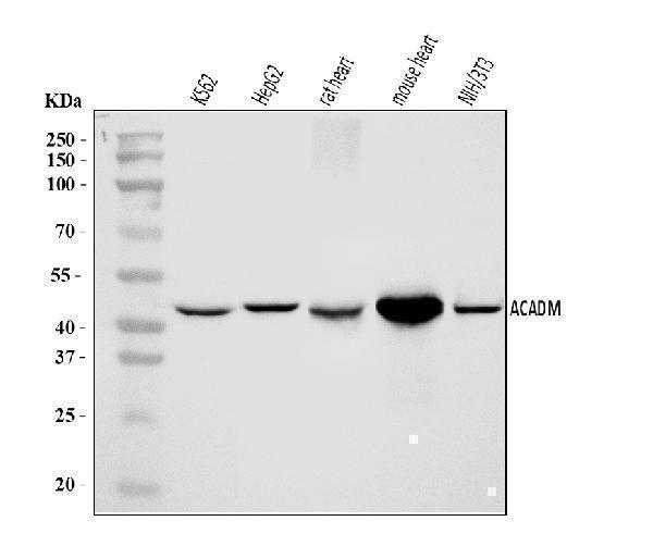
Western blot analysis of ACADM/MCAD using anti-ACADM/MCAD antibody (A02383-2).
Electrophoresis was performed on a 5-20% SDS-PAGE gel at 70V (Stacking gel) / 90V (Resolving gel) for 2-3 hours. The sample well of each lane was loaded with 30 ug of sample under reducing conditions.
Lane 1: human K562 whole cell lysates,
Lane 2: human HepG2 whole cell lysates,
Lane 3: rat heart tissue lysates,
Lane 4: mouse heart tissue lysates,
Lane 5: mouse NIH/3T3 whole cell lysates.
After electrophoresis, proteins were transferred to a nitrocellulose membrane at 150 mA for 50-90 minutes. Blocked the membrane with 5% non-fat milk/TBS for 1.5 hour at RT. The membrane was incubated with rabbit anti-ACADM/MCAD antigen affinity purified polyclonal antibody (Catalog # A02383-2) at 0.25 μg/mL overnight at 4°C, then washed with TBS-0.1%Tween 3 times with 5 minutes each and probed with a goat anti-rabbit IgG-HRP secondary antibody at a dilution of 1:5000 for 1.5 hour at RT. The signal is developed using an Enhanced Chemiluminescent detection (ECL) kit (Catalog # EK1002) with Tanon 5200 system. A specific band was detected for ACADM/MCAD at approximately 47 kDa. The expected band size for ACADM/MCAD is at 47 kDa.

Click image to see more details
IHC analysis of ACADM/MCAD using anti-ACADM/MCAD antibody (A02383-2).
ACADM/MCAD was detected in a paraffin-embedded section of human bladder epithelial carcinoma tissue. Heat mediated antigen retrieval was performed in EDTA buffer (pH 8.0, epitope retrieval solution). The tissue section was blocked with 10% goat serum. The tissue section was then incubated with 2 μg/ml rabbit anti-ACADM/MCAD Antibody (A02383-2) overnight at 4°C. Biotinylated goat anti-rabbit IgG was used as secondary antibody and incubated for 30 minutes at 37°C. The tissue section was developed using Strepavidin-Biotin-Complex (SABC) (Catalog # SA1022) with DAB as the chromogen.

Click image to see more details
IHC analysis of ACADM/MCAD using anti-ACADM/MCAD antibody (A02383-2).
ACADM/MCAD was detected in a paraffin-embedded section of human breast carcinoma tissue. Heat mediated antigen retrieval was performed in EDTA buffer (pH 8.0, epitope retrieval solution). The tissue section was blocked with 10% goat serum. The tissue section was then incubated with 2 μg/ml rabbit anti-ACADM/MCAD Antibody (A02383-2) overnight at 4°C. Biotinylated goat anti-rabbit IgG was used as secondary antibody and incubated for 30 minutes at 37°C. The tissue section was developed using Strepavidin-Biotin-Complex (SABC) (Catalog # SA1022) with DAB as the chromogen.

Click image to see more details
IHC analysis of ACADM/MCAD using anti-ACADM/MCAD antibody (A02383-2).
ACADM/MCAD was detected in a paraffin-embedded section of human Classic Hodgkin's lymphoma tissue. Heat mediated antigen retrieval was performed in EDTA buffer (pH 8.0, epitope retrieval solution). The tissue section was blocked with 10% goat serum. The tissue section was then incubated with 2 μg/ml rabbit anti-ACADM/MCAD Antibody (A02383-2) overnight at 4°C. Biotinylated goat anti-rabbit IgG was used as secondary antibody and incubated for 30 minutes at 37°C. The tissue section was developed using Strepavidin-Biotin-Complex (SABC) (Catalog # SA1022) with DAB as the chromogen.

Click image to see more details
IHC analysis of ACADM/MCAD using anti-ACADM/MCAD antibody (A02383-2).
ACADM/MCAD was detected in a paraffin-embedded section of human hepatocellular carcinoma tissue. Heat mediated antigen retrieval was performed in EDTA buffer (pH 8.0, epitope retrieval solution). The tissue section was blocked with 10% goat serum. The tissue section was then incubated with 2 μg/ml rabbit anti-ACADM/MCAD Antibody (A02383-2) overnight at 4°C. Biotinylated goat anti-rabbit IgG was used as secondary antibody and incubated for 30 minutes at 37°C. The tissue section was developed using Strepavidin-Biotin-Complex (SABC) (Catalog # SA1022) with DAB as the chromogen.

Click image to see more details
IHC analysis of ACADM/MCAD using anti-ACADM/MCAD antibody (A02383-2).
ACADM/MCAD was detected in a paraffin-embedded section of human Metaplasia of squamous cells of the renal pelvis tissue. Heat mediated antigen retrieval was performed in EDTA buffer (pH 8.0, epitope retrieval solution). The tissue section was blocked with 10% goat serum. The tissue section was then incubated with 2 μg/ml rabbit anti-ACADM/MCAD Antibody (A02383-2) overnight at 4°C. Biotinylated goat anti-rabbit IgG was used as secondary antibody and incubated for 30 minutes at 37°C. The tissue section was developed using Strepavidin-Biotin-Complex (SABC) (Catalog # SA1022) with DAB as the chromogen.

Click image to see more details
IHC analysis of ACADM/MCAD using anti-ACADM/MCAD antibody (A02383-2).
ACADM/MCAD was detected in a paraffin-embedded section of human Moderately differentiated adenocarcinoma of the rectum tissue. Heat mediated antigen retrieval was performed in EDTA buffer (pH 8.0, epitope retrieval solution). The tissue section was blocked with 10% goat serum. The tissue section was then incubated with 2 μg/ml rabbit anti-ACADM/MCAD Antibody (A02383-2) overnight at 4°C. Biotinylated goat anti-rabbit IgG was used as secondary antibody and incubated for 30 minutes at 37°C. The tissue section was developed using Strepavidin-Biotin-Complex (SABC) (Catalog # SA1022) with DAB as the chromogen.

Click image to see more details
IF analysis of ACADM/MCAD using anti-ACADM/MCAD antibody (A02383-2).
ACADM/MCAD was detected in an immunocytochemical section of Hela cells. Enzyme antigen retrieval was performed using IHC enzyme antigen retrieval reagent (AR0022) for 15 mins. The cells were blocked with 10% goat serum. And then incubated with 5 μg/mL rabbit anti-ACADM/MCAD Antibody (A02383-2) overnight at 4°C. DyLight®488 Conjugated Goat Anti-Rabbit IgG (BA1127) was used as secondary antibody at 1:100 dilution and incubated for 30 minutes at 37°C. The section was counterstained with DAPI. Visualize using a fluorescence microscope and filter sets appropriate for the label used.

Click image to see more details
Flow Cytometry analysis of Hela cells using anti-ACADM/MCAD antibody (A02383-2).
Overlay histogram showing Hela cells stained with A02383-2 (Blue line). To facilitate intracellular staining, cells were fixed with 4% paraformaldehyde and permeabilized with permeabilization buffer. The cells were blocked with 10% normal goat serum. And then incubated with rabbit anti-ACADM/MCAD Antibody (A02383-2, 1 μg/1x106 cells) for 30 min at 20°C. DyLight®488 conjugated goat anti-rabbit IgG (BA1127, 5-10 μg/1x106 cells) was used as secondary antibody for 30 minutes at 20°C. Isotype control antibody (Green line) was rabbit IgG (1 μg/1x106) used under the same conditions. Unlabelled sample without incubation with primary antibody and secondary antibody (Red line) was used as a blank control.
Specific Publications For Anti-ACADM/MCAD Antibody Picoband® (A02383-2)
Loading publications
Recommended Resources
Here are featured tools and databases that you might find useful.
- Boster's Pathways Library
- Protein Databases
- Bioscience Research Protocol Resources
- Data Processing & Analysis Software
- Photo Editing Software
- Scientific Literature Resources
- Research Paper Management Tools
- Molecular Biology Software
- Primer Design Tools
- Bioinformatics Tools
- Phylogenetic Tree Analysis
Customer Reviews
Have you used Anti-ACADM/MCAD Antibody Picoband®?
Share your experimental results or join a short interview to earn up to $1,000 in product credits or other rewards.
0 Reviews For Anti-ACADM/MCAD Antibody Picoband®
Customer Q&As
Have a question?
Find answers in Q&As, reviews.
Can't find your answer?
Submit your question
