Click image to see more details
-

-
-
-
-
+5
Product Info Summary
| SKU: | A00756 |
|---|---|
| Size: | 0.1 mg |
| Reactive Species: | Human, Mouse, Rat |
| Host: | Rabbit |
| Application: | ELISA, IF, IHC-P, WB |
Customers Who Bought This Also Bought

Product info
Product Name
Anti-ACE2 Antibody
SKU/Catalog Number
A00756
Size
0.1 mg
Form
Liquid
Description
Boster Bio Anti-ACE2 Antibody (Catalog # A00756). Tested in ELISA, WB, IHC-P, IF applications. This antibody reacts with Human, Mouse, Rat.
Storage & Handling
ACE2 antibody can be stored at 4°C for three months and -20°C, stable for up to one year. Avoid repeated freeze-thaw cycles. Antibodies should not be exposed to prolonged high temperatures.
Cite This Product
Anti-ACE2 Antibody (Boster Biological Technology, Pleasanton CA, USA, Catalog # A00756)
Host
Rabbit
Contents
ACE2 Antibody is supplied in PBS containing 0.02% sodium azide.
Clonality
Polyclonal
Isotype
IgG
Immunogen
Anti-ACE2 antibody was raised against a peptide corresponding to 17 amino acids near the center of human ACE2. The immunogen is located within amino acids 180 - 230 of ACE2.
Cross-reactivity
Anti-ACE2 has no cross response to ACE1.
Reactive Species
A00756 is reactive to ACE2 in Human, Mouse, Rat
Observed Molecular Weight
68 kDa
Calculated molecular weight
92.5 kDa
Background of ACE2
Angiotensin-converting enzyme 2 (ACE2) plays a central role in vascular, renal, and myocardial physiology. In contrast to its homolog ACE, ACE2 expression is restricted to heart, kidney, and testis. Recently. ACE2 has also been shown to be a functional receptor of the SARS coronavirus. Homology modeling shows 2019-nCoV has a similar receptor-binding domain structure as SARS-CoV, which suggests COVID-19 (2019-nCoV) may use ACE2 as a receptor in humans for infection. The normal function of ACE2 is to convert the inactive vasoconstrictor angiotensin I (AngI) to Ang1-9 and the active form AngII to Ang1-7, unlike ACE, which converts AngI to AngII. While the role of these vasoactive peptides is not well understood, lack of ACE2 expression in ace2-/ace2- mice leads to severely reduced cardiac contractility, indicating its importance in regulating heart function.
Antibody Validation
Boster validates all antibodies on WB, IHC, ICC, Immunofluorescence, and ELISA with known positive control and negative samples to ensure specificity and high affinity, including thorough antibody incubations.
Application & Images
Applications
A00756 is guaranteed for ELISA, IF, IHC-P, WB Boster Guarantee
Recommend Dilution
| Application | Dilution | Species |
|---|---|---|
| Antibody validated: Western Blot in human | mouse and rat samples; Immunohistochemistry in human samples; Immunofluorescence in human and rat samples. All other applications and species not yet tested. Optimal dilutions for each application should be determined by the researcher. |
Validation Images & Assay Conditions
Click image to see more details
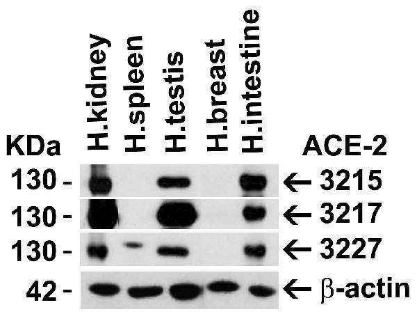
Independent Antibody Validation (IAV) via Protein Expression Profile in Human Tissues
Loading: 15 μg of lysates per lane.
Antibodies: ACE2, A00756 (2 μg/mL), ACE2, 3217 (2 μg/mL), ACE2, 3227 (2 μg/mL) and beta-actin 3779 (1 μg/mL), 1h incubation at RT in 5% NFDM/TBST.
Secondary: Goat anti-rabbit IgG HRP conjugate at 1:10000 dilution.

Click image to see more details
Western Blot Validation in Human Tissues
Loading: 15 μg of lysates per lane.
Antibodies: ACE2, A00756 (2 μg/mL), 1h incubation at RT in 5% NFDM/TBST.
Secondary: Goat anti-rabbit IgG HRP conjugate at 1:10000 dilution.

Click image to see more details
Western Blot Validation in Mouse Tissues
Loading: 15 μg of lysates per lane.
Antibodies: ACE2, A00756 (2 μg/mL), 1h incubation at RT in 5% NFDM/TBST.
Secondary: Goat anti-rabbit IgG HRP conjugate at 1:10000 dilution.

Click image to see more details
Western Blot Validation in Rat Skin Tissue
Loading: 15 μg of lysates per lane.
Antibodies: ACE2, A00756 (2 μg/mL), 1h incubation at RT in 5% NFDM/TBST.
Secondary: Goat anti-rabbit IgG HRP conjugate at 1:10000 dilution.

Click image to see more details
Immunohistochemistry Validation of ACE2 in Human Kidney Tissue
Immunohistochemical analysis of paraffin-embedded human kidney tissue using anti-ACE2 antibody (A00756) at 2 μg/ml. Tissue was fixed with formaldehyde and blocked with 10% serum for 1 h at RT; antigen retrieval was by heat mediation with a citrate buffer (pH6). Samples were incubated with primary antibody overnight at 4˚C. A goat anti-rabbit IgG H&L (HRP) at 1/250 was used as secondary. Counter stained with Hematoxylin.

Click image to see more details
Immunofluorescence Validation of ACE2 in Human Testis Tissue
Immunofluorescent analysis of 4% paraformaldehyde-fixed human testis tissue labeling ACE-2 with A00756 at 20 ug/mL, followed by goat anti-rabbit IgG secondary antibody at 1/500 dilution (green) and DAPI staining (blue).

Click image to see more details
Immunofluorescence Validation of ACE2 in Human Lung Tissue
Immunofluorescent analysis of 4% paraformaldehyde-fixed human lung tissue labeling ACE-2 with A00756 at 20 ug/mL, followed by goat anti-rabbit IgG secondary antibody at 1/500 dilution (green) and DAPI staining (blue).

Click image to see more details
Immunofluorescence Validation of ACE2 in Rat Lung Tissue
Immunofluorescent analysis of 4% paraformaldehyde-fixed rat lung tissue labeling ACE-2 with A00756 at 20 ug/mL, followed by goat anti-rabbit IgG secondary antibody at 1/500 dilution (green) and DAPI staining (blue).

Click image to see more details
Immunofluorescence Validation of ACE2 In Caco2 Cells
Immunofluorescent analysis of 4% paraformaldehyde-fixed Caco2 cells labeling ACE2 with A00756 at 20 ug/mL, followed by goat anti-rabbit IgG secondary antibody at 1/500 dilution (green) and DAPI staining (blue). Image showing membrane staining on Caco2 cells.
Specific Publications For Anti-ACE2 Antibody (A00756)
Loading publications
Recommended Resources
Here are featured tools and databases that you might find useful.
- Boster's Pathways Library
- Protein Databases
- Bioscience Research Protocol Resources
- Data Processing & Analysis Software
- Photo Editing Software
- Scientific Literature Resources
- Research Paper Management Tools
- Molecular Biology Software
- Primer Design Tools
- Bioinformatics Tools
- Phylogenetic Tree Analysis
Customer Reviews
Have you used Anti-ACE2 Antibody?
Share your experimental results or join a short interview to earn up to $1,000 in product credits or other rewards.
0 Reviews For Anti-ACE2 Antibody
Customer Q&As
Have a question?
Find answers in Q&As, reviews.
Can't find your answer?
Submit your question
