Click image to see more details
-

-
-
-
-
+4
Product Info Summary
| SKU: | A05948 |
|---|---|
| Size: | 100uL |
| Reactive Species: | Human, Mouse |
| Host: | Rabbit |
| Application: | ELISA, IHC, WB |
Customers Who Bought This Also Bought

Product info
Product Name
Anti-AHSP/Eraf Antibody
SKU/Catalog Number
A05948
Size
100uL
Form
Lyophilized
Description
Boster Bio Anti-AHSP/Eraf Antibody (Catalog # A05948). Tested in IHC, WB applications. This antibody reacts with Human, Mouse.
Storage & Handling
Store vial at 4°C prior to restoration. For extended storage aliquot contents and freeze at -20°C or below. Avoid cycles of freezing and thawing. Centrifuge product if not completely clear after standing at room temperature. This product is stable for several weeks at 4°C as an undiluted liquid. Dilute only prior to immediate use. Expiration date is one (1) year from date of opening. (Ship at ambient temperature.)
Cite This Product
Anti-AHSP/Eraf Antibody (Boster Biological Technology, Pleasanton CA, USA, Catalog # A05948)
Host
Rabbit
Contents
0.02 M Potassium Phosphate, 0.15 M Sodium Chloride, pH 7.2, 0.01% (w/v) Sodium Azide
Clonality
Polyclonal
Isotype
Antiserum
Immunogen
Anti-AHSP Antibody was produced from whole rabbit serum prepared by repeated immunizations with the full length human AHSP protein.
Reactive Species
A05948 is reactive to AHSP in Human, Mouse
Calculated molecular weight
11.8 kDa
Background of AHSP
AHSP Antibody detects Alpha hemoglobin stabilizing protein (AHSP). AHSP acts as a chaperone to prevent the harmful aggregation of alpha-hemoglobin during normal erythroid cell development. AHSP binds free a-globin to promote its folding and inhibit its ability to produce damaging reactive oxygen species. Reduced AHSP levels correlate with increased severity of b-thalassemia in some human cohorts.
Antibody Validation
Boster validates all antibodies on WB, IHC, ICC, Immunofluorescence, and ELISA with known positive control and negative samples to ensure specificity and high affinity, including thorough antibody incubations.
Application & Images
Applications
A05948 is guaranteed for ELISA, IHC, WB Boster Guarantee
Recommend Dilution
| Application | Dilution | Species |
|---|---|---|
| ELISA: 1:2 | 000 - 1:10 | 000 |
Validation Images & Assay Conditions
Click image to see more details
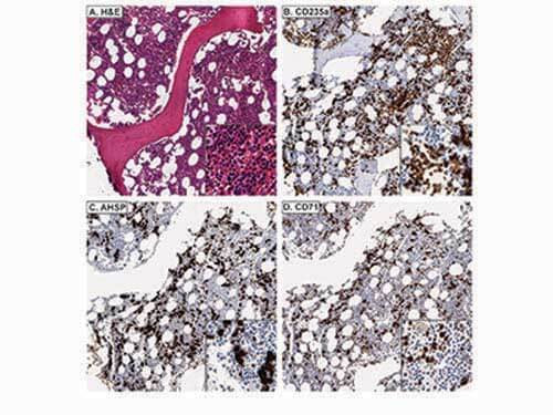
Immunohistochemistry of Rabbit anti-AHSP antibody. Tissue: A.) Normal bone marrow, H&E. B.) CD235a stains both nucleated EPs and mature, anucleate RBCs. C.) AHSP stains nucleated EPs, but not mature, anucleate RBCs. D.) CD71 stains nucleated EPs, but not mature, anucleate RBCs. Fixation: acetic acid-zinc-formalin and formalin fixation, embedded in paraffin. Antigen retrieval: TRIS-EDTA pH9.0. Primary antibody: AHSP antibody at 1:8,000 for overnight at 4°C. Secondary antibody: anti-rabbit secondary at 1:10,000 for 45 min at RT. Localization: Anti-AHSP is cytoplasmic. Staining: AHSP antibody as precipitated brown signal with a purple nuclear counterstain using Bond-max™ – fully automated for IHC.

Click image to see more details
Immunohistochemistry of Rabbit anti-AHSP antibody. Tissue: A.) Acute erythroleukemia, H&E. B.) CD235a stains erythroid blasts and mature, anucleate RBCs. C.) AHSP stains erythroid blasts. D.) CD71 stains erythroid blasts. Fixation: acetic acid-zinc-formalin and formalin fixation, embedded in paraffin. Antigen retrieval: TRIS-EDTA pH9.0. Primary antibody: AHSP antibody at 1:8,000 for overnight at 4°C. Secondary antibody: anti-rabbit secondary at 1:10,000 for 45 min at RT. Localization: Anti-AHSP is cytoplasmic. Staining: AHSP antibody as precipitated brown signal with a purple nuclear counterstain using Bond-max™ – fully automated for IHC.

Click image to see more details
Immunohistochemistry of Rabbit anti-AHSP antibody. CD71 stains myeloid blasts in acute myeloid leukemia, whereas AHSP does not. AHSP stains residual EPs and not myeloid blasts in acute myeloid leukemia with minimal differentiation. (A), whereas CD71 stains both myeloid blasts and EPs (B). AHSP does not stain myeloid blasts in acute myelomonocytic leukemia (D), whereas CD71 does (E). C and F are corresponding H&Es, respectively. Fixation: acetic acid-zinc-formalin and formalin fixation, embedded in paraffin. Antigen retrieval: TRIS-EDTA pH9.0. Primary antibody: AHSP antibody at 1:8,000 for overnight at 4°C Secondary antibody: anti-rabbit secondary at 1:10,000 for 45 min at RT. Localization: Anti-AHSP is cytoplasmic. Staining: AHSP antibody as precipitated brown signal with a purple nuclear counterstain using Bond-max™ – fully automated for IHC.

Click image to see more details
Immunohistochemistry of Rabbit anti-AHSP antibody. AHSP and CD71 stain megakaryocytes in primary myelofibrosis. Tissue: A.) Primary myelofibrosis, H&E. B.) CD235a stains both nucleated EPs and mature, anucleate RBCs. AHSP C.) and CD71. D.) variably stain megakaryocytes and also stain nucleated EPs. Fixation: acetic acid-zinc-formalin and formalin fixation, embedded in paraffin. Antigen retrieval: TRIS-EDTA pH9.0. Primary antibody: AHSP antibody at 1:8,000 for overnight at 4°C Secondary antibody: anti-rabbit secondary at 1:10,000 for 45 min at RT. Localization: Anti-AHSP is cytoplasmic. Staining: AHSP antibody as precipitated brown signal with a purple nuclear counterstain using Bond-max™ – fully automated for IHC.

Click image to see more details
Immunohistochemistry of Rabbit anti-AHSP antibody. AHSP (A) stains residual EPs and not lymphoma cells in DLBCL, whereas CD71 (B) stains both lymphoma cells and EPs. C. Corresponding H&E. AHSP (D) does not metastatic carcinoma, whereas CD71 (E) does. F. Corresponding H&E. Fixation: acetic acid-zinc-formalin and formalin fixation, embedded in paraffin. Antigen retrieval: TRIS-EDTA pH9.0. Primary antibody: AHSP antibody at 1:8,000 for overnight at 4°C Secondary antibody: anti-rabbit secondary at 1:10,000 for 45 min at RT. Localization: Anti-AHSP is cytoplasmic. Staining: AHSP antibody as precipitated brown signal with a purple nuclear counterstain using Bond-max™ – fully automated for IHC.

Click image to see more details
Immunohistochemistry of Rabbit anti-AHSP antibody. Tissue: A.) Giant pronormoblasts are evident in parvoviral infection, H&E. B.) CD235a does not stain giant pronormoblasts. C.) AHSP and D.) CD71 stain giant pronormoblasts. Fixation: acetic acid-zinc-formalin and formalin fixation, embedded in paraffin. Antigen retrieval: TRIS-EDTA pH9.0. Primary antibody: AHSP antibody at 1:8,000 for overnight at 4°C Secondary antibody: anti-rabbit secondary at 1:10,000 for 45 min at RT. Localization: Anti-AHSP is cytoplasmic. Staining: AHSP antibody as precipitated brown signal with a purple nuclear counterstain using Bond-max™ – fully automated for IHC.

Click image to see more details
Western Blot of Rabbit anti-AHSP antibody. Lane 1: Recombinant hAHSP (10ng). Lane 2: Recombinant mAHSP (10ng). Lane 3: mice Spleen cells transfected with TNS9.2-hAHSP. Lane 4: mice Spleen cells transfected with TNS9.2 control vector. Lane 5: mice Spleen cells transfected with TNS9.2-hAHSP fractionated by MACS using Ter119+ microbeads. Lane 6: mice Spleen cells transfected with TNS9.2 control vector fractionated by Ter119+. Lane 7: mice Spleen cells transfected with TNS9.2-hAHSP fractionated by Ter119-. Lane 8: Spleen cells from mice transduced with TNS9.2 control vector fractionated by Ter119-. Load: 10 ng per lane. Primary antibody: AHSP antibody at 1:1,000 for overnight at 4°C. Secondary antibody: HRP Streptavidin secondary antibody at 1:40,000 for 30 min at RT. Block: 5% dry milk 30 min at RT. Predicted/Observed size: ~12kDa. Other band(s): none.

Click image to see more details
Western Blot of Rabbit anti-AHSP antibody. Lane 1: Recombinant hAHSP (2 ng). Lane 2: Recombinant mAHSP (2 ng). Lane 3: RBC Lysates Mouse #24 - GFP. Lane 4: RBC Lysates Mouse #25 - GFP. Lane 5: RBC Lysates Mouse #16 - hAHSP. Lane 6: RBC Lysates Mouse #17 - hAHSP. Lane 7: RBC Lysates Mouse #22 - hAHSP. Lane 8: RBC Lysates Mouse #19 - hAHSP. Lane 9: RBC Lysates Mouse #20 - hAHSP. Load: if not described differently, 10 ng per lane. Primary antibody: hAHSP antibody, Beta-Actin antibody at 1:1,000 for overnight at 4°C. Secondary antibody: HRP Streptavidin secondary antibody at 1:40,000 for 30 min at RT. Block: 5% dry milk 30 min at RT. Predicted/Observed size: ~12kDa. Other band(s): none.
Specific Publications For Anti-AHSP/Eraf Antibody (A05948)
Loading publications
Recommended Resources
Here are featured tools and databases that you might find useful.
- Boster's Pathways Library
- Protein Databases
- Bioscience Research Protocol Resources
- Data Processing & Analysis Software
- Photo Editing Software
- Scientific Literature Resources
- Research Paper Management Tools
- Molecular Biology Software
- Primer Design Tools
- Bioinformatics Tools
- Phylogenetic Tree Analysis
Customer Reviews
Have you used Anti-AHSP/Eraf Antibody?
Share your experimental results or join a short interview to earn up to $1,000 in product credits or other rewards.
0 Reviews For Anti-AHSP/Eraf Antibody
Customer Q&As
Have a question?
Find answers in Q&As, reviews.
Can't find your answer?
Submit your question
3 Customer Q&As for Anti-AHSP/Eraf Antibody
Question
What kind of buffer and how much to use in the reconstitution of A05948?
Verified customer
Asked: 2020-11-13
Answer
The Anti-AHSP/Eraf Antibody (A05948) may be restored with deionized water (or equivalent) to 100ul volume.
Boster Scientific Support
Answered: 2020-11-13
Question
We have the sample size of A05948. How much buffer should we use? Can you tell me the final concentration after the reconstitution?
Verified customer
Asked: 2020-11-13
Answer
The sample size of the Anti-AHSP/Eraf Antibody (A05948) may be reconstituted in deionized water to a final volume of 25ul. This will allow to use antibody at dilutions listed on the datasheet. Alternatively, our company also proposes that this vial contains a relatively low volume of reagent (25 µL). To minimize loss of volume dilute 1:10 by adding 225 µL of the buffer stated above directly to the vial. Recap, mix thoroughly and briefly centrifuge to collect the volume at the bottom of the vial. Use this intermediate dilution when calculating final dilutions.
Boster Scientific Support
Answered: 2020-11-13
Question
We are currently using anti-AHSP/Eraf antibody A05948 for human tissue, and we are happy with the IHC results. The species of reactivity given in the datasheet says human. Is it possible that the antibody can work on goat tissues as well?
B. Wu
Verified customer
Asked: 2020-02-05
Answer
The anti-AHSP/Eraf antibody (A05948) has not been tested for cross reactivity specifically with goat tissues, though there is a good chance of cross reactivity. We have an innovator award program that if you test this antibody and show it works in goat you can get your next antibody for free. Please contact me if I can help you with anything.
Boster Scientific Support
Answered: 2020-02-05
