Click image to see more details
Product Info Summary
| SKU: | A01820-1 |
|---|---|
| Size: | 100 μg/vial |
| Reactive Species: | Human |
| Host: | Rabbit |
| Application: | Flow Cytometry, WB |
Customers Who Bought This Also Bought

Product info
Product Name
Anti-AKR1C3 Antibody Picoband®
SKU/Catalog Number
A01820-1
Size
100 μg/vial
Form
Lyophilized
Description
Boster Bio Anti-AKR1C3 Antibody Picoband® catalog # A01820-1. Tested in Flow Cytometry, WB applications. This antibody reacts with Human. The brand Picoband indicates this is a premium antibody that guarantees superior quality, high affinity, and strong signals with minimal background in Western blot applications. Only our best-performing antibodies are designated as Picoband, ensuring unmatched performance.
Storage & Handling
At -20°C for one year from date of receipt. After reconstitution, at 4°C for one month. It can also be aliquotted and stored frozen at -20°C for six months. Avoid repeated freezing and thawing.
Cite This Product
Anti-AKR1C3 Antibody Picoband® (Boster Biological Technology, Pleasanton CA, USA, Catalog # A01820-1)
Host
Rabbit
Contents
Each vial contains 4 mg Trehalose, 0.9 mg NaCl, 0.2 mg Na2HPO4.
Clonality
Polyclonal
Isotype
Rabbit IgG
Immunogen
A synthetic peptide corresponding to a sequence at the C-terminus of human AKR1C3.
Cross-reactivity
No cross-reactivity with other proteins.
Reactive Species
A01820-1 is reactive to AKR1C3 in Human
Observed Molecular Weight
37 kDa
Calculated molecular weight
36.9 kDa
Background of AKR1C3
Aldo-keto reductase family 1 member C3 (AKR1C3), also known as 17β-hydroxysteroid dehydrogenase type 5 (17β-HSD5, HSD17B5) is a key steroidogenic enzyme that in humans is encoded by the AKR1C3 gene. This gene encodes a member of the aldo/keto reductase superfamily, which consists of more than 40 known enzymes and proteins. These enzymes catalyze the conversion of aldehydes and ketones to their corresponding alcohols by utilizing NADH and/or NADPH as cofactors. The enzymes display overlapping but distinct substrate specificity. This enzyme catalyzes the reduction of prostaglandin (PG) D2, PGH2 and phenanthrenequinone (PQ), and the oxidation of 9alpha,11beta-PGF2 to PGD2. It may play an important role in the pathogenesis of allergic diseases such as asthma, and may also have a role in controlling cell growth and/or differentiation. This gene shares high sequence identity with three other gene members and is clustered with those three genes at chromosome 10p15-p14. Three transcript variants encoding different isoforms have been found for this gene.
Antibody Validation
Boster validates all antibodies on WB, IHC, ICC, Immunofluorescence, and ELISA with known positive control and negative samples to ensure specificity and high affinity, including thorough antibody incubations.
Application & Images
Applications
A01820-1 is guaranteed for Flow Cytometry, WB Boster Guarantee
Recommend Dilution
| Application | Dilution | Species |
|---|---|---|
| Western blot | 0.25-0.5 μg/ml | Human |
| Flow Cytometry (Fixed) | 1-3 μg/1x106 cells | Human |
Tested application
Suggested blocking solution with 5% non-fat milk or BSA; (*)Recommended protein loading: 20-40 µg per lane
Validation Images & Assay Conditions
Click image to see more details
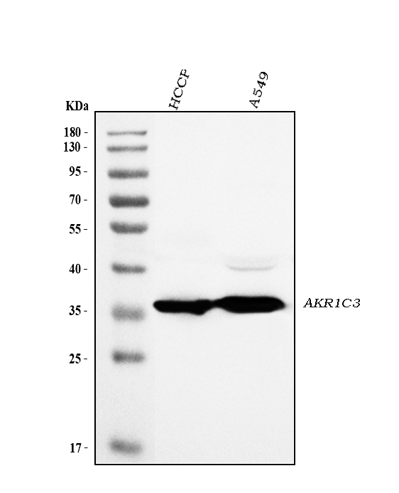
Western blot analysis of AKR1C3 using anti-AKR1C3 antibody (A01820-1).
Electrophoresis was performed on a 5-20% SDS-PAGE gel at 70V (Stacking gel) / 90V (Resolving gel) for 2-3 hours. The sample well of each lane was loaded with 30 ug of sample under reducing conditions.
Lane 1: human HCCP tissue lysates,
Lane 2: human A549 whole cell lysates.
After electrophoresis, proteins were transferred to a nitrocellulose membrane at 150 mA for 50-90 minutes. Blocked the membrane with 5% non-fat milk/TBS for 1.5 hour at RT. The membrane was incubated with rabbit anti-AKR1C3 antigen affinity purified polyclonal antibody (Catalog # A01820-1) at 0.5 μg/mL overnight at 4°C, then washed with TBS-0.1%Tween 3 times with 5 minutes each and probed with a goat anti-rabbit IgG-HRP secondary antibody at a dilution of 1:5000 for 1.5 hour at RT. The signal is developed using an Enhanced Chemiluminescent detection (ECL) kit (Catalog # EK1002) with Tanon 5200 system. A specific band was detected for AKR1C3 at approximately 37 kDa. The expected band size for AKR1C3 is at 37 kDa.

Click image to see more details
Flow Cytometry analysis of HepG2 cells using anti-AKR1C3 antibody (A01820-1).
Specific Publications For Anti-AKR1C3 Antibody Picoband® (A01820-1)
Loading publications
Recommended Resources
Here are featured tools and databases that you might find useful.
- Boster's Pathways Library
- Protein Databases
- Bioscience Research Protocol Resources
- Data Processing & Analysis Software
- Photo Editing Software
- Scientific Literature Resources
- Research Paper Management Tools
- Molecular Biology Software
- Primer Design Tools
- Bioinformatics Tools
- Phylogenetic Tree Analysis
Customer Reviews
Have you used Anti-AKR1C3 Antibody Picoband®?
Share your experimental results or join a short interview to earn up to $1,000 in product credits or other rewards.
0 Reviews For Anti-AKR1C3 Antibody Picoband®
Customer Q&As
Have a question?
Find answers in Q&As, reviews.
Can't find your answer?
Submit your question
