Click image to see more details
-

-
-
-
-
+2
Product Info Summary
| SKU: | PB9472 |
|---|---|
| Size: | 100 μg/vial |
| Reactive Species: | Human, Mouse, Rat |
| Host: | Rabbit |
| Application: | Flow Cytometry, IHC, WB |
Customers Who Bought This Also Bought

Product info
Product Name
Anti-ALDH2 Antibody Picoband®
SKU/Catalog Number
PB9472
PB0494 is an alternative SKU for this antibody, used in previous lots.
Size
100 μg/vial
Form
Lyophilized
Description
Boster Bio Anti-ALDH2 Antibody Picoband® catalog # PB9472. Tested in Flow Cytometry, IHC, WB applications. This antibody reacts with Human, Mouse, Rat. The brand Picoband indicates this is a premium antibody that guarantees superior quality, high affinity, and strong signals with minimal background in Western blot applications. Only our best-performing antibodies are designated as Picoband, ensuring unmatched performance.
Storage & Handling
Store at -20˚C for one year from date of receipt. After reconstitution, at 4˚C for one month. It can also be aliquotted and stored frozen at -20˚C for six months. Avoid repeated freeze-thaw cycles.
Cite This Product
Anti-ALDH2 Antibody Picoband® (Boster Biological Technology, Pleasanton CA, USA, Catalog # PB9472)
Host
Rabbit
Contents
Each vial contains 4 mg Trehalose, 0.9 mg NaCl and 0.2 mg Na2HPO4.
Clonality
Polyclonal
Isotype
Rabbit IgG
Immunogen
A synthetic peptide corresponding to a sequence at the N-terminus of human ALDH2, different from the related mouse sequence by two amino acids, and from the related rat sequence by one amino acid.
Cross-reactivity
No cross-reactivity with other proteins
Reactive Species
PB9472 is reactive to ALDH2 in Human, Mouse, Rat
Observed Molecular Weight
56 kDa
Calculated molecular weight
56.4 kDa
Background of ALDH2
ALDH2 (Aldehyde Dehydrogenase 2 Family) is a human gene. The enzyme encoded by this gene belongs to the aldehyde dehydrogenase family of enzymes that catalyze the chemical transformation from acetaldehyde to acetic acid. Aldehyde dehydrogenase is the second enzyme of the major oxidative pathway of alcohol metabolism. Hsu et al. (1985) assigned the ALDH2 locus to chromosome 12 by means of a cDNA probe and Southern blot analysis of somatic cell hybrids. Using an unbiased proteomic search, Chen et al. (2008) identified mitochondrial ALDH2 as an enzyme whose activation correlated with reduced ischemic heart damage in rodent models. A high-throughput screen identified a small molecule activator of ALDH2, which they called Alda-1, that, when administered to rats before an ischemic event, reduced infarct size by 60%, most likely through its inhibitory effect on the formation of cytotoxic aldehydes.
Antibody Validation
Boster validates all antibodies on WB, IHC, ICC, Immunofluorescence, and ELISA with known positive control and negative samples to ensure specificity and high affinity, including thorough antibody incubations.
Application & Images
Applications
PB9472 is guaranteed for Flow Cytometry, IHC, WB Boster Guarantee
Recommend Dilution
| Application | Dilution | Species |
|---|---|---|
| Western blot | 0.1-0.5μg/ml | Human, Mouse, Rat |
| Immunohistochemistry (Paraffin-embedded Section) | 2-5μg/ml | Human |
| Flow Cytometry(Fixed) | 1-3 μg/1x106 cells | Human |
Tested application
Suggested blocking solution with 5% non-fat milk or BSA; (*)Recommended protein loading: 20-40 µg per lane
Use TE buffer pH 9.0 for antigen retrieval; (*) citrate buffer pH 6.0 is an alternative.
Validation Images & Assay Conditions
Click image to see more details
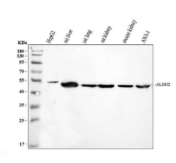
Western blot analysis of ALDH2 using anti-ALDH2 antibody (PB9472).
Electrophoresis was performed on a 10% SDS-PAGE gel at 80V (Stacking gel) / 120V (Resolving gel) for 2 hours. The sample well of each lane was loaded with 30 ug of sample under reducing conditions.
Lane 1: human HepG2 whole cell lysates,
Lane 2: rat liver tissuelysates,
Lane 3: rat lung tissue lysates,
Lane 4: rat kidney tissue lysates,
Lane 5: mouse kidney tissue lysates,
Lane 6: mouse ANA-1 whole cell lysates.
After electrophoresis, proteins were transferred to a nitrocellulose membrane at 150 mA for 50-90 minutes. Blocked the membrane with 5% non-fat milk/TBS for 1.5 hour at RT. The membrane was incubated with rabbit anti-ALDH2 antigen affinity purified polyclonal antibody (PB9472) at 0.5 μg/mL overnight at 4°C, then washed with TBS-0.1%Tween 3 times with 5 minutes each and probed with a goat anti-rabbit IgG-HRP secondary antibody (Catalog # BA1054) at a dilution of 1:5000 for 1.5 hour at RT. The signal is developed using an ECL Plus Western Blotting Substrate (Catalog # AR1196-200) with Tanon 5200 system. A specific band was detected for ALDH2 at approximately 56 kDa. The expected band size for ALDH2 is at 56 kDa.

Click image to see more details
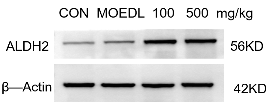
Western blot analysis of ALDH2 using anti-ALDH2 antibody (PB9472).
Electrophoresis was performed on a 5-20% SDS-PAGE gel at 80V (Stacking gel) / 120V (Resolving gel) for 2 hours. The sample well of each lane was loaded with 30 ug of sample under reducing conditions.
Lane 1: Control group-mouse hippocampus tissue,
Lane 2: Model group-mouse hippocampus tissue,
Lane 3: Drug treatment (100 mg/kg) – Mouse hippocampus tissue,
Lane 4: : Drug treatment (100 mg/kg) – Mouse hippocampus tissue.
After electrophoresis, proteins were transferred to a nitrocellulose membrane at 150 mA for 50-90 minutes. Blocked the membrane with 5% non-fat milk/TBS for 1.5 hour at RT. The membrane was incubated with rabbit anti-ALDH2 antigen affinity purified polyclonal antibody (PB9472) overnight at 4°C, then washed with TBS-0.1%Tween 3 times with 5 minutes each and probed with a goat anti-rabbit IgG-HRP secondary antibody (Catalog # BA1054) for 1 hour at RT. The signal is developed using an ECL Plus Western Blotting Substrate (Catalog # AR1196-200) with ChemiDoc MP system. A specific band was detected for ALDH2 at approximately 56 kDa. The expected band size for ALDH2 is at 56 kDa.

Click image to see more details
Western blot analysis of ALDH2 using anti-ALDH2 antibody (PB9472).
Electrophoresis was performed on a 10% SDS-PAGE gel at 80V (Stacking gel) / 120V (Resolving gel) for 2 hours. The sample well of each lane was loaded with 30 ug of sample under reducing conditions.
Lane 1: human A549- WT whole cell lysates,
Lane 2: human A549-ALDH2 KO whole cell lysates.
After electrophoresis, proteins were transferred to a nitrocellulose membrane at 150 mA for 50-90 minutes. Blocked the membrane with 5% non-fat milk/TBS for 1.5 hour at RT. The membrane was incubated with rabbit anti-ALDH2 antigen affinity purified polyclonal antibody (PB9472) at 0.5 μg/mL overnight at 4°C, then washed with TBS-0.1%Tween 3 times with 5 minutes each and probed with a goat anti-rabbit IgG-HRP secondary antibody at a dilution of 1:5000 for 1.5 hour at RT. The signal is developed using an ECL Plus Western Blotting Substrate (Catalog # AR1196-200) with Tanon 5200 system. A specific band was detected for ALDH2 at approximately 56 kDa. The expected band size for ALDH2 is at 56 kDa.

Click image to see more details
IHC analysis of ALDH2 using anti-ALDH2 antibody (PB9473).
ALDH2 was detected in a paraffin-embedded section of human liver cancer tissue. Heat mediated antigen retrieval was performed in EDTA buffer (pH 8.0, epitope retrieval solution). The tissue section was blocked with 10% goat serum. The tissue section was then incubated with 2 μg/ml rabbit anti-ALDH2 Antibody (PB9473) overnight at 4°C. Peroxidase Conjugated Goat Anti-rabbit IgG was used as secondary antibody and incubated for 30 minutes at 37°C. The tissue section was developed using HRP Conjugated Rabbit IgG Super Vision Assay Kit (Catalog # SV0002) with DAB as the chromogen.

Click image to see more details
Flow Cytometry analysis of HepG2 cells using anti-ALDH2 antibody (PB9472).
Overlay histogram showing HepG2 cells stained with PB9472 (Blue line). To facilitate intracellular staining, cells were fixed with 4% paraformaldehyde and permeabilized with permeabilization buffer. The cells were blocked with 10% normal goat serum. And then incubated with rabbit anti-ALDH2 Antibody (PB9472, 1 μg/1x106 cells) for 30 min at 20°C. DyLight®488 conjugated goat anti-rabbit IgG (BA1127, 5-10 μg/1x106 cells) was used as secondary antibody for 30 minutes at 20°C. Isotype control antibody (Green line) was rabbit IgG (1 μg/1x106) used under the same conditions. Unlabelled sample without incubation with primary antibody and secondary antibody (Red line) was used as a blank control.

Click image to see more details
Effects of WEATs on ALDH2 expression in the liver of mice. Representative immunoblots (A) and IHC images (B) of ALDH2 expression in the liver. Quantification of ALDH2 expression by western blotting (C) and IHC (D) . Each value represents the mean ± SEM ( n = 9). * p < 0.05 vs. MOD; ** p < 0.01 vs. MOD.
Index in PubMed under a CC BY license. PMID: 35677547
Specific Publications For Anti-ALDH2 Antibody Picoband® (PB9472)
Loading publications
Recommended Resources
Here are featured tools and databases that you might find useful.
- Boster's Pathways Library
- Protein Databases
- Bioscience Research Protocol Resources
- Data Processing & Analysis Software
- Photo Editing Software
- Scientific Literature Resources
- Research Paper Management Tools
- Molecular Biology Software
- Primer Design Tools
- Bioinformatics Tools
- Phylogenetic Tree Analysis
Customer Reviews
Have you used Anti-ALDH2 Antibody Picoband®?
Share your experimental results or join a short interview to earn up to $1,000 in product credits or other rewards.
1 Reviews For Anti-ALDH2 Antibody Picoband®
The antibody was used to perform WB quantitative detection of ALDH2 protein in mouse hippocampus. The experimental results showed clear and distinct bands, indicating that it is suitable for WB detection of ALDH2 protein in mouse tissues.
Excellent
| SKU | PB9472 |
|---|---|
| Application | Western Blot |
| Sample | Mouse hippocampus tissue |
| Sample Processing Description | The mouse hippocampus tissue was lysed using RIPA lysis buffer supplemented with a protease inhibitor cocktail. Protein concentration was determined, and samples were mixed with 5× protein loading buffer and denatured by heating at 100°C for 10 minutes. Five microliters of each protein sample were loaded per lane onto SDS-PAGE. |
| Primary Antibody | Anti-ALDH2 Antibody Picoband® |
| Primary Incubation | overnight at 4 ℃ |
| Secondary Antibody | HRP-conjugated Anti-Rabbit IgG Secondary Antibody |
| Secondary Incubation | 1 hour in room temperature |
| Detection | Substrate: Ultra-sensitive ECL luminescent reagent (Cat# AR1191), Imaging system: ChemiDoc MP (Bio-Rad) |
| Results Summary | The antibody was used for quantitative WB detection of ALDH2 protein in mouse hippocampus. The results showed clear and distinct bands, indicating its suitability for WB detection of ALDH2 protein in mouse tissues. |
Changbin Yuan, LNUTCM
Verified customer
Submitted 2025-11-06
Customer Q&As
Have a question?
Find answers in Q&As, reviews.
Can't find your answer?
Submit your question
5 Customer Q&As for Anti-ALDH2 Antibody Picoband®
Question
Would anti-ALDH2 antibody PB9472 work on feline IHC with brain cajal-retzius cell?
D. Singh
Verified customer
Asked: 2019-02-18
Answer
Our lab technicians have not tested anti-ALDH2 antibody PB9472 on feline. You can run a BLAST between feline and the immunogen sequence of anti-ALDH2 antibody PB9472 to see if they may cross-react. If the sequence homology is close, then you can perform a pilot test. Keep in mind that since we have not validated feline samples, this use of the antibody is not covered by our guarantee. However we have an innovator award program that if you test this antibody and show it works in feline brain cajal-retzius cell in IHC, you can get your next antibody for free.
Boster Scientific Support
Answered: 2019-02-18
Question
We are currently using anti-ALDH2 antibody PB9472 for human tissue, and we are happy with the IHC results. The species of reactivity given in the datasheet says human, mouse, rat. Is it possible that the antibody can work on horse tissues as well?
G. Singh
Verified customer
Asked: 2018-10-19
Answer
The anti-ALDH2 antibody (PB9472) has not been validated for cross reactivity specifically with horse tissues, though there is a good chance of cross reactivity. We have an innovator award program that if you test this antibody and show it works in horse you can get your next antibody for free. Please contact me if I can help you with anything.
Boster Scientific Support
Answered: 2018-10-19
Question
I see that the anti-ALDH2 antibody PB9472 works with WB, what is the protocol used to produce the result images on the product page?
Verified Customer
Verified customer
Asked: 2018-10-12
Answer
You can find protocols for WB on the "support/technical resources" section of our navigation menu. If you have any further questions, please send an email to support@bosterbio.com
Boster Scientific Support
Answered: 2018-10-12
Question
I appreciate helping with my inquiry over the phone. Here are the WB image, lot number and protocol we used for brain cajal-retzius cell using anti-ALDH2 antibody PB9472. Let me know if you need anything else.
Verified Customer
Verified customer
Asked: 2018-01-02
Answer
I appreciate the data. You have provided everything we needed. Our lab team are working to resolve your inquiry as quickly as possible, and we appreciate your patience and understanding! Please let me know if there is anything you need in the meantime.
Boster Scientific Support
Answered: 2018-01-02
Question
Does PB9472 anti-ALDH2 antibody work on parafin embedded sections? If so, which fixation method do you recommend we use (PFA, paraformaldehyde, other)?
A. Parker
Verified customer
Asked: 2015-01-19
Answer
It shows on the product datasheet, PB9472 anti-ALDH2 antibody as been tested on WB. It is best to use PFA for fixation because it has better tissue penetration ability. PFA needs to be prepared fresh before use. Long term stored PFA turns into formalin, as the PFA molecules congregate and become formalin.
Boster Scientific Support
Answered: 2015-01-19
