Click image to see more details
Product Info Summary
| SKU: | A06146 |
|---|---|
| Size: | 100 μg/vial |
| Reactive Species: | Human, Mouse, Rat |
| Host: | Rabbit |
| Application: | WB |
Customers Who Bought This Also Bought

Product info
Product Name
Anti-AMD1 Antibody Picoband®
SKU/Catalog Number
A06146
Size
100 μg/vial
Form
Lyophilized
Description
Boster Bio Anti-AMD1 Antibody Picoband® catalog # A06146. Tested in WB applications. This antibody reacts with Human, Mouse, Rat. The brand Picoband indicates this is a premium antibody that guarantees superior quality, high affinity, and strong signals with minimal background in Western blot applications. Only our best-performing antibodies are designated as Picoband, ensuring unmatched performance.
Storage & Handling
Store at -20˚C for one year from date of receipt. After reconstitution, at 4˚C for one month. It can also be aliquotted and stored frozen at -20˚C for six months. Avoid repeated freeze-thaw cycles.
Cite This Product
Anti-AMD1 Antibody Picoband® (Boster Biological Technology, Pleasanton CA, USA, Catalog # A06146)
Host
Rabbit
Contents
Each vial contains 4 mg Trehalose, 0.9 mg NaCl and 0.2 mg Na2HPO4.
Clonality
Polyclonal
Isotype
Rabbit IgG
Immunogen
A synthetic peptide corresponding to a sequence in the middle region of human AMD1, identical to the related mouse and rat sequences.
Cross-reactivity
No cross-reactivity with other proteins.
Reactive Species
A06146 is reactive to AMD1 in Human, Mouse, Rat
Observed Molecular Weight
40-42 kDa
Calculated molecular weight
38.3 kDa
Background of AMD1
S-adenosylmethionine decarboxylase (AdoMet-DC), also known as S-adenosylmethionine decarboxylase proenzyme (SAMDC) or AMD1, is a key enzyme in polyamine biosynthesis. It is localized to chromosome region 6q21-q22. SAMDC has an unusual distribution in polysomes from cells of T lymphocyte origin. It associates predominantly with monosomes and small polysomes with none located in the preribosomal or ribonucleoprotein pool. SAMDC is a critical regulatory enzyme of the polyamine synthetic pathway, and a well-studied drug target. Since SAMDC is a key regulatory enzyme in the synthesis of spermidine and spermine, the marked increase in SAMDC activity in the neonate and the sustained high enzyme levels throughout adulthood, imply a role for these polyamines in both development and mature brain function.
Antibody Validation
Boster validates all antibodies on WB, IHC, ICC, Immunofluorescence, and ELISA with known positive control and negative samples to ensure specificity and high affinity, including thorough antibody incubations.
Application & Images
Applications
A06146 is guaranteed for WB Boster Guarantee
Recommend Dilution
| Application | Dilution | Species |
|---|---|---|
| Western blot | 0.1-0.5μg/ml | Human, Mouse, Rat |
Tested application
Suggested blocking solution with 5% non-fat milk or BSA; (*)Recommended protein loading: 20-40 µg per lane
Validation Images & Assay Conditions
Click image to see more details
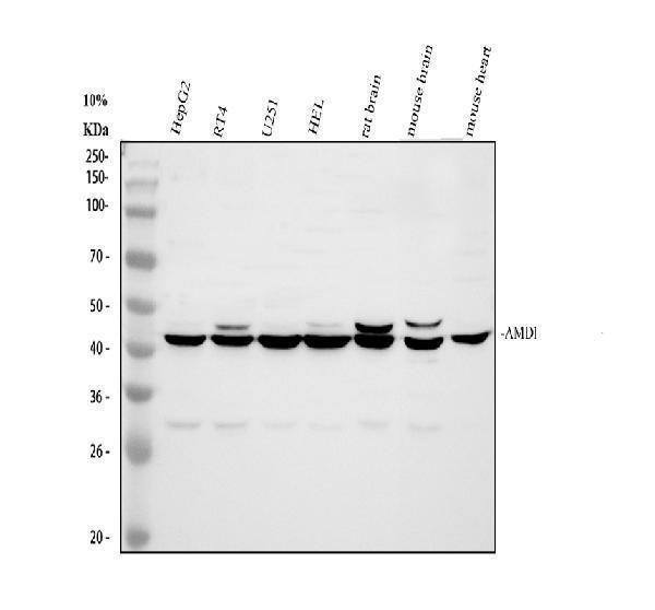
Western blot analysis of AMD1 using anti-AMD1 antibody (A06146).
Electrophoresis was performed on a 10% SDS-PAGE gel at 80V (Stacking gel) / 120V (Resolving gel) for 2 hours. The sample well of each lane was loaded with 30 ug of sample under reducing conditions.
Lane 1: human HepG2 whole cell lysates,
Lane 2: human RT4 whole cell lysates,
Lane 3: human U251 whole cell lysates,
Lane 4: human HEL whole cell lysates,
Lane 5: rat brain tissue lysates,
Lane 6: mouse brain tissue lysates,
Lane 7: mouse heart tissue lysates.
After electrophoresis, proteins were transferred to a nitrocellulose membrane at 150 mA for 50-90 minutes. Blocked the membrane with 5% non-fat milk/TBS for 1.5 hour at RT. The membrane was incubated with rabbit anti-AMD1 antigen affinity purified polyclonal antibody (A06146) at 0.5 μg/mL overnight at 4°C, then washed with TBS-0.1%Tween 3 times with 5 minutes each and probed with a goat anti-rabbit IgG-HRP secondary antibody (Catalog # BA1054) at a dilution of 1:5000 for 1.5 hour at RT. The signal is developed using an ECL Plus Western Blotting Substrate (Catalog # AR1196-200) with Tanon 5200 system. A specific band was detected for AMD1 at approximately 40-42 kDa. The expected band size for AMD1 is at 38 kDa.
Specific Publications For Anti-AMD1 Antibody Picoband® (A06146)
Loading publications
Recommended Resources
Here are featured tools and databases that you might find useful.
- Boster's Pathways Library
- Protein Databases
- Bioscience Research Protocol Resources
- Data Processing & Analysis Software
- Photo Editing Software
- Scientific Literature Resources
- Research Paper Management Tools
- Molecular Biology Software
- Primer Design Tools
- Bioinformatics Tools
- Phylogenetic Tree Analysis
Customer Reviews
Have you used Anti-AMD1 Antibody Picoband®?
Share your experimental results or join a short interview to earn up to $1,000 in product credits or other rewards.
0 Reviews For Anti-AMD1 Antibody Picoband®
Customer Q&As
Have a question?
Find answers in Q&As, reviews.
Can't find your answer?
Submit your question
7 Customer Q&As for Anti-AMD1 Antibody Picoband®
Question
Is there a BSA free version of anti-AMD1 antibody A06146 available?
Verified Customer
Verified customer
Asked: 2020-02-10
Answer
I appreciate your recent telephone inquiry. I can confirm that some lots of this anti-AMD1 antibody A06146 are BSA free. For now, these lots are available and we can make a BSA free formula for you free of charge. It will take 3 extra days to prepare. If you require this antibody BSA free again in future, please do not hesitate to contact me and I will be pleased to check which lots we have in stock that are BSA free.
Boster Scientific Support
Answered: 2020-02-10
Question
Is a blocking peptide available for product anti-AMD1 antibody (A06146)?
Verified Customer
Verified customer
Asked: 2019-10-15
Answer
We do provide the blocking peptide for product anti-AMD1 antibody (A06146). If you would like to place an order for it please contact support@bosterbio.com and make a special request.
Boster Scientific Support
Answered: 2019-10-15
Question
I am interested in to test anti-AMD1 antibody A06146 on rat placenta for research purposes, then I may be interested in using anti-AMD1 antibody A06146 for diagnostic purposes as well. Is the antibody suitable for diagnostic purposes?
Verified Customer
Verified customer
Asked: 2019-10-14
Answer
The products we sell, including anti-AMD1 antibody A06146, are only intended for research use. They would not be suitable for use in diagnostic work. If you have the means to develop a product into diagnostic use, and are interested in collaborating with us and develop our product into an IVD product, please contact us for more discussions.
Boster Scientific Support
Answered: 2019-10-14
Question
We appreciate helping with my inquiry over the phone. Here are the WB image, lot number and protocol we used for placenta using anti-AMD1 antibody A06146. Let me know if you need anything else.
Verified Customer
Verified customer
Asked: 2019-09-17
Answer
Thanks for the data. You have provided everything we needed. Our lab team are working to resolve your inquiry as quickly as possible, and we appreciate your patience and understanding! Please let me know if there is anything you need in the meantime.
Boster Scientific Support
Answered: 2019-09-17
Question
See below the WB image, lot number and protocol we used for placenta using anti-AMD1 antibody A06146. Please let me know if you require anything else.
F. Dhar
Verified customer
Asked: 2018-05-10
Answer
Thank you very much for the data. Our lab team are working to resolve this as quickly as possible, and we appreciate your patience and understanding! You have provided everything we needed. Please let me know if there is anything you need in the meantime.
Boster Scientific Support
Answered: 2018-05-10
Question
I see that the anti-AMD1 antibody A06146 works with IHC, what is the protocol used to produce the result images on the product page?
Verified Customer
Verified customer
Asked: 2017-09-28
Answer
You can find protocols for IHC on the "support/technical resources" section of our navigation menu. If you have any further questions, please send an email to support@bosterbio.com
Boster Scientific Support
Answered: 2017-09-28
Question
Would A06146 anti-AMD1 antibody work on parafin embedded sections? If so, which fixation method do you recommend we use (PFA, paraformaldehyde, other)?
Verified Customer
Verified customer
Asked: 2017-08-16
Answer
You can see on the product datasheet, A06146 anti-AMD1 antibody as been validated on IHC. It is best to use PFA for fixation because it has better tissue penetration ability. PFA needs to be prepared fresh before use. Long term stored PFA turns into formalin, as the PFA molecules congregate and become formalin.
Boster Scientific Support
Answered: 2017-08-16
