Click image to see more details
-

-
-
-
-
+1
Product Info Summary
| SKU: | A01787-1 |
|---|---|
| Size: | 100 μg/vial |
| Reactive Species: | Mouse, Rat |
| Host: | Rabbit |
| Application: | Flow Cytometry, IHC, WB, ELISA (Cap) |
Customers Who Bought This Also Bought

Product info
Product Name
Anti-Amphiregulin/Areg Antibody Picoband®
SKU/Catalog Number
A01787-1
Size
100 μg/vial
Form
Lyophilized
Description
Amphiregulin (AREG) is an EGFR ligand (EGF family) that supports autocrine/paracrine mitogenic signaling in multiple cell types and is commonly discussed in epithelial repair and inflammation-associated remodeling contexts (context dependent). Assay context: Mouse/Rat-reactive antibody tested for ELISA, flow cytometry, IHC, and WB; AREG readouts are often compared with other growth/angiogenesis mediators such as FGF2 (cross-factor interpretation is putative and study-design dependent).
Storage & Handling
Store at -20˚C for one year from date of receipt. After reconstitution, at 4˚C for one month. It can also be aliquotted and stored frozen at -20˚C for six months. Avoid repeated freeze-thaw cycles.
Cite This Product
Anti-Amphiregulin/Areg Antibody Picoband® (Boster Biological Technology, Pleasanton CA, USA, Catalog # A01787-1)
Host
Rabbit
Contents
Each vial contains 4 mg Trehalose, 0.9 mg NaCl and 0.2 mg Na2HPO4.
Clonality
Polyclonal
Isotype
Rabbit IgG
Immunogen
E. coli-derived mouse Amphiregulin recombinant protein (Position:V100-K191).
Cross-reactivity
No cross-reactivity with other proteins.
Reactive Species
A01787-1 is reactive to Areg in Mouse, Rat
Observed Molecular Weight
45 kDa
Calculated molecular weight
27.5 kDa
Background of Areg
Amphiregulin, also known as AREG, is a protein that in humans is encoded by the AREG gene. The protein encoded by this gene is a member of the epidermal growth factor (EGF) family. It is an autocrine growth factor as well as a mitogen for astrocytes, Schwann cells, fibroblasts. It is related to epidermal growth factor (EGF) and transforming growth factor alpha (TGF-alpha). This protein interacts with the Epidermal growth factor receptor (EGFR) to promote the growth of normal epithelial cells. It is mapped to 9q32. It has been shown to play a role in immunity, inflammation, tissue repair, and lung and mammary gland development. Homozygous knockout mice for this gene exhibit impaired immune system regulation in the skin and gene expression changes characteristic of chronic liver damage.
Antibody Validation
Boster validates all antibodies on WB, IHC, ICC, Immunofluorescence, and ELISA with known positive control and negative samples to ensure specificity and high affinity, including thorough antibody incubations.
Application & Images
Applications
A01787-1 is guaranteed for Flow Cytometry, IHC, WB, ELISA (Cap) Boster Guarantee
Assay Dilutions Recommendation
The recommendations below provide a starting point for assay optimization. The actual working concentration varies and should be decided by the user.
Western blot, 0.1-0.5μg/ml, Mouse, Rat
Immunohistochemistry (Paraffin-embedded Section), 2-5μg/ml, Mouse, Rat
Flow Cytometry(Fixed), 1-3 μg/1x106 cells, Rat
ELISA (Cap), 1-5μg/ml
Positive Control
WB: rat brain tissue, rat thymus tissue, rat spleen tissue, rat PC-12 whole cell, mouse brain tissue, mouse thymus tissue, mouse spleen tissue, mouse RAW264.7 whole cell
IHC: mouse brain tissue, rat brain tissue
FCM: PC-12 cell
Validation Images & Assay Conditions
Click image to see more details
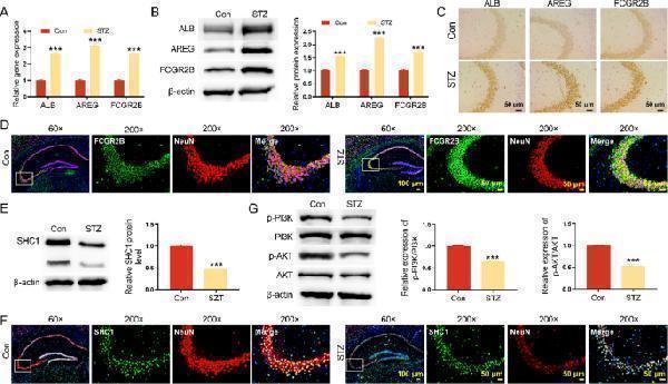
FCGR2B were up-regulated in hippocampus of DM mice. A qRT-PCR was performed to detect the expression of ALB, AREG and FCGR2B mRNA expression in hippocampus of mice. B Western blot was conducted to detect the ALB, AREG and FCGR2B protein expression in hippocampus of mice. C IHC assay was employed to examine the ALB, AREG and FCGR2B protein expression in hippocampus of mice. D IF staining was utilized to detect the expression of FCGR2B and NeuN in hippocampus of mice. E Western blot was performed to detect the SHC1 protein expression in hippocampus of mice. F IF staining was performed to detect the expression of SHC1 and NeuN in hippocampus of mice. G Western blot was used to detect the p-PI3K and p-AKT protein expression in hippocampus of mice. *** P < 0.001 Full size image
Index in PubMed under a CC BY license. PMID: 40537751

Click image to see more details
Western blot analysis of Amphiregulin using anti-Amphiregulin antibody (A01787-1).
Electrophoresis was performed on a 10% SDS-PAGE gel at 80V (Stacking gel) / 120V (Resolving gel) for 2 hours. The sample well of each lane was loaded with 30 ug of sample under reducing conditions.
Lane 1: rat brain tissue lysates,
Lane 2: rat thymus tissue lysates,
Lane 3: rat spleen tissue lysates,
Lane 4: rat PC-12 whole cell lysates,
Lane 5: mouse brain tissue lysates,
Lane 6: mouse thymus tissue lysates,
Lane 7: mouse spleen tissue lysates,
Lane 8: mouse RAW264.7 whole cell lysates.
After electrophoresis, proteins were transferred to a nitrocellulose membrane at 150 mA for 50-90 minutes. Blocked the membrane with 5% non-fat milk/TBS for 1.5 hour at RT. The membrane was incubated with rabbit anti-Amphiregulin antigen affinity purified polyclonal antibody (A01787-1) at 0.5 μg/mL overnight at 4°C, then washed with TBS-0.1%Tween 3 times with 5 minutes each and probed with a goat anti-rabbit IgG-HRP secondary antibody (Catalog # BA1054) at a dilution of 1:5000 for 1.5 hour at RT. The signal is developed using an ECL Plus Western Blotting Substrate (Catalog # AR1196-200) with Tanon 5200 system. A specific band was detected for Amphiregulin at approximately 45 kDa. The expected band size for Amphiregulin is at 28 kDa.

Click image to see more details
IHC analysis of Amphiregulin using anti-Amphiregulin antibody (A01787-1).
Amphiregulin was detected in a paraffin-embedded section of mouse brain tissue. Heat mediated antigen retrieval was performed in EDTA buffer (pH 8.0, epitope retrieval solution). The tissue section was blocked with 10% goat serum. The tissue section was then incubated with 2 μg/ml rabbit anti-Amphiregulin Antibody (A01787-1) overnight at 4°C. Peroxidase Conjugated Goat Anti-rabbit IgG was used as secondary antibody and incubated for 30 minutes at 37°C. The tissue section was developed using HRP Conjugated Rabbit IgG Super Vision Assay Kit (Catalog # SV0002) with DAB as the chromogen.

Click image to see more details
IHC analysis of Amphiregulin using anti-Amphiregulin antibody (A01787-1).
Amphiregulin was detected in a paraffin-embedded section of rat brain tissue. Heat mediated antigen retrieval was performed in EDTA buffer (pH 8.0, epitope retrieval solution). The tissue section was blocked with 10% goat serum. The tissue section was then incubated with 2 μg/ml rabbit anti-Amphiregulin Antibody (A01787-1) overnight at 4°C. Peroxidase Conjugated Goat Anti-rabbit IgG was used as secondary antibody and incubated for 30 minutes at 37°C. The tissue section was developed using HRP Conjugated Rabbit IgG Super Vision Assay Kit (Catalog # SV0002) with DAB as the chromogen.

Click image to see more details
Flow Cytometry analysis of PC-12 cells using anti-Amphiregulin antibody (A01787-1).
Overlay histogram showing PC-12 cells stained with A01787-1 (Blue line). The cells were fixed with 4% paraformaldehyde and blocked with 10% normal goat serum. And then incubated with rabbit anti-Amphiregulin Antibody (A01787-1, 1 μg/1x106 cells) for 30 min at 20°C. DyLight®488 conjugated goat anti-rabbit IgG (BA1127, 5-10 μg/1x106 cells) was used as secondary antibody for 30 minutes at 20°C. Isotype control antibody (Green line) was rabbit IgG (1 μg/1x106) used under the same conditions. Unlabelled sample without incubation with primary antibody and secondary antibody (Red line) was used as a blank control.
Specific Publications For Anti-Amphiregulin/Areg Antibody Picoband® (A01787-1)
Loading publications
Recommended Resources
Here are featured tools and databases that you might find useful.
- Boster's Pathways Library
- Protein Databases
- Bioscience Research Protocol Resources
- Data Processing & Analysis Software
- Photo Editing Software
- Scientific Literature Resources
- Research Paper Management Tools
- Molecular Biology Software
- Primer Design Tools
- Bioinformatics Tools
- Phylogenetic Tree Analysis
Customer Reviews
Have you used Anti-Amphiregulin/Areg Antibody Picoband®?
Share your experimental results or join a short interview to earn up to $1,000 in product credits or other rewards.
0 Reviews For Anti-Amphiregulin/Areg Antibody Picoband®
Customer Q&As
Have a question?
Find answers in Q&As, reviews.
Can't find your answer?
Submit your question
5 Customer Q&As for Anti-Amphiregulin/Areg Antibody Picoband®
Question
We are currently using anti-Amphiregulin/Areg antibody A01787-1 for rat tissue, and we are well pleased with the ELISA results. The species of reactivity given in the datasheet says mouse, rat. Is it likely that the antibody can work on zebrafish tissues as well?
S. Miller
Verified customer
Asked: 2017-11-21
Answer
The anti-Amphiregulin/Areg antibody (A01787-1) has not been tested for cross reactivity specifically with zebrafish tissues, but there is a good chance of cross reactivity. We have an innovator award program that if you test this antibody and show it works in zebrafish you can get your next antibody for free. Please contact me if I can help you with anything.
Boster Scientific Support
Answered: 2017-11-21
Question
See below the WB image, lot number and protocol we used for brain using anti-Amphiregulin/Areg antibody A01787-1. Please let me know if you require anything else.
Verified Customer
Verified customer
Asked: 2017-08-30
Answer
Thank you very much for the data. Our lab team are working to resolve this as quickly as possible, and we appreciate your patience and understanding! You have provided everything we needed. Please let me know if there is anything you need in the meantime.
Boster Scientific Support
Answered: 2017-08-30
Question
Does anti-Amphiregulin/Areg antibody A01787-1 work on bovine WB with brain?
Verified Customer
Verified customer
Asked: 2017-06-06
Answer
Our lab technicians have not tested anti-Amphiregulin/Areg antibody A01787-1 on bovine. You can run a BLAST between bovine and the immunogen sequence of anti-Amphiregulin/Areg antibody A01787-1 to see if they may cross-react. If the sequence homology is close, then you can perform a pilot test. Keep in mind that since we have not validated bovine samples, this use of the antibody is not covered by our guarantee. However we have an innovator award program that if you test this antibody and show it works in bovine brain in WB, you can get your next antibody for free.
Boster Scientific Support
Answered: 2017-06-06
Question
Would anti-Amphiregulin/Areg antibody A01787-1 work for WB with brain?
Z. Brown
Verified customer
Asked: 2017-02-14
Answer
According to the expression profile of brain, AREG is highly expressed in brain. So, it is likely that anti-Amphiregulin/Areg antibody A01787-1 will work for WB with brain.
Boster Scientific Support
Answered: 2017-02-14
Question
Is a blocking peptide available for product anti-Amphiregulin/Areg antibody (A01787-1)?
B. Wu
Verified customer
Asked: 2016-08-12
Answer
We do provide the blocking peptide for product anti-Amphiregulin/Areg antibody (A01787-1). If you would like to place an order for it please contact support@bosterbio.com and make a special request.
Boster Scientific Support
Answered: 2016-08-12
