Click image to see more details
-

-
-
-
-
+6
Product Info Summary
| SKU: | PA2260 |
|---|---|
| Size: | 100 μg/vial |
| Reactive Species: | Human, Mouse, Rat |
| Host: | Rabbit |
| Application: | Flow Cytometry, IF, IHC, ICC, WB |
Customers Who Bought This Also Bought

Product info
Product Name
Anti-APG5L/ATG5 Antibody Picoband®
SKU/Catalog Number
PA2260
BA3525-2 is an alternative SKU for this antibody, used in previous lots.
Size
100 μg/vial
Form
Lyophilized
Description
Boster Bio Anti-APG5L/ATG5 Antibody catalog # PA2260. Tested in Flow Cytometry, ICC/IF, IHC, WB applications. This antibody reacts with Human, Mouse, Rat. The brand Picoband indicates this is a premium antibody that guarantees superior quality, high affinity, and strong signals with minimal background in Western blot applications. Only our best-performing antibodies are designated as Picoband, ensuring unmatched performance. ATG5 (autophagy protein 5; also termed APG5/ASP) is described as necessary for autophagy due to its role in autophagosome elongation and as an E3 ubiquitin ligase; knockdown in hepatocytes is reported to increase triglyceride levels and lipid-droplet number/size under stimulatory conditions. Assay context: antibody validated for Flow Cytometry, ICC/IF, IHC, and WB; reactive to ATG5 in human/mouse/rat; reported observed MW ~50–52 kDa with stated no cross-reactivity. Frequently interpreted within cellular stress–survival programs with proteostasis markers such as CRYAB and apoptosis/mitosis regulators such as BIRC5 (Survivin) (putative); tissue-level compartmentalization questions are commonly addressed by IHC, while abundance changes are often confirmed via western blotting (putative).
Storage & Handling
Store at -20˚C for one year from date of receipt. After reconstitution, at 4˚C for one month. It can also be aliquotted and stored frozen at -20˚C for six months. Avoid repeated freeze-thaw cycles.
Cite This Product
Anti-APG5L/ATG5 Antibody Picoband® (Boster Biological Technology, Pleasanton CA, USA, Catalog # PA2260)
Host
Rabbit
Contents
Each vial contains 4 mg Trehalose, 0.9 mg NaCl and 0.2 mg Na2HPO4.
Clonality
Polyclonal
Isotype
Rabbit IgG
Immunogen
A synthetic peptide corresponding to a sequence in the middle region of human APG5L, identical to the related mouse sequence, and different from the related rat sequence by one amino acid.
Cross-reactivity
No cross-reactivity with other proteins
Reactive Species
PA2260 is reactive to ATG5 in Human, Mouse, Rat
Observed Molecular Weight
50-52 kDa
Calculated molecular weight
32.4 kDa
Background of ATG5
Autophagy protein 5 is a protein that in humans is encoded by the ATG5 gene. It is also known as APG5 or ASP, and this gene is mapped to 6q21. It is found that knockdown of ATG5 in hepatocytes increased triglyceride levels with oleate or a second endogenous stimulus for triglyceride formation. These hepatocytes with ATG5 knockdown also had increased lipid droplet number and size. ATG5 is an E3 ubiquitin ligase which is necessary for autophagy due to its role in autophagosome elongation. It is activated by ATG7 and forms a complex with ATG12 and ATG16L1. This complex is necessary for LC3-1 conjugation to PE to form LC3-II.
Antibody Validation
Boster validates all antibodies on WB, IHC, ICC, Immunofluorescence, and ELISA with known positive control and negative samples to ensure specificity and high affinity, including thorough antibody incubations.
Application & Images
Applications
PA2260 is guaranteed for Flow Cytometry, IF, IHC, ICC, WB Boster Guarantee
Recommend Dilution
| Application | Dilution | Species |
|---|---|---|
| Western blot | 0.1-0.5μg/ml | Human, Mouse, Rat |
| Immunohistochemistry (Paraffin-embedded Section) | 2-5ug/ml | Human |
| Immunocytochemistry/Immunofluorescence | 5 μg/ml | Human |
| Flow Cytometry(Fixed) | 1-3 μg/1x106 cells | Human |
Tested application
Suggested blocking solution with 5% non-fat milk or BSA; (*)Recommended protein loading: 20-40 µg per lane
Use TE buffer pH 9.0 for antigen retrieval; (*) citrate buffer pH 6.0 is an alternative.
Validation Images & Assay Conditions
Click image to see more details
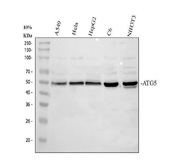
Western blot analysis of ATG5 using anti-ATG5 antibody (PA2260).
Electrophoresis was performed on a 10% SDS-PAGE gel at 80V (Stacking gel) / 120V (Resolving gel) for 2 hours. The sample well of each lane was loaded with 30 ug of sample under reducing conditions.
Lane 1: Lane 1: human A549 whole cell lysates,
Lane 2: human Hela whole cell lysates,
Lane 3: human HepG2 whole cell lysates,
Lane 4: rat C6 whole cell lysates,
Lane 5: mouse NIH/3T3 whole cell lysates.
After electrophoresis, proteins were transferred to a nitrocellulose membrane at 150 mA for 50-90 minutes. Blocked the membrane with 5% non-fat milk/TBS for 1.5 hour at RT. The membrane was incubated with rabbit anti-ATG5 antigen affinity purified polyclonal antibody (PA2260) at 0.5 μg/mL overnight at 4°C, then washed with TBS-0.1%Tween 3 times with 5 minutes each and probed with a goat anti-rabbit IgG-HRP secondary antibody at a dilution of 1:5000 for 1.5 hour at RT. The signal is developed using an ECL Plus Western Blotting Substrate (Catalog # AR1196-200) with Tanon 5200 system. A specific band was detected for ATG5 at approximately 50-52 kDa. The expected band size for ATG5 is at 32 kDa.

Click image to see more details
IHC analysis of ATG5 using anti-ATG5 antibody (PA2260).
ATG5 was detected in a paraffin-embedded section of human ovarian cancer tissue. Heat mediated antigen retrieval was performed in EDTA buffer (pH 8.0, epitope retrieval solution). The tissue section was blocked with 10% goat serum. The tissue section was then incubated with 2 μg/ml rabbit anti-ATG5 Antibody (PA2260) overnight at 4°C. Peroxidase Conjugated Goat Anti-rabbit IgG was used as secondary antibody and incubated for 30 minutes at 37°C. The tissue section was developed using HRP Conjugated Rabbit IgG Super Vision Assay Kit (Catalog # SV0002) with DAB as the chromogen.

Click image to see more details
Hypoxia leads to the transfer of ZNF423 from the nucleus to the cytoplasm, where it bound BCAT1 to promote autophagy activity. a Bioinformatics analysis of proteins associated with BCAT1. Upside: According to the JASPAR database and LASAGNA-Search 2.0 database, there was 28 genes that may bind to bcat1, and the binding ability of ZNF423, STAT1, Pou5f1, STAT3, SP1, SOX9, and TEAD1 was strong. Underside: RT-PCR analysis of the mRNA levels of ZNF423, STAT1, Pou5f1, STAT3, SP1, SOX9, and TEAD1 with rat β-actin serving as the standard in PASMCs under NOR or HYP for 24 h ( n = 5). b Western blot analysis of the expression of ZNF423 in PASMCs under NOR or HYP for 24 h ( n = 6). c ZNF423 protein levels were assayed in pulmonary arterial tissues of hypoxic model rats ( n = 4). d Coimmunoprecipitation of whole-cell lysates of PASMCs exposed to normoxia or hypoxia for 24 h with anti-ZNF423, followed by probing with anti-BCAT1 ( n = 3). e Western blot analysis of BCAT1 expression in PASMCs transfected with ZNF423 siRNA under NOR or HYP for 24 h ( n = 4). f PASMCs were exposed to HYP for 24 h, and the colocalization between BCAT1 and ZNF423 was determined by immunofluorescence. GFP-BCAT1 (green), ZNF423 (red), and DAPI (blue). Scale bar = 50 μm ( n = 3). g The translocation of ZNF423 between the nucleus and cytoplasm in PASMCs transfected with BCAT1 siRNA or gabapentin ( n = 3). h Western blot analysis of the expression of BECN1 and Atg5 in PASMCs transfected with ZNF423 siRNA under HYP for 24 h ( n = 4). i Autophagic flux of PASMCs cotransfected with eGFP-mRFP-LC3 plasmid and control siRNA or ZNF423 siRNA under HYP for 24 h. Scale bar = 50 μm ( n = 5). Nor normoxia, Hyp hypoxia, H + G hypoxia plus gabapentin, H + NC hypoxia plus control siRNA, H + SI hypoxia plus BCAT1 siRNA, H + si-ZNF423 hypoxia plus ZNF423 siRNA, IP immunoprecipitation, IB immunoblotting. Statistical analysis was performed with one-way ANOVA or the Student’s t test. All values are presented as the mean ± SEM. * p < 0.05; ** p < 0.01; *** p < 0.001.
Index in PubMed under a CC BY license. PMID: 32938905

Click image to see more details
BCAT1 regulates autophagy during hypoxia by activating ERs via the IRE1-XBP1-RIDD axis. a Western blot analysis of BECN1 and Atg5 in PASMCs cotransfected with BCAT1 and IRE1 siRNA ( n = 5). b Autophagic flux was monitored in PASMCs cotransfected with eGFP-mRFP-LC3 plasmid and control siRNA or IRE1 siRNA that were then exposed to HYP for 24 h. Scale bar = 50 μm ( n = 3). c , d RT-PCR analysis of the mRNA levels of XBP1-s, sparc, pmp2, and Scara3 with rat β-actin serving as the standard ( n = 5). e The formation of autophagosomes was detected, and autophagic activity was estimated in cells in which the expression of XBP1 was knocked down with XBP1 siRNA under HYP for 24 h. Scale bar = 50 µm ( n = 5). Nor normoxia, Hyp hypoxia, H + G hypoxia plus gabapentin, H + NC hypoxia plus control siRNA, H + SI hypoxia plus BCAT1 siRNA, H + SI-IRE1 hypoxia plus IRE1 siRNA, H + SI-XBP1 hypoxia plus XBP1 siRNA, H + Con hypoxia plus control vector, H + B hypoxia plus BCAT1 plasmid, H + Con+NC hypoxia plus control vector plus control siRNA, H + B + Si-IRE hypoxia plus BCAT1 plasmid plus IRE1 siRNA. Statistical analysis was performed with one-way ANOVA. All values are presented as the mean ± SEM. * p < 0.05; ** p < 0.01; *** p < 0.001.
Index in PubMed under a CC BY license. PMID: 32938905

Click image to see more details
BCAT1 regulates autophagy through the endoplasmic reticulum stress pathway. a Expression of BCAT1 and ER-Tracker Red staining in PASMCs exposed to NOR or HYP for 24 h. Scale bar = 50 μm ( n = 3). b Western blot analysis of PERK, IRE1, ATF6, and GRP78 protein expression in the ERs pathway in PASMCs treated with gabapentin ( n = 8). c Western blot analysis of IRE1, PERK, ATF6, and GRP78 expression in PASMCs transfected with BCAT1 siRNA ( n = 8). d Western blot analysis of BECN1 and Atg5 in PASMCs treated with the ERs pathway inhibitor 4-PBA and BCAT1 plasmid ( n = 8). e Coimmunoprecipitation of the whole-cell lysates of PASMCs exposed to normoxia or hypoxia for 24 h with anti-IRE1, followed by probing with anti-BCAT1 ( n = 3). Nor normoxia, Hyp hypoxia, H + G hypoxia plus gabapentin, H + NC hypoxia plus control siRNA, H + SI hypoxia plus BCAT1 siRNA, N + Con normoxia plus control vector, H + Con hypoxia plus control vector, H + B hypoxia plus BCAT1 plasmid, H + Con+4 hypoxia plus control vector plus 4-phenylbutyric acid, H + B + 4 hypoxia plus BCAT1 plasmid plus 4-phenylbutyric acid, IP immunoprecipitation, IB immunoblotting. Statistical analysis was performed with one-way ANOVA. All values are presented as the mean ± SEM. ** p < 0.01; *** p < 0.001.
Index in PubMed under a CC BY license. PMID: 32938905

Click image to see more details
Iron-overloaded EMFF induced ferritinophagy-dependent ferroptosis in granulosa cells. A – C Levels of total iron, hepcidin, and transferrin in EMFF ( n = 15) and COFF ( n = 15). Data are expressed as means ± SD and analyzed by Student’s t test. D Results of mouse granulosa cells proliferation under different intervention conditions (each group in the figure is compared with COFF group). DFO, iron chelators; FER, ferroptosis inhibitor; NEC, necrosis inhibitor; ZDF, apoptosis inhibitor; ME, autophagy inhibitor. Data are expressed as means ± SD and analyzed by one-way ANOVA. E – H Comparison of ferritinophagy-related proteins FTH1, NCOA4, and ATG5 between human granulosa cells of infertile patients with EMs (EMGC) and of control group (COGC). The expression of β-actin was used as an internal control. Data are expressed as means ± SD and analyzed by Student’s t test. I – L Detection of ferroptosis-related indicators iron, GSH, GPX4, and MDA in COGC and EMGC. Data are expressed as means ± SD and analyzed by Student’s t test. M Representative images of the mitochondrial morphology of mouse granulosa cells intervened by COFF and EMFF were observed under TEM. Yellow arrows indicate mitochondrion. Scale bar = 1.0 µm. Scale bar = 5.0 µm. N Representative images of ROS and ferrous ion fluorescence staining after COFF and EMFF intervention in mouse granulosa cells. Scale bar = 100 µm. * P < 0.05, ** P < 0.01, *** P < 0.001, **** P < 0.0001, and ns, no significance.
Index in PubMed under a CC BY license. PMID: 35787614

Click image to see more details
Upregulation of BCAT1 expression induced by hypoxia leads to PASMC autophagy. a Western blot analysis of BECN1 and Atg5 protein expression in PASMCs treated with the inhibitor gabapentin (20 µM) ( n = 8). b Western blot analysis of BECN1 and Atg5 protein expression in PASMCs transfected with BCAT1 siRNA or BCAT1 plasmid ( n = 8). c , d Immunofluorescence staining for BECN1 and Atg5 in PASMCs. BECN1 and Atg5 (green), α-SMA (red), and DAPI (blue). Scale bar = 50 μm ( n = 3). e Western blot analysis of BECN1 and Atg5 expression in the pulmonary arterial tissues of hypoxia model rats treated with gabapentin ( n = 7). f Measurement of autophagic flux in PASMCs transfected with eGFP-mRFP-LC3 plasmid and exposed under NOR or HYP for 24 h treated with BCAT1 siRNA or the BCAT1 inhibitor gabapentin. Yellow and red dots indicate autolysosomes and autophagosomes, respectively. Scale bar = 50 μm ( n = 6). Nor normoxia, Hyp hypoxia, Mct monocrotaline, H + G hypoxia plus gabapentin, M + G monocrotaline plus gabapentin, H + NC hypoxia plus control siRNA, H + SI hypoxia plus BCAT1 siRNA, H + Con hypoxia plus control vector, H + B hypoxia plus BCAT1 plasmid. Statistical analysis was performed with one-way ANOVA. All values are presented as the mean ± SEM. * p < 0.05; ** p < 0.01; *** p < 0.001.
Index in PubMed under a CC BY license. PMID: 32938905

Click image to see more details
Construction of an iron overload mouse model. A – C Serum levels of E 2 , FSH, and LH in standard iron (STD), low iron (LID), and high iron (HID) diet feeding groups ( n = 8). D – F Total iron, GSH, and MDA levels in the ovary tissues of mice in each group ( n = 8). G Representative images of ROS fluorescence staining of ovarian mouse granulosa cells in three groups of mice. Scale bar = 20 µm. H – K Western blot analysis of ferritinophagy-related proteins, FTH1, NCOA4, and ATG5 in mouse ovary tissues in the STD, LID, and HID group. The expression of β-actin was used as an internal control. All data are expressed as means ± SD and analyzed by one-way ANOVA. * P < 0.05, ** P < 0.01, *** P < 0.001, **** P < 0.0001, and ns, no significance.
Index in PubMed under a CC BY license. PMID: 35787614

Click image to see more details
IF analysis of ATG5 using anti-ATG5 antibody (PA2260).
ATG5 was detected in an immunocytochemical section of A549 cells. Enzyme antigen retrieval was performed using IHC enzyme antigen retrieval reagent (AR0022) for 15 mins. The cells were blocked with 10% goat serum. And then incubated with 5 μg/mL rabbit anti-ATG5 Antibody (PA2260) overnight at 4°C. Fluoro488 Conjugated Goat Anti-Rabbit IgG (BA1127) was used as secondary antibody at 1:500 dilution and incubated for 30 minutes at 37°C. The section was counterstained with DAPI. Visualize using a fluorescence microscope and filter sets appropriate for the label used.

Click image to see more details
Flow Cytometry analysis of HepG2 cells using anti-ATG5 antibody (PA2260).
Overlay histogram showing CACO-2 cells stained with PA2260 (Blue line). To facilitate intracellular staining, cells were fixed with 4% paraformaldehyde and permeabilized with permeabilization buffer. The cells were blocked with 10% normal goat serum. And then incubated with rabbit anti-ATG5 Antibody (PA2260, 1 μg/1x106 cells) for 30 min at 20°C. DyLight®488 conjugated goat anti-rabbit IgG (BA1127, 5-10 μg/1x106 cells) was used as secondary antibody for 30 minutes at 20°C. Isotype control antibody (Green line) was rabbit IgG (1 μg/1x106) used under the same conditions. Unlabelled sample without incubation with primary antibody and secondary antibody (Red line) was used as a blank control.
Specific Publications For Anti-APG5L/ATG5 Antibody Picoband® (PA2260)
Loading publications
Recommended Resources
Here are featured tools and databases that you might find useful.
- Boster's Pathways Library
- Protein Databases
- Bioscience Research Protocol Resources
- Data Processing & Analysis Software
- Photo Editing Software
- Scientific Literature Resources
- Research Paper Management Tools
- Molecular Biology Software
- Primer Design Tools
- Bioinformatics Tools
- Phylogenetic Tree Analysis
Customer Reviews
Have you used Anti-APG5L/ATG5 Antibody Picoband®?
Share your experimental results or join a short interview to earn up to $1,000 in product credits or other rewards.
0 Reviews For Anti-APG5L/ATG5 Antibody Picoband®
Customer Q&As
Have a question?
Find answers in Q&As, reviews.
Can't find your answer?
Submit your question
15 Customer Q&As for Anti-APG5L/ATG5 Antibody Picoband®
Question
My question regards to test anti-APG5L/ATG5 antibody PA2260 on mouse endometrium placenta for research purposes, then I may be interested in using anti-APG5L/ATG5 antibody PA2260 for diagnostic purposes as well. Is the antibody suitable for diagnostic purposes?
Verified Customer
Verified customer
Asked: 2020-04-10
Answer
The products we sell, including anti-APG5L/ATG5 antibody PA2260, are only intended for research use. They would not be suitable for use in diagnostic work. If you have the means to develop a product into diagnostic use, and are interested in collaborating with us and develop our product into an IVD product, please contact us for more discussions.
Boster Scientific Support
Answered: 2020-04-10
Question
Thanks for helping with my inquiry over the phone. Here are the WB image, lot number and protocol we used for endometrium placenta using anti-APG5L/ATG5 antibody PA2260. Let me know if you need anything else.
Verified Customer
Verified customer
Asked: 2019-10-31
Answer
I appreciate the data. You have provided everything we needed. Our lab team are working to resolve your inquiry as quickly as possible, and we appreciate your patience and understanding! Please let me know if there is anything you need in the meantime.
Boster Scientific Support
Answered: 2019-10-31
Question
Will PA2260 anti-APG5L/ATG5 antibody work on parafin embedded sections? If so, which fixation method do you recommend we use (PFA, paraformaldehyde, other)?
Verified Customer
Verified customer
Asked: 2019-09-26
Answer
As indicated on the product datasheet, PA2260 anti-APG5L/ATG5 antibody as been tested on WB. It is best to use PFA for fixation because it has better tissue penetration ability. PFA needs to be prepared fresh before use. Long term stored PFA turns into formalin, as the PFA molecules congregate and become formalin.
Boster Scientific Support
Answered: 2019-09-26
Question
Our lab were happy with the WB result of your anti-APG5L/ATG5 antibody. However we have observed positive staining in colonic mucosa cytoplasm. using this antibody. Is that expected? Could you tell me where is ATG5 supposed to be expressed?
Verified Customer
Verified customer
Asked: 2019-08-15
Answer
From literature, colonic mucosa does express ATG5. Generally ATG5 expresses in cytoplasm. Regarding which tissues have ATG5 expression, here are a few articles citing expression in various tissues:
Embryonic stem cell, Pubmed ID: 11266458
Endometrium, and Placenta, Pubmed ID: 15489334
Fetal brain, Pubmed ID: 17974005
Fetal liver, Pubmed ID: 9563500
Boster Scientific Support
Answered: 2019-08-15
Question
Is a blocking peptide available for product anti-APG5L/ATG5 antibody (PA2260)?
Verified Customer
Verified customer
Asked: 2019-07-26
Answer
We do provide the blocking peptide for product anti-APG5L/ATG5 antibody (PA2260). If you would like to place an order for it please contact support@bosterbio.com and make a special request.
Boster Scientific Support
Answered: 2019-07-26
Question
Can you help my question with product PA2260, anti-APG5L/ATG5 antibody. I was wondering if it would be possible to conjugate this antibody with biotin. I would need it to be without BSA or sodium azide. I am planning on using a buffer exchange of sodium azide with PBS only. Would there be problems for me to conjugate the antibody and store it in -20 degrees in small aliquots?
Verified Customer
Verified customer
Asked: 2019-05-28
Answer
We suggest not storing this antibody with PBS buffer only in -20 degrees. If you want to store it in -20 degrees it is best to add some cryoprotectant like glycerol. If you want carrier free PA2260 anti-APG5L/ATG5 antibody, we can provide it to you in a special formula with trehalose and/or glycerol. These molecules will not interfere with conjugation chemistry and provide a good level of protection for the antibody from degradation. Please be sure to specify this in your purchase order.
Boster Scientific Support
Answered: 2019-05-28
Question
Is there a BSA free version of anti-APG5L/ATG5 antibody PA2260 available?
Verified Customer
Verified customer
Asked: 2018-09-24
Answer
We appreciate your recent telephone inquiry. I can confirm that some lots of this anti-APG5L/ATG5 antibody PA2260 are BSA free. For now, these lots are available and we can make a BSA free formula for you free of charge. It will take 3 extra days to prepare. If you require this antibody BSA free again in future, please do not hesitate to contact me and I will be pleased to check which lots we have in stock that are BSA free.
Boster Scientific Support
Answered: 2018-09-24
Question
We are currently using anti-APG5L/ATG5 antibody PA2260 for mouse tissue, and we are well pleased with the WB results. The species of reactivity given in the datasheet says human, mouse, rat. Is it true that the antibody can work on monkey tissues as well?
Verified Customer
Verified customer
Asked: 2017-11-30
Answer
The anti-APG5L/ATG5 antibody (PA2260) has not been validated for cross reactivity specifically with monkey tissues, though there is a good chance of cross reactivity. We have an innovator award program that if you test this antibody and show it works in monkey you can get your next antibody for free. Please contact me if I can help you with anything.
Boster Scientific Support
Answered: 2017-11-30
Question
My question regards using your anti-APG5L/ATG5 antibody for negative stranded viral rna replication studies. Has this antibody been tested with western blotting on nih whole cell lysate? We would like to see some validation images before ordering.
J. Bhatt
Verified customer
Asked: 2017-10-20
Answer
We appreciate your inquiry. This PA2260 anti-APG5L/ATG5 antibody is validated on rat liver tissue, tissue lysate, spleen tissue, kidney tissue, hela whole cell lysate, raji whole cell lysate, nih whole cell lysate, hepg2 whole cell lysate, pc12 whole cell lysate, nrk whole cell lysate. It is guaranteed to work for WB in human, mouse, rat. Our Boster guarantee will cover your intended experiment even if the sample type has not been be directly tested.
Boster Scientific Support
Answered: 2017-10-20
Question
I see that the anti-APG5L/ATG5 antibody PA2260 works with WB, what is the protocol used to produce the result images on the product page?
J. Zhang
Verified customer
Asked: 2017-04-21
Answer
You can find protocols for WB on the "support/technical resources" section of our navigation menu. If you have any further questions, please send an email to support@bosterbio.com
Boster Scientific Support
Answered: 2017-04-21
Question
We have seen staining in human endometrium placenta. Are there any suggestions? Is anti-APG5L/ATG5 antibody supposed to stain endometrium placenta positively?
R. Bhatt
Verified customer
Asked: 2016-05-16
Answer
Based on literature endometrium placenta does express ATG5. Based on Uniprot.org, ATG5 is expressed in colonic mucosa, fetal liver, fetal brain, endometrium placenta, embryonic stem cell, among other tissues. Regarding which tissues have ATG5 expression, here are a few articles citing expression in various tissues:
Embryonic stem cell, Pubmed ID: 11266458
Endometrium, and Placenta, Pubmed ID: 15489334
Fetal brain, Pubmed ID: 17974005
Fetal liver, Pubmed ID: 9563500
Boster Scientific Support
Answered: 2016-05-16
Question
I was wanting to use your anti-APG5L/ATG5 antibody for WB for mouse endometrium placenta on frozen tissues, but I want to know if it has been tested for this particular application. Has this antibody been tested and is this antibody a good choice for mouse endometrium placenta identification?
M. Collins
Verified customer
Asked: 2014-12-18
Answer
It shows on the product datasheet, PA2260 anti-APG5L/ATG5 antibody has been tested for WB on human, mouse, rat tissues. We have an innovator award program that if you test this antibody and show it works in mouse endometrium placenta in IHC-frozen, you can get your next antibody for free.
Boster Scientific Support
Answered: 2014-12-18
Question
Is this PA2260 anti-APG5L/ATG5 antibody reactive to the isotypes of ATG5?
J. Collins
Verified customer
Asked: 2013-10-03
Answer
The immunogen of PA2260 anti-APG5L/ATG5 antibody is A synthetic peptide corresponding to a sequence in the middle region of human APG5L(82-97aa DRFDQFWAINRKLMEY), identical to the related mouse sequence, and different from the related rat sequence by one amino acid. Could you tell me which isotype you are interested in so I can help see if the immunogen is part of this isotype?
Boster Scientific Support
Answered: 2013-10-03
Question
Would anti-APG5L/ATG5 antibody PA2260 work for WB with endometrium placenta?
D. Brown
Verified customer
Asked: 2013-08-23
Answer
According to the expression profile of endometrium placenta, ATG5 is highly expressed in endometrium placenta. So, it is likely that anti-APG5L/ATG5 antibody PA2260 will work for WB with endometrium placenta.
Boster Scientific Support
Answered: 2013-08-23
Question
See attached the WB image, lot number and protocol we used for endometrium placenta using anti-APG5L/ATG5 antibody PA2260. Please let me know if you require anything else.
L. Jones
Verified customer
Asked: 2013-01-21
Answer
Thank you very much for the data. Our lab team are working to resolve this as quickly as possible, and we appreciate your patience and understanding! You have provided everything we needed. Please let me know if there is anything you need in the meantime.
Boster Scientific Support
Answered: 2013-01-21
