Click image to see more details
-

-
-
-
-
+1
Product Info Summary
| SKU: | M00865 |
|---|---|
| Size: | 100 μl |
| Reactive Species: | Human, Mouse, Rat |
| Host: | Rabbit |
| Application: | IF, IHC, ICC, WB |
Customers Who Bought This Also Bought

Product info
Product Name
Anti-AQP1 Rabbit Monoclonal Antibody
SKU/Catalog Number
M00865
BM5035 is an alternative SKU for this antibody, used in previous lots.
Size
100 μl
Form
Liquid
Description
Boster Bio Anti-AQP1 Rabbit Monoclonal Antibody catalog # M00865. Tested in WB, IHC, ICC/IF applications. This antibody reacts with Human, Mouse, Rat.
Storage & Handling
Store at -20°C for one year. For short term storage and frequent use, store at 4°C for up to one month. Avoid repeated freeze-thaw cycles.
Cite This Product
Anti-AQP1 Rabbit Monoclonal Antibody (Boster Biological Technology, Pleasanton CA, USA, Catalog # M00865)
Host
Rabbit
Contents
Rabbit IgG in stabilizing components, phosphate buffered saline, pH 7.4, 150mM NaCl, 0.02% sodium azide and 50% glycerol.
*This antibody is supplied in a stabilized formulation.
Compatibility with conjugation reactions depends on the chemistry of the conjugation method used.
For conjugation methods that are not compatible with the stabilizing components present in this formulation, a carrier-free antibody format is required.
Clonality
Monoclonal
Clone Number
AAEE-1
Isotype
Rabbit IgG
Immunogen
A synthesized peptide derived from human AQP1
Reactive Species
M00865 is reactive to AQP1 in Human, Mouse, Rat
Observed Molecular Weight
25 kDa
Calculated molecular weight
28.5 kDa
Antibody Validation
Boster validates all antibodies on WB, IHC, ICC, Immunofluorescence, and ELISA with known positive control and negative samples to ensure specificity and high affinity, including thorough antibody incubations.
Application & Images
Applications
M00865 is guaranteed for IF, IHC, ICC, WB Boster Guarantee
Recommend Dilution
WB 1:500-2000
IHC 1:50-200
ICC/IF 1:50-200
Tested application
Suggested blocking solution with 5% non-fat milk or BSA; (*)Recommended protein loading: 20-40 µg per lane
Use TE buffer pH 9.0 for antigen retrieval; (*) citrate buffer pH 6.0 is an alternative.
Validation Images & Assay Conditions
Click image to see more details
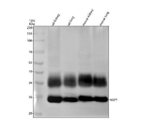
Western blot analysis of AQP1 using anti-AQP1 antibody (M00865).
Electrophoresis was performed on a 10% SDS-PAGE gel at 80V (Stacking gel) / 120V (Resolving gel) for 2 hours. The sample well of each lane was loaded with 30 ug of sample under reducing conditions.
Lane 1: rat kidney tissue lysates,
Lane 2: rat lung tissue lysates,
Lane 3: mouse kidney tissue lysates,
Lane 4: mouse lung tissue lysates.
After electrophoresis, proteins were transferred to a nitrocellulose membrane at 150 mA for 50-90 minutes. Blocked the membrane with 5% non-fat milk/TBS for 1.5 hour at RT. The membrane was incubated with rabbit anti-AQP1 antigen affinity purified monoclonal antibody (M00865) at 1: 500 overnight at 4°C, then washed with TBS-0.1%Tween 3 times with 5 minutes each and probed with a goat anti-rabbit IgG-HRP secondary antibody at a dilution of 1:5000 for 1.5 hour at RT. The signal is developed using an ECL Plus Western Blotting Substrate (Catalog # AR1196-200) with Tanon 5200 system. A specific band was detected for AQP1 at approximately 25 kDa. The expected band size for AQP1 is at 25 kDa.

Click image to see more details
IHC analysis of AQP1 using anti-AQP1 antibody (M00865).
AQP1 was detected in a paraffin-embedded section of human kidney tissue. Heat mediated antigen retrieval was performed in EDTA buffer (pH 8.0, epitope retrieval solution). The tissue section was blocked with 10% goat serum. The tissue section was then incubated with 1:50 rabbit anti-AQP1 Antibody (M00865) overnight at 4°C. Peroxidase Conjugated Goat Anti-rabbit IgG was used as secondary antibody and incubated for 30 minutes at 37°C. The tissue section was developed using HRP Conjugated Rabbit IgG Super Vision Assay Kit (Catalog # SV0002) with DAB as the chromogen.

Click image to see more details
IHC analysis of AQP1 using anti-AQP1 antibody (M00865).
AQP1 was detected in a paraffin-embedded section of mouse lung tissue. Heat mediated antigen retrieval was performed in EDTA buffer (pH 8.0, epitope retrieval solution). The tissue section was blocked with 10% goat serum. The tissue section was then incubated with 1:50 rabbit anti-AQP1 Antibody (M00865) overnight at 4°C. Peroxidase Conjugated Goat Anti-rabbit IgG was used as secondary antibody and incubated for 30 minutes at 37°C. The tissue section was developed using HRP Conjugated Rabbit IgG Super Vision Assay Kit (Catalog # SV0002) with DAB as the chromogen.

Click image to see more details
IHC analysis of AQP1 using anti-AQP1 antibody (M00865).
AQP1 was detected in a paraffin-embedded section of rat lung tissue. Heat mediated antigen retrieval was performed in EDTA buffer (pH 8.0, epitope retrieval solution). The tissue section was blocked with 10% goat serum. The tissue section was then incubated with 1:50 rabbit anti-AQP1 Antibody (M00865) overnight at 4°C. Peroxidase Conjugated Goat Anti-rabbit IgG was used as secondary antibody and incubated for 30 minutes at 37°C. The tissue section was developed using HRP Conjugated Rabbit IgG Super Vision Assay Kit (Catalog # SV0002) with DAB as the chromogen.

Click image to see more details
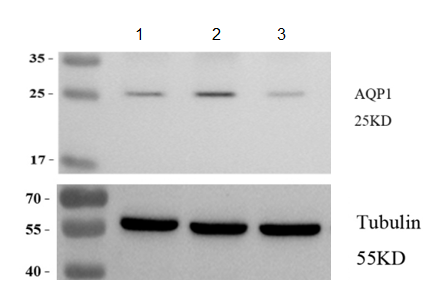
Western blot analysis of AQP1 using anti-AQP1 antibody (M00865).
Electrophoresis was performed on a 10% SDS-PAGE gel at 80V (Stacking gel) / 120V (Resolving gel) for 2 hours. The sample well of each lane was loaded with 30 ug of sample under reducing conditions.
Lane 1: MADB106 cells under normal culture conditions,
Lane 2: MADB106 cells treated with agonist,
Lane 3: MADB106 cells treated with inhibitor.
After electrophoresis, proteins were transferred to a nitrocellulose membrane at 150 mA for 50-90 minutes. Blocked the membrane with 5% non-fat milk/TBS for 1.5 hour at RT. The membrane was incubated with rabbit anti-AQP1 antigen affinity purified monoclonal antibody (M00865) at 1: 3000 overnight at 4°C, then washed with TBS-0.1%Tween 3 times with 5 minutes each and probed with a goat anti-rabbit IgG-HRP secondary antibody at a dilution of 1:10000 for 1 hour at RT. The signal is developed using an ECL Plus Western Blotting Substrate (Catalog # AR1196-200) with ChemiDoc MP system. A specific band was detected for AQP1 at approximately 25 kDa. The expected band size for AQP1 is at 25 kDa.
Specific Publications For Anti-AQP1 Rabbit Monoclonal Antibody (M00865)
Loading publications
Recommended Resources
Here are featured tools and databases that you might find useful.
- Boster's Pathways Library
- Protein Databases
- Bioscience Research Protocol Resources
- Data Processing & Analysis Software
- Photo Editing Software
- Scientific Literature Resources
- Research Paper Management Tools
- Molecular Biology Software
- Primer Design Tools
- Bioinformatics Tools
- Phylogenetic Tree Analysis
Customer Reviews
Have you used Anti-AQP1 Rabbit Monoclonal Antibody?
Share your experimental results or join a short interview to earn up to $1,000 in product credits or other rewards.
1 Reviews For Anti-AQP1 Rabbit Monoclonal Antibody
In this Western blot experiment using Anti-AQP1 antibody (Cat# BM5035) on MADB106 rat mammary carcinoma cells, AQP1 protein levels were markedly increased in cells treated with the agonist and significantly decreased in cells treated with the inhibitor.
Excellent
| SKU | M00865 |
|---|---|
| Application | Western blot |
| Sample | MADB106 rat mammary carcinoma cells |
| Sample Processing Description | MADB106 cells under normal culture conditions , MADB106 cells treated with agonist , MADB106 cells treated with inhibitor |
| Other Reagents | RIPA Lysis Buffer, Protease Inhibitor, Resolving Gel Solution ,Transfer Buffer ,Blocking Buffer |
| Primary Antibody | AQP1 Rabbit Monoclonal Antibody |
| Primary Incubation | 1:3000, overnight at 4 ℃ |
| Secondary Antibody | 1:10,000, HRP-conjugated Goat Anti-Rabbit IgG |
| Secondary Incubation | 1h at RT |
| Detection | Substrate: ECL substrate, Imaging system:ChemiDoc MP |
| Results Summary | AQP1 (Aquaporin 1) is the first discovered water channel protein, whose primary function is efficient water transport. Recent studies have revealed that AQP1 plays a critical role in cancer progression, promoting tumor growth in breast cancer by enhancing angiogenesis and cell migration. In this experiment, AQP1 protein levels were markedly increased in cells treated with the agonist and significantly decreased in cells treated with the inhibitor. |
Xinshuo Wang, Xi’an Jiaotong University
Verified customer
Submitted 2026-02-25
Customer Q&As
Have a question?
Find answers in Q&As, reviews.
Can't find your answer?
Submit your question
15 Customer Q&As for Anti-AQP1 Rabbit Monoclonal Antibody
Question
Is a blocking peptide available for product anti-AQP1 Rabbit Monoclonal antibody (M00865)?
Verified Customer
Verified customer
Asked: 2020-04-22
Answer
We do provide the blocking peptide for product anti-AQP1 Rabbit Monoclonal antibody (M00865). If you would like to place an order for it please contact support@bosterbio.com and make a special request.
Boster Scientific Support
Answered: 2020-04-22
Question
I have a question about product M00865, anti-AQP1 Rabbit Monoclonal antibody. I was wondering if it would be possible to conjugate this antibody with biotin. I would need it to be without BSA or sodium azide. I am planning on using a buffer exchange of sodium azide with PBS only. Would there be problems for me to conjugate the antibody and store it in -20 degrees in small aliquots?
Verified Customer
Verified customer
Asked: 2020-04-14
Answer
We suggest not storing this antibody with PBS buffer only in -20 degrees. If you want to store it in -20 degrees it is best to add some cryoprotectant like glycerol. If you want carrier free M00865 anti-AQP1 Rabbit Monoclonal antibody, we can provide it to you in a special formula with trehalose and/or glycerol. These molecules will not interfere with conjugation chemistry and provide a good level of protection for the antibody from degradation. Please be sure to specify this in your purchase order.
Boster Scientific Support
Answered: 2020-04-14
Question
We have observed staining in rat mesangial cell. What should we do? Is anti-AQP1 Rabbit Monoclonal antibody supposed to stain mesangial cell positively?
Verified Customer
Verified customer
Asked: 2020-02-20
Answer
From what I have seen in literature mesangial cell does express AQP1. From what I have seen in Uniprot.org, AQP1 is expressed in right lung, retinal pigment epithelium, uterus, mesangial cell, brain, articular cartilage, among other tissues. Regarding which tissues have AQP1 expression, here are a few articles citing expression in various tissues:
Articular cartilage, Pubmed ID: 2007592, 1373524
Brain, Pubmed ID: 15489334
Mesangial cell, Pubmed ID: 14702039
Retinal pigment epithelium, Pubmed ID: 8703970
Uterus, Pubmed ID: 7517253
Boster Scientific Support
Answered: 2020-02-20
Question
See below the WB image, lot number and protocol we used for uterus using anti-AQP1 Rabbit Monoclonal antibody M00865. Please let me know if you require anything else.
Verified Customer
Verified customer
Asked: 2019-12-19
Answer
Thank you very much for the data. Our lab team are working to resolve this as quickly as possible, and we appreciate your patience and understanding! You have provided everything we needed. Please let me know if there is anything you need in the meantime.
Boster Scientific Support
Answered: 2019-12-19
Question
I see that the anti-AQP1 Rabbit Monoclonal antibody M00865 works with WB, what is the protocol used to produce the result images on the product page?
Verified Customer
Verified customer
Asked: 2019-11-28
Answer
You can find protocols for WB on the "support/technical resources" section of our navigation menu. If you have any further questions, please send an email to support@bosterbio.com
Boster Scientific Support
Answered: 2019-11-28
Question
My colleagues were happy with the WB result of your anti-AQP1 Rabbit Monoclonal antibody. However we have been able to see positive staining in articular cartilage cell membrane using this antibody. Is that expected? Could you tell me where is AQP1 supposed to be expressed?
Verified Customer
Verified customer
Asked: 2019-09-02
Answer
From literature, articular cartilage does express AQP1. Generally AQP1 expresses in cell membrane. Regarding which tissues have AQP1 expression, here are a few articles citing expression in various tissues:
Articular cartilage, Pubmed ID: 2007592, 1373524
Brain, Pubmed ID: 15489334
Mesangial cell, Pubmed ID: 14702039
Retinal pigment epithelium, Pubmed ID: 8703970
Uterus, Pubmed ID: 7517253
Boster Scientific Support
Answered: 2019-09-02
Question
Do you have a BSA free version of anti-AQP1 Rabbit Monoclonal antibody M00865 available?
Verified Customer
Verified customer
Asked: 2019-06-24
Answer
I appreciate your recent telephone inquiry. I can confirm that some lots of this anti-AQP1 Rabbit Monoclonal antibody M00865 are BSA free. For now, these lots are available and we can make a BSA free formula for you free of charge. It will take 3 extra days to prepare. If you require this antibody BSA free again in future, please do not hesitate to contact me and I will be pleased to check which lots we have in stock that are BSA free.
Boster Scientific Support
Answered: 2019-06-24
Question
We appreciate helping with my inquiry over the phone. Here are the WB image, lot number and protocol we used for uterus using anti-AQP1 Rabbit Monoclonal antibody M00865. Let me know if you need anything else.
Verified Customer
Verified customer
Asked: 2018-12-06
Answer
Thank you for the data. You have provided everything we needed. Our lab team are working to resolve your inquiry as quickly as possible, and we appreciate your patience and understanding! Please let me know if there is anything you need in the meantime.
Boster Scientific Support
Answered: 2018-12-06
Question
Is this M00865 anti-AQP1 Rabbit Monoclonal antibody reactive to the isotypes of AQP1?
Verified Customer
Verified customer
Asked: 2017-12-12
Answer
The immunogen of M00865 anti-AQP1 Rabbit Monoclonal antibody is A synthesized peptide derived from human AQP1. Could you tell me which isotype you are interested in so I can help see if the immunogen is part of this isotype?
Boster Scientific Support
Answered: 2017-12-12
Question
We bought anti-AQP1 Rabbit Monoclonal antibody for WB on right lung in a previous project. I am using human, and We are going to use the antibody for IF next. We are interested in examining right lung as well as mesangial cell in our next experiment. Could you please give me some suggestion on which antibody would work the best for IF?
Verified Customer
Verified customer
Asked: 2017-09-21
Answer
I took a look at the website and datasheets of our anti-AQP1 Rabbit Monoclonal antibody and it seems that M00865 has been validated on human in both WB and IF. Thus M00865 should work for your application. Our Boster satisfaction guarantee will cover this product for IF in human even if the specific tissue type has not been validated. We do have a comprehensive range of products for IF detection and you can check out our website bosterbio.com to find out more information about them.
Boster Scientific Support
Answered: 2017-09-21
Question
We need to test anti-AQP1 Rabbit Monoclonal antibody M00865 on human uterus for research purposes, then I may be interested in using anti-AQP1 Rabbit Monoclonal antibody M00865 for diagnostic purposes as well. Is the antibody suitable for diagnostic purposes?
P. Zhang
Verified customer
Asked: 2017-08-30
Answer
The products we sell, including anti-AQP1 Rabbit Monoclonal antibody M00865, are only intended for research use. They would not be suitable for use in diagnostic work. If you have the means to develop a product into diagnostic use, and are interested in collaborating with us and develop our product into an IVD product, please contact us for more discussions.
Boster Scientific Support
Answered: 2017-08-30
Question
We are currently using anti-AQP1 Rabbit Monoclonal antibody M00865 for rat tissue, and we are happy with the IF results. The species of reactivity given in the datasheet says human, mouse, rat. Is it likely that the antibody can work on monkey tissues as well?
Verified Customer
Verified customer
Asked: 2017-06-30
Answer
The anti-AQP1 Rabbit Monoclonal antibody (M00865) has not been validated for cross reactivity specifically with monkey tissues, but there is a good chance of cross reactivity. We have an innovator award program that if you test this antibody and show it works in monkey you can get your next antibody for free. Please contact me if I can help you with anything.
Boster Scientific Support
Answered: 2017-06-30
Question
I was wanting to use your anti-AQP1 Rabbit Monoclonal antibody for WB for human uterus on frozen tissues, but I want to know if it has been validated for this particular application. Has this antibody been validated and is this antibody a good choice for human uterus identification?
D. Dhar
Verified customer
Asked: 2015-08-11
Answer
As indicated on the product datasheet, M00865 anti-AQP1 Rabbit Monoclonal antibody has been validated for IF, WB on human, mouse, rat tissues. We have an innovator award program that if you test this antibody and show it works in human uterus in IHC-frozen, you can get your next antibody for free.
Boster Scientific Support
Answered: 2015-08-11
Question
Would M00865 anti-AQP1 Rabbit Monoclonal antibody work on parafin embedded sections? If so, which fixation method do you recommend we use (PFA, paraformaldehyde, other)?
E. Kulkarni
Verified customer
Asked: 2015-08-06
Answer
As indicated on the product datasheet, M00865 anti-AQP1 Rabbit Monoclonal antibody as been validated on WB. It is best to use PFA for fixation because it has better tissue penetration ability. PFA needs to be prepared fresh before use. Long term stored PFA turns into formalin, as the PFA molecules congregate and become formalin.
Boster Scientific Support
Answered: 2015-08-06
Question
Will anti-AQP1 Rabbit Monoclonal antibody M00865 work for WB with uterus?
J. Huang
Verified customer
Asked: 2015-03-17
Answer
According to the expression profile of uterus, AQP1 is highly expressed in uterus. So, it is likely that anti-AQP1 Rabbit Monoclonal antibody M00865 will work for WB with uterus.
Boster Scientific Support
Answered: 2015-03-17
