Click image to see more details
-

-
-
-
-
+8
Product Info Summary
| SKU: | PB9473 |
|---|---|
| Size: | 100 μg/vial |
| Reactive Species: | Human, Mouse, Rat |
| Host: | Rabbit |
| Application: | Flow Cytometry, IF, IHC, WB |
Customers Who Bought This Also Bought

Product info
Product Name
Anti-Aquaporin 1/AQP1 Antibody Picoband®
SKU/Catalog Number
PB9473
Size
100 μg/vial
Form
Lyophilized
Description
Boster Bio Anti-Aquaporin 1/AQP1 Antibody Picoband® catalog # PB9473. Tested in Flow Cytometry, IF, IHC, WB applications. This antibody reacts with Human, Mouse, Rat. The brand Picoband indicates this is a premium antibody that guarantees superior quality, high affinity, and strong signals with minimal background in Western blot applications. Only our best-performing antibodies are designated as Picoband, ensuring unmatched performance.
Storage & Handling
Store at -20˚C for one year from date of receipt. After reconstitution, at 4˚C for one month. It can also be aliquotted and stored frozen at -20˚C for six months. Avoid repeated freeze-thaw cycles.
Cite This Product
Anti-Aquaporin 1/AQP1 Antibody Picoband® (Boster Biological Technology, Pleasanton CA, USA, Catalog # PB9473)
Host
Rabbit
Contents
Each vial contains 4 mg Trehalose, 0.9 mg NaCl and 0.2 mg Na2HPO4.
Clonality
Polyclonal
Isotype
Rabbit IgG
Immunogen
A synthetic peptide corresponding to a sequence at the C-terminus of human Aquaporin 1, different from the related mouse and rat sequences by one amino acid.
Cross-reactivity
No cross-reactivity with other proteins
Reactive Species
PB9473 is reactive to AQP1 in Human, Mouse, Rat
Observed Molecular Weight
25, 35-38 kDa
Calculated molecular weight
28.5 kDa
Background of AQP1
Aquaporin 1 is a 28-kD integral protein thought at first to be a breakdown product of the Rh polypeptide but was later shown to be a unique molecule that is abundant in erythrocytes and renal tubules. AQP1 is also expressed by the choroid plexus and various other tissues. It forms a water-specific channel that provides the plasma membranes of red cells and kidney proximal tubules with high permeability to water, thereby permitting water to move in the direction of an osmotic gradient.
Antibody Validation
Boster validates all antibodies on WB, IHC, ICC, Immunofluorescence, and ELISA with known positive control and negative samples to ensure specificity and high affinity, including thorough antibody incubations.
Application & Images
Applications
PB9473 is guaranteed for Flow Cytometry, IF, IHC, WB Boster Guarantee
Recommend Dilution
| Application | Dilution | Species |
|---|---|---|
| Western blot | 0.1-0.5μg/ml | Mouse, Rat |
| Immunohistochemistry (Paraffin-embedded Section) | 2-5μg/ml | Human, Mouse, Rat |
| Immunofluorescence | 5μg/ml | Rat |
| Flow Cytometry(Fixed) | 1-3μg/1x106 cells | Human |
Tested application
Suggested blocking solution with 5% non-fat milk or BSA; (*)Recommended protein loading: 20-40 µg per lane
Use TE buffer pH 9.0 for antigen retrieval; (*) citrate buffer pH 6.0 is an alternative.
Validation Images & Assay Conditions
Click image to see more details
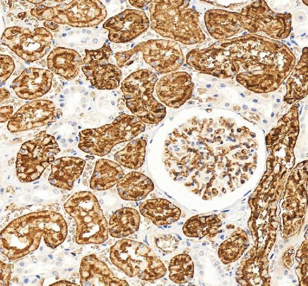
IHC analysis of AQP1 using anti-AQP1 antibody (PB9473).
AQP1 was detected in a paraffin-embedded section of human kidney tissue. Heat mediated antigen retrieval was performed in EDTA buffer (pH 8.0, epitope retrieval solution). The tissue section was blocked with 10% goat serum. The tissue section was then incubated with 2 μg/ml rabbit anti-AQP1 Antibody (PB9473) overnight at 4°C. Peroxidase Conjugated Goat Anti-rabbit IgG was used as secondary antibody and incubated for 30 minutes at 37°C. The tissue section was developed using HRP Conjugated Rabbit IgG Super Vision Assay Kit (Catalog # SV0002) with DAB as the chromogen.

Click image to see more details
Immunoprecipitation determinations of HMGB1 and AQP1 (A) Western blots of HMGB1 in plasmids. (B) Western blots of AQP1 in plasmids. (C) CO-IP assay results. 293T: 293T-null cells; 293T-E5061-E5077: 293T- E5061 HA empty control plasmid transfection- E5077 negative control CON238 plasmid; 293T-E5062-E5078: 293T- E5062 HA-Aqp1 overexpression plasmid transfection- E5078 Hmgb1-3flag overexpression plasmid. Flag: HMGB1; HA:AQP1.
Index in PubMed under a CC BY license. PMID: 39359252

Click image to see more details
Changes in AQP1 expression after HMGB1 knockdown (A) Protein expression levels of HMGB1 and AQP1 in the spinal cords of rats in each group. (B) Protein expression levels of HMGB1 and AQP1 in the LPS inflammatory cell model in each group. (C) mRNA levels of HMGB1 and AQP1 in the spinal cords of rats in each group. (D) mRNA levels of HMGB1 and AQP1 in the LPS inflammatory cell model in each group. N = 3 per group **** p < 0.0001 *** p < 0.001 ** p < 0.01* p < 0.05.
Index in PubMed under a CC BY license. PMID: 39359252

Click image to see more details
Changes in AQP1 expression after TAK-242 and FPS-ZM1 treatments (A) Protein expression levels of TLR4 and AQP1 in the spinal cords of rats in each group. (B) Protein expression levels of TLR4 and AQP1 in the LPS inflammatory cell model in each group. (C) Protein expression levels of RAGE and AQP1 in the spinal cords of rats in each group. (D) Protein expression levels of RAGE and AQP1 in the LPS inflammatory cell model in each group. (E) mRNA levels of TLR4 and AQP1 in each group. (F) mRNA levels of RAGE and AQP1 in each group. N = 3 per group **** p < 0.0001 *** p < 0.001 ** p < 0.01* p < 0.05.
Index in PubMed under a CC BY license. PMID: 39359252

Click image to see more details
Changes in NF-κB expression following knockdown of HMGB1 or AQP1 (A) Protein expression levels of HMGB1 and NF-κB in the spinal cords of rats in each group. (B) mRNA levels of HMGB1 and NF-κB in the spinal cords of rats in each group. (C) Protein expression levels of AQP1 and NF-κB in the spinal cords of rats in each group. (D) mRNA levels of AQP1 and NF-κB in the spinal cords of rats in each group. (E) Protein expression levels of NF-κB and AQP1 in the LPS inflammatory cell model in each group. (F) mRNA levels of NF-κB and AQP1 in the LPS inflammatory cell model in each group. N = 3 per group **** p < 0.0001 *** p < 0.001 ** p < 0.01* p < 0.05.
Index in PubMed under a CC BY license. PMID: 39359252

Click image to see more details
Western blot analysis of AQP1 using anti-AQP1 antibody (PB9473).
Electrophoresis was performed on a 10% SDS-PAGE gel at 80V (Stacking gel) / 120V (Resolving gel) for 2 hours. The sample well of each lane was loaded with 30 ug of sample under reducing conditions.
Lane 1: rat heart tissue lysates,
Lane 2: rat kidney tissue lysates,
Lane 3: rat lung tissue lysates,
Lane 4: mouse heart tissue lysates,
Lane 5: mouse kidney tissue lysates,
Lane 6: mouse lung tissue lysates.
After electrophoresis, proteins were transferred to a nitrocellulose membrane at 150 mA for 50-90 minutes. Blocked the membrane with 5% non-fat milk/TBS for 1.5 hour at RT. The membrane was incubated with rabbit anti-AQP1 antigen affinity purified polyclonal antibody (PB9473) at 0.5 μg/mL overnight at 4°C, then washed with TBS-0.1%Tween 3 times with 5 minutes each and probed with a goat anti-rabbit IgG-HRP secondary antibody (Catalog # BA1054) at a dilution of 1:5000 for 1.5 hour at RT. The signal is developed using an ECL Plus Western Blotting Substrate (Catalog # AR1196-200) with Tanon 5200 system. A specific band was detected for AQP1 at approximately 25, 35-38 kDa. The expected band size for AQP1 is at 28 kDa.

Click image to see more details
IHC analysis of AQP1 using anti-AQP1 antibody (PB9473).
AQP1 was detected in a paraffin-embedded section of human kidney tissue. Heat mediated antigen retrieval was performed in EDTA buffer (pH 8.0, epitope retrieval solution). The tissue section was blocked with 10% goat serum. The tissue section was then incubated with 2 μg/ml rabbit anti-AQP1 Antibody (PB9473) overnight at 4°C. Peroxidase Conjugated Goat Anti-rabbit IgG was used as secondary antibody and incubated for 30 minutes at 37°C. The tissue section was developed using HRP Conjugated Rabbit IgG Super Vision Assay Kit (Catalog # SV0002) with DAB as the chromogen.

Click image to see more details
IHC analysis of AQP1 using anti-AQP1 antibody (PB9473).
AQP1 was detected in a paraffin-embedded section of mouse kidney tissue. Heat mediated antigen retrieval was performed in EDTA buffer (pH 8.0, epitope retrieval solution). The tissue section was blocked with 10% goat serum. The tissue section was then incubated with 2 μg/ml rabbit anti-AQP1 Antibody (PB9473) overnight at 4°C. Peroxidase Conjugated Goat Anti-rabbit IgG was used as secondary antibody and incubated for 30 minutes at 37°C. The tissue section was developed using HRP Conjugated Rabbit IgG Super Vision Assay Kit (Catalog # SV0002) with DAB as the chromogen.

Click image to see more details
IHC analysis of AQP1 using anti-AQP1 antibody (PB9473).
AQP1 was detected in a paraffin-embedded section of rat kidney tissue. Heat mediated antigen retrieval was performed in EDTA buffer (pH 8.0, epitope retrieval solution). The tissue section was blocked with 10% goat serum. The tissue section was then incubated with 2 μg/ml rabbit anti-AQP1 Antibody (PB9473) overnight at 4°C. Peroxidase Conjugated Goat Anti-rabbit IgG was used as secondary antibody and incubated for 30 minutes at 37°C. The tissue section was developed using HRP Conjugated Rabbit IgG Super Vision Assay Kit (Catalog # SV0002) with DAB as the chromogen.

Click image to see more details
IF analysis of AQP1 using anti-AQP1 antibody (PB9473).
AQP1 was detected in a paraffin-embedded section of rat kidney tissue. Heat mediated antigen retrieval was performed in EDTA buffer (pH 8.0, epitope retrieval solution). The tissue section was blocked with 10% goat serum. The tissue section was then incubated with 5 μg/mL rabbit anti-AQP1 Antibody (PB9473) overnight at 4°C. Cy3 Conjugated Goat Anti-Rabbit IgG (BA1032) was used as secondary antibody at 1:500 dilution and incubated for 30 minutes at 37°C. The section was counterstained with DAPI. Visualize using a fluorescence microscope and filter sets appropriate for the label used.

Click image to see more details
Flow Cytometry analysis of U2OS cells using anti-AQP1 antibody (PB9473).
Overlay histogram showing U2OS cells stained with PB9473 (Blue line). The cells were fixed with 4% paraformaldehyde and blocked with 10% normal goat serum. And then incubated with rabbit anti-AQP1 Antibody (PB9473, 1 μg/1x106 cells) for 30 min at 20°C. DyLight®488 conjugated goat anti-rabbit IgG (BA1127, 5-10 μg/1x106 cells) was used as secondary antibody for 30 minutes at 20°C. Isotype control antibody (Green line) was rabbit IgG (1 μg/1x106) used under the same conditions. Unlabelled sample without incubation with primary antibody and secondary antibody (Red line) was used as a blank control.
Specific Publications For Anti-Aquaporin 1/AQP1 Antibody Picoband® (PB9473)
Loading publications
Recommended Resources
Here are featured tools and databases that you might find useful.
- Boster's Pathways Library
- Protein Databases
- Bioscience Research Protocol Resources
- Data Processing & Analysis Software
- Photo Editing Software
- Scientific Literature Resources
- Research Paper Management Tools
- Molecular Biology Software
- Primer Design Tools
- Bioinformatics Tools
- Phylogenetic Tree Analysis
Customer Reviews
Have you used Anti-Aquaporin 1/AQP1 Antibody Picoband®?
Share your experimental results or join a short interview to earn up to $1,000 in product credits or other rewards.
0 Reviews For Anti-Aquaporin 1/AQP1 Antibody Picoband®
Customer Q&As
Have a question?
Find answers in Q&As, reviews.
Can't find your answer?
Submit your question
6 Customer Q&As for Anti-Aquaporin 1/AQP1 Antibody Picoband®
Question
We are currently using anti-Aquaporin 1/AQP1 antibody PB9473 for mouse tissue, and we are content with the IHC results. The species of reactivity given in the datasheet says human, mouse, rat. Is it true that the antibody can work on horse tissues as well?
Verified Customer
Verified customer
Asked: 2019-08-26
Answer
The anti-Aquaporin 1/AQP1 antibody (PB9473) has not been validated for cross reactivity specifically with horse tissues, but there is a good chance of cross reactivity. We have an innovator award program that if you test this antibody and show it works in horse you can get your next antibody for free. Please contact me if I can help you with anything.
Boster Scientific Support
Answered: 2019-08-26
Question
My colleagues were satisfied with the WB result of your anti-Aquaporin 1/AQP1 antibody. However we have seen positive staining in retinal pigment epithelium cell membrane using this antibody. Is that expected? Could you tell me where is AQP1 supposed to be expressed?
Verified Customer
Verified customer
Asked: 2019-08-21
Answer
From what I have seen in literature, retinal pigment epithelium does express AQP1. Generally AQP1 expresses in cell membrane. Regarding which tissues have AQP1 expression, here are a few articles citing expression in various tissues:
Articular cartilage, Pubmed ID: 2007592, 1373524
Brain, Pubmed ID: 15489334
Mesangial cell, Pubmed ID: 14702039
Retinal pigment epithelium, Pubmed ID: 8703970
Uterus, Pubmed ID: 7517253
Boster Scientific Support
Answered: 2019-08-21
Question
We have observed staining in rat retinal pigment epithelium. What should we do? Is anti-Aquaporin 1/AQP1 antibody supposed to stain retinal pigment epithelium positively?
Verified Customer
Verified customer
Asked: 2019-06-10
Answer
From what I have seen in literature retinal pigment epithelium does express AQP1. From what I have seen in Uniprot.org, AQP1 is expressed in right lung, retinal pigment epithelium, uterus, mesangial cell, brain, articular cartilage, among other tissues. Regarding which tissues have AQP1 expression, here are a few articles citing expression in various tissues:
Articular cartilage, Pubmed ID: 2007592, 1373524
Brain, Pubmed ID: 15489334
Mesangial cell, Pubmed ID: 14702039
Retinal pigment epithelium, Pubmed ID: 8703970
Uterus, Pubmed ID: 7517253
Boster Scientific Support
Answered: 2019-06-10
Question
We purchased anti-Aquaporin 1/AQP1 antibody for IHC on retinal pigment epithelium in the past. I am using mouse, and We want to use the antibody for WB next. Our lab want to know about examining retinal pigment epithelium as well as uterus in our next experiment. Could give a recommendation on which antibody would work the best for WB?
Verified Customer
Verified customer
Asked: 2019-05-22
Answer
I have checked the website and datasheets of our anti-Aquaporin 1/AQP1 antibody and it seems that PB9473 has been tested on mouse in both IHC and WB. Thus PB9473 should work for your application. Our Boster satisfaction guarantee will cover this product for WB in mouse even if the specific tissue type has not been validated. We do have a comprehensive range of products for WB detection and you can check out our website bosterbio.com to find out more information about them.
Boster Scientific Support
Answered: 2019-05-22
Question
Has this antibody been used to image erythrocytes by fluorescence confocal microscopy?
Verified Customer
Verified customer
Asked: 2019-01-02
Answer
No. This antibody has not been validated with erythrocytes.
Boster Scientific Support
Answered: 2019-01-02
Question
I am looking for using your anti-Aquaporin 1/AQP1 antibody for vasopressin regulates renal water homeostasis via aquaporins studies. Has this antibody been tested with western blotting on intestinal cancer tissue? We would like to see some validation images before ordering.
W. Kulkarni
Verified customer
Asked: 2016-04-04
Answer
Thank you for your inquiry. This PB9473 anti-Aquaporin 1/AQP1 antibody is tested on rat lung tissue, kidney tissue, tissue lysate, cardiac muscle tissue, hepa whole cell lysate, mouse kidney tissue, intestinal cancer tissue. It is guaranteed to work for IHC, WB in human, mouse, rat. Our Boster guarantee will cover your intended experiment even if the sample type has not been be directly tested.
Boster Scientific Support
Answered: 2016-04-04
