Click image to see more details
Product Info Summary
| SKU: | A00071-2 |
|---|---|
| Size: | 100 μg/vial |
| Reactive Species: | Human, Mouse, Rat |
| Host: | Rabbit |
| Application: | ELISA, WB |
Customers Who Bought This Also Bought

Product info
Product Name
Anti-Aromatase/Cyp19a1 Antibody Picoband®
SKU/Catalog Number
A00071-2
Size
100 μg/vial
Form
Lyophilized
Description
Boster Bio Anti-Aromatase/Cyp19a1 Antibody Picoband® catalog # A00071-2. Tested in ELISA, WB applications. This antibody reacts with Human, Mouse, Rat. The brand Picoband indicates this is a premium antibody that guarantees superior quality, high affinity, and strong signals with minimal background in Western blot applications. Only our best-performing antibodies are designated as Picoband, ensuring unmatched performance.
Storage & Handling
At -20°C for one year from date of receipt. After reconstitution, at 4°C for one month. It can also be aliquotted and stored frozen at -20°C for six months. Avoid repeated freezing and thawing.
Cite This Product
Anti-Aromatase/Cyp19a1 Antibody Picoband® (Boster Biological Technology, Pleasanton CA, USA, Catalog # A00071-2)
Host
Rabbit
Contents
Each vial contains 4 mg Trehalose, 0.9 mg NaCl, 0.2 mg Na2HPO4.
Clonality
Polyclonal
Isotype
Rabbit IgG
Immunogen
E.coli-derived mouse Aromatase/Cyp19a1 recombinant protein (Position: N75-Q428).
Cross-reactivity
No cross-reactivity with other proteins.
Reactive Species
A00071-2 is reactive to Cyp19a1 in Human, Mouse, Rat
Observed Molecular Weight
49-58 kDa
Calculated molecular weight
58.0 kDa
Background of Cyp19a1
CYP19A1, also called Aromatase, is an enzyme responsible for a key step in the biosynthesis of estrogens. It is a member of the cytochrome P450 superfamily, which are monooxygenases that catalyze many reactions involved in steroidogenesis. In particular, aromatase is responsible for the aromatization of androgens into estrogens. The CYP19 gene spans at least 70 kb of genomic DNA and contains 10 exons. By in situ hybridization, the ARO gene is mapped to 15q21.1. The aromatase enzyme can be found in many tissues including gonads, brain, adipose tissue, placenta, blood vessels, skin, bone, and endometrium, as well as in tissue of endometriosis, uterine fibroids, breast cancer, and endometrial cancer. It is an important factor in sexual development. Some bodybuilders taking steroids also take antiaromatase supplements to prevent excess testosterone conversion into estrogens, which can cause gynecomastia.
Antibody Validation
Boster validates all antibodies on WB, IHC, ICC, Immunofluorescence, and ELISA with known positive control and negative samples to ensure specificity and high affinity, including thorough antibody incubations.
Application & Images
Applications
A00071-2 is guaranteed for ELISA, WB Boster Guarantee
Recommend Dilution
| Application | Dilution | Species |
|---|---|---|
| Western blot | 0.25-0.5 μg/ml | Human, Mouse, Rat |
| ELISA | 0.1-0.5 μg/ml | - |
Tested application
Suggested blocking solution with 5% non-fat milk or BSA; (*)Recommended protein loading: 20-40 µg per lane
Validation Images & Assay Conditions
Click image to see more details
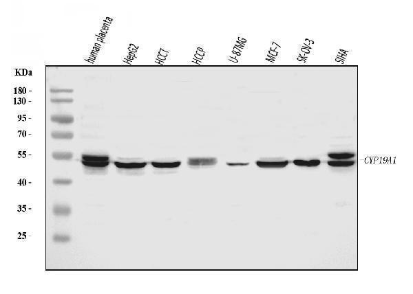
Western blot analysis of Aromatase/Cyp19a1 using anti-Aromatase/Cyp19a1 antibody (A00071-2).
Electrophoresis was performed on a 5-20% SDS-PAGE gel at 70V (Stacking gel) / 90V (Resolving gel) for 2-3 hours. The sample well of each lane was loaded with 30 ug of sample under reducing conditions.
Lane 1: human placenta tissue lysates,
Lane 2: human HepG2 whole cell lysates,
Lane 3: human HCCT tissue lysates,
Lane 4: human HCCP tissue lysates,
Lane 5: human U-87MG whole cell lysates,
Lane 6: human MCF-7 whole cell lysates,
Lane 7: human SK-OV-3 whole cell lysates,
Lane 8: human SiHa whole cell lysates.
After electrophoresis, proteins were transferred to a nitrocellulose membrane at 150 mA for 50-90 minutes. Blocked the membrane with 5% non-fat milk/TBS for 1.5 hour at RT. The membrane was incubated with rabbit anti-Aromatase/Cyp19a1 antigen affinity purified polyclonal antibody (Catalog # A00071-2) at 0.5 μg/mL overnight at 4°C, then washed with TBS-0.1%Tween 3 times with 5 minutes each and probed with a goat anti-rabbit IgG-HRP secondary antibody at a dilution of 1:5000 for 1.5 hour at RT. The signal is developed using an Enhanced Chemiluminescent detection (ECL) kit (Catalog # EK1002) with Tanon 5200 system. A specific band was detected for Aromatase/Cyp19a1 at approximately 49-58 kDa. The expected band size for Aromatase/Cyp19a1 is at 58 kDa.

Click image to see more details
Western blot analysis of liver P450arom expressions of the rats. The upper picture shows the Western blot analysis of the liver aromatase P450. Densitometric analysis of the protein concentration using aromatase/β-actin expressed as the mean with SEM bar in each column indicated in the lower panel. * p < 0.05 vs corresponding intact controls, # p < 0.05 vs OVX1M.
Index in PubMed under a CC BY license. PMID: 15661083

Click image to see more details
Western blot analysis of SA adipose P450arom expressions of the rats. The upper picture shows the Western blot analysis of the SA adipose aromatase P450. Densitometric analysis of the protein concentration using aromatase/β-actin expressed as the mean with SEM bar in each column indicated in the lower panel. * p < 0.05 vs corresponding intact controls, # p < 0.05 vs OVX1M.
Index in PubMed under a CC BY license. PMID: 15661083

Click image to see more details
Western blot analysis of Aromatase/Cyp19a1 using anti-Aromatase/Cyp19a1 antibody (A00071-2).
Electrophoresis was performed on a 5-20% SDS-PAGE gel at 70V (Stacking gel) / 90V (Resolving gel) for 2-3 hours. The sample well of each lane was loaded with 30 ug of sample under reducing conditions.
Lane 1: rat testis tissue lysates,
Lane 2: rat ovary tissue lysates,
Lane 3: rat brain tissue lysates,
Lane 4: rat C6 whole cell lysates,
Lane 5: mouse testis tissue lysates,
Lane 6: mouse ovary tissue lysates,
Lane 7: mouse brain tissue lysates,
Lane 8: mouse NIH/3T3 whole cell lysates.
After electrophoresis, proteins were transferred to a nitrocellulose membrane at 150 mA for 50-90 minutes. Blocked the membrane with 5% non-fat milk/TBS for 1.5 hour at RT. The membrane was incubated with rabbit anti-Aromatase/Cyp19a1 antigen affinity purified polyclonal antibody (Catalog # A00071-2) at 0.5 μg/mL overnight at 4°C, then washed with TBS-0.1%Tween 3 times with 5 minutes each and probed with a goat anti-rabbit IgG-HRP secondary antibody at a dilution of 1:5000 for 1.5 hour at RT. The signal is developed using an Enhanced Chemiluminescent detection (ECL) kit (Catalog # EK1002) with Tanon 5200 system. A specific band was detected for Aromatase/Cyp19a1 at approximately 49-58 kDa. The expected band size for Aromatase/Cyp19a1 is at 58 kDa.
Specific Publications For Anti-Aromatase/Cyp19a1 Antibody Picoband® (A00071-2)
Loading publications
Recommended Resources
Here are featured tools and databases that you might find useful.
- Boster's Pathways Library
- Protein Databases
- Bioscience Research Protocol Resources
- Data Processing & Analysis Software
- Photo Editing Software
- Scientific Literature Resources
- Research Paper Management Tools
- Molecular Biology Software
- Primer Design Tools
- Bioinformatics Tools
- Phylogenetic Tree Analysis
Customer Reviews
Have you used Anti-Aromatase/Cyp19a1 Antibody Picoband®?
Share your experimental results or join a short interview to earn up to $1,000 in product credits or other rewards.
0 Reviews For Anti-Aromatase/Cyp19a1 Antibody Picoband®
Customer Q&As
Have a question?
Find answers in Q&As, reviews.
Can't find your answer?
Submit your question
