Click image to see more details
Product Info Summary
| SKU: | PB9468 |
|---|---|
| Size: | 100 μg/vial |
| Reactive Species: | Human, Rat |
| Host: | Rabbit |
| Application: | WB |
Customers Who Bought This Also Bought

Product info
Product Name
Anti-c Abl/ABL1 Antibody Picoband®
SKU/Catalog Number
PB9468
PB0491 is an alternative SKU for this antibody, used in previous lots.
Size
100 μg/vial
Form
Lyophilized
Description
Boster Bio Anti-c Abl/ABL1 Antibody Picoband® catalog # PB9468. Tested in WB applications. This antibody reacts with Human, Rat. The brand Picoband indicates this is a premium antibody that guarantees superior quality, high affinity, and strong signals with minimal background in Western blot applications. Only our best-performing antibodies are designated as Picoband, ensuring unmatched performance.
Storage & Handling
Store at -20˚C for one year from date of receipt. After reconstitution, at 4˚C for one month. It can also be aliquotted and stored frozen at -20˚C for six months. Avoid repeated freeze-thaw cycles.
Cite This Product
Anti-c Abl/ABL1 Antibody Picoband® (Boster Biological Technology, Pleasanton CA, USA, Catalog # PB9468)
Host
Rabbit
Contents
Each vial contains antibody formulated with stabilizing components, 0.9 mg NaCl, 0.2 mg Na2HPO4, and 0.05 mg NaN3.
*This antibody is supplied in a stabilized formulation.
Compatibility with conjugation reactions depends on the chemistry of the conjugation method used.
For conjugation methods that are not compatible with the stabilizing components present in this formulation, a carrier-free antibody format is required.
Clonality
Polyclonal
Isotype
Rabbit IgG
Immunogen
A synthetic peptide corresponding to a sequence at the C-terminus of human c Abl, different from the related mouse sequence by one amino acid.
Cross-reactivity
No cross-reactivity with other proteins
Reactive Species
PB9468 is reactive to ABL1 in Human, Rat
Observed Molecular Weight
150 kDa
Calculated molecular weight
122.9 kDa
Background of ABL1
c Abl is also called as ABL1. This gene is a protooncogene that encodes a protein tyrosine kinase involved in a variety of cellular processes, including cell division, adhesion, differentiation, and response to stress. The activity of the protein is negatively regulated by its SH3 domain, whereby deletion of the region encoding this domain results in an oncogene. The ubiquitously expressed protein has DNA-binding activity that is regulated by CDC2-mediated phosphorylation, suggesting a cell cycle function. This gene has been found fused to a variety of translocation partner genes in various leukemias, most notably the t (9;22) translocation that results in a fusion with the 5' end of the breakpoint cluster region gene. Alternative splicing of this gene results in two transcript variants, which contain alternative first exons that are spliced to the remaining common exons.
Antibody Validation
Boster validates all antibodies on WB, IHC, ICC, Immunofluorescence, and ELISA with known positive control and negative samples to ensure specificity and high affinity, including thorough antibody incubations.
Application & Images
Applications
PB9468 is guaranteed for WB Boster Guarantee
Recommend Dilution
| Application | Dilution | Species |
|---|---|---|
| Western blot | 0.1-0.5μg/ml | Human, Rat |
Tested application
Suggested blocking solution with 5% non-fat milk or BSA; (*)Recommended protein loading: 20-40 µg per lane
Validation Images & Assay Conditions
Click image to see more details
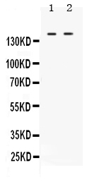
Western blot analysis of cABI using anti-cABI antibody (PB9468).
Electrophoresis was performed on a 5-20% SDS-PAGE gel at 70V (Stacking gel) / 90V (Resolving gel) for 2-3 hours.
Lane 1: Rat Brain Tissue Lysate at 50ug,
Lane 2: HELA Whole Cell Lysate at 40ug.
After electrophoresis, proteins were transferred to a nitrocellulose membrane at 150 mA for 50-90 minutes. Blocked the membrane with 5% non-fat milk/TBS for 1.5 hour at RT. The membrane was incubated with rabbit anti-cABI antigen affinity purified polyclonal antibody (Catalog # PB9468) at 0.5 μg/mL overnight at 4°C, then washed with TBS-0.1%Tween 3 times with 5 minutes each and probed with a goat anti-rabbit IgG-HRP secondary antibody at a dilution of 1:5000 for 1.5 hour at RT. The signal is developed using an Enhanced Chemiluminescent detection (ECL) kit (Catalog # EK1002) with Tanon 5200 system. A specific band was detected for cABI at approximately 150 kDa. The expected band size for cABI is at 123 kDa.
Specific Publications For Anti-c Abl/ABL1 Antibody Picoband® (PB9468)
Loading publications
Recommended Resources
Here are featured tools and databases that you might find useful.
- Boster's Pathways Library
- Protein Databases
- Bioscience Research Protocol Resources
- Data Processing & Analysis Software
- Photo Editing Software
- Scientific Literature Resources
- Research Paper Management Tools
- Molecular Biology Software
- Primer Design Tools
- Bioinformatics Tools
- Phylogenetic Tree Analysis
Customer Reviews
Have you used Anti-c Abl/ABL1 Antibody Picoband®?
Share your experimental results or join a short interview to earn up to $1,000 in product credits or other rewards.
0 Reviews For Anti-c Abl/ABL1 Antibody Picoband®
Customer Q&As
Have a question?
Find answers in Q&As, reviews.
Can't find your answer?
Submit your question
6 Customer Q&As for Anti-c Abl/ABL1 Antibody Picoband®
Question
Does anti-c Abl/ABL1 antibody PB9468 work on canine WB with cervix carcinoma erythroleukemia?
Verified Customer
Verified customer
Asked: 2020-04-22
Answer
Our lab technicians have not validated anti-c Abl/ABL1 antibody PB9468 on canine. You can run a BLAST between canine and the immunogen sequence of anti-c Abl/ABL1 antibody PB9468 to see if they may cross-react. If the sequence homology is close, then you can perform a pilot test. Keep in mind that since we have not validated canine samples, this use of the antibody is not covered by our guarantee. However we have an innovator award program that if you test this antibody and show it works in canine cervix carcinoma erythroleukemia in WB, you can get your next antibody for free.
Boster Scientific Support
Answered: 2020-04-22
Question
See below the WB image, lot number and protocol we used for lung using anti-c Abl/ABL1 antibody PB9468. Please let me know if you require anything else.
Verified Customer
Verified customer
Asked: 2019-11-25
Answer
Thank you very much for the data. Our lab team are working to resolve this as quickly as possible, and we appreciate your patience and understanding! You have provided everything we needed. Please let me know if there is anything you need in the meantime.
Boster Scientific Support
Answered: 2019-11-25
Question
Is a blocking peptide available for product anti-c Abl/ABL1 antibody (PB9468)?
Verified Customer
Verified customer
Asked: 2019-10-21
Answer
We do provide the blocking peptide for product anti-c Abl/ABL1 antibody (PB9468). If you would like to place an order for it please contact support@bosterbio.com and make a special request.
Boster Scientific Support
Answered: 2019-10-21
Question
We are currently using anti-c Abl/ABL1 antibody PB9468 for human tissue, and we are well pleased with the WB results. The species of reactivity given in the datasheet says human, rat. Is it true that the antibody can work on dog tissues as well?
E. Parker
Verified customer
Asked: 2019-03-04
Answer
The anti-c Abl/ABL1 antibody (PB9468) has not been tested for cross reactivity specifically with dog tissues, though there is a good chance of cross reactivity. We have an innovator award program that if you test this antibody and show it works in dog you can get your next antibody for free. Please contact me if I can help you with anything.
Boster Scientific Support
Answered: 2019-03-04
Question
Would anti-c Abl/ABL1 antibody PB9468 work for WB with lung?
Verified Customer
Verified customer
Asked: 2018-11-12
Answer
According to the expression profile of lung, ABL1 is highly expressed in lung. So, it is likely that anti-c Abl/ABL1 antibody PB9468 will work for WB with lung.
Boster Scientific Support
Answered: 2018-11-12
Question
Would PB9468 anti-c Abl/ABL1 antibody work on parafin embedded sections? If so, which fixation method do you recommend we use (PFA, paraformaldehyde, other)?
T. Singh
Verified customer
Asked: 2018-09-28
Answer
It shows on the product datasheet, PB9468 anti-c Abl/ABL1 antibody as been validated on WB. It is best to use PFA for fixation because it has better tissue penetration ability. PFA needs to be prepared fresh before use. Long term stored PFA turns into formalin, as the PFA molecules congregate and become formalin.
Boster Scientific Support
Answered: 2018-09-28
