Click image to see more details
-

-
-
-
-
+2
Product Info Summary
| SKU: | PA1318 |
|---|---|
| Size: | 100 μg/vial |
| Reactive Species: | Human, Mouse, Rat |
| Host: | Rabbit |
| Application: | WB |
Customers Who Bought This Also Bought

Product info
Product Name
Anti-c-Fos Antibody Picoband®
SKU/Catalog Number
PA1318
BA0207-2 is an alternative SKU for this antibody, used in previous lots.
Size
100 μg/vial
Form
Lyophilized
Description
Boster Bio Anti-c-Fos Antibody catalog # PA1318. Tested in WB applications. This antibody reacts with Human, Mouse, Rat. The brand Picoband indicates this is a premium antibody that guarantees superior quality, high affinity, and strong signals with minimal background in Western blot applications. Only our best-performing antibodies are designated as Picoband, ensuring unmatched performance.
Storage & Handling
Store at -20˚C for one year from date of receipt. After reconstitution, at 4˚C for one month. It can also be aliquotted and stored frozen at -20˚C for six months. Avoid repeated freeze-thaw cycles.
Cite This Product
Anti-c-Fos Antibody Picoband® (Boster Biological Technology, Pleasanton CA, USA, Catalog # PA1318)
Host
Rabbit
Contents
Each vial contains antibody formulated with stabilizing components, 0.9mg NaCl, 0.2mg Na2HPO4, 0.05mg Thimerosal, 0.05mg NaN3.
*This antibody is supplied in a stabilized formulation.
Compatibility with conjugation reactions depends on the chemistry of the conjugation method used.
For conjugation methods that are not compatible with the stabilizing components present in this formulation, a carrier-free antibody format is required.
Clonality
Polyclonal
Isotype
Rabbit IgG
Immunogen
A synthetic peptide corresponding to a sequence in the middle region of human c-Fos, identical to the related rat and mouse sequences.
Cross-reactivity
No cross-reactivity with other proteins
Reactive Species
PA1318 is reactive to FOS in Human, Mouse, Rat
Observed Molecular Weight
37 kDa
Calculated molecular weight
40.7 kDa
Background of FOS
The human oncogene c-fos is cellular homolog of the transforming gene of Finkel-Biskis-Jinkins (FBJ) murine osteosarcoma virus which was mapped to a single human chromosome. c-Fos is encoded by the FOS gene. FOS was the first transcription factor identified that has a critical function in regulating the development of cells destined to form and maintain the skeleton. FOS is also a major component of the activator protein-1 (AP-1) transcription factor complex, which includes members of the JUN family. c-fos is a major nuclear target for signal transduction pathways involved in the regulation of cell growth, differentiation, and transformation. Using transgenic and knockout mice, Grigoriadis et al. (1995) established a unique role for the proto-oncogene and nuclear transcription factor, Fos, in regulating the differentiation and activity of specific bone cell populations, both during normal development and in bone disease.
Antibody Validation
Boster validates all antibodies on WB, IHC, ICC, Immunofluorescence, and ELISA with known positive control and negative samples to ensure specificity and high affinity, including thorough antibody incubations.
Application & Images
Applications
PA1318 is guaranteed for WB Boster Guarantee
Recommend Dilution
| Application | Dilution | Species |
|---|---|---|
| Western blot | 0.1-0.5μg/ml | Human, Rat, Mouse |
Tested application
Suggested blocking solution with 5% non-fat milk or BSA; (*)Recommended protein loading: 20-40 µg per lane
Validation Images & Assay Conditions
Click image to see more details
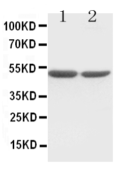
Anti-c-Fos antibody, PA1318, Western blotting
Lane 1: HT1080 Cell Lysate
Lane 2: COLO320 Cell Lysate

Click image to see more details
DFSC-EVs regulated tooth eruption by inhibiting osteoclast differentiation. (A) Schematic illustration of RAW264.7 and DFSC co-culture system. (B) Representative images of TRAP staining. Scale bar = 200 μm. (C) Quantitative analysis of TRAP-positive area. (D) The mRNA level of ACP5 , CTSK and CFOS in RAW264.7 cultured with DFSC. (E) The protein level of ACP5, CTSK and CFOS in RAW264.7 cultured with DFSC. (F) Western blotting quantification. (G) Schematic illustration of RAW264.7 and DFSC-EVs co-culture system. (H) Representative images of TRAP staining. Scale bar = 200 μm. (I) Quantitative analysis of TRAP-positive area. (J) The mRNA level of ACP5 , CTSK and CFOS in RAW264.7 cultured with DFSC-EVs. (K) The protein level of ACP5, CTSK and CFOS in RAW264.7 cultured with DFSC-EVs. (L) Western blotting quantification. ns, not significant. * p < 0.05, ** p < 0.01, *** p < 0.001, **** p < 0.0001. n = 3.
Index in PubMed under a CC BY license. PMID: 39834384

Click image to see more details
ANXA1 was the core factor of DFSC-EVs regulating osteoclast differentiation. (A) Gene ontology enrichment analysis of DFSC-EVs protein profiles. (B) The top proteins of Cadherin related to regulating osteoblast differentiation based on expression level. (C) The mRNA level of ANXA1 . (D) The protein level of ANXA1. (E) Western blotting quantification. (F) Schematic illustration of RAW264.7 and siANXA1-EVs co-culture system. (G) Representative images of TRAP staining. Scale bar = 200 μm. (H) Quantitative analysis of TRAP-positive area. (I) The mRNA level of ACP5 , CTSK and CFOS in RAW264.7 cultured with siANXA1-EVs. (J) The protein level of ACP5, CTSK and CFOS in RAW264.7 cultured with siANXA1-EVs. (K) Western blotting quantification. * p < 0.05, ** p < 0.01, *** p < 0.001, **** p < 0.0001. n = 3.
Index in PubMed under a CC BY license. PMID: 39834384

Click image to see more details
ANXA1 mediated PPARγ-CEBPα pathway to regulate osteoclast differentiation (A) The mRNA level of PPARγ in RAW264.7 cultured with siANXA1-EVs. (B) The mRNA level of CEBPα in RAW264.7 cultured with siANXA1-EVs. (C) The protein level of PPARγ and CEBPα in RAW264.7 cultured with siANXA1-EVs. (D) Quantitative analysis of PPARγ protein expression. (E) Quantitative analysis of CEBPα protein expression. (F) Schematic illustration of PPARγ inhibited RAW264.7 and DFSC-EVs co-culture system. (G) Representative images of TRAP staining. Scale bar = 200 μm. (H) Quantitative analysis of TRAP-positive area. (I) PPARγ inhibited RAW264.7 construction. (J) The mRNA level of CEBPα in PPARγ inhibited RAW264.7. (K) The protein level of PPARγ and CEBPα in PPARγ inhibited RAW264.7. (L) Quantitative analysis of PPARγ protein expression. (M) Quantitative analysis of CEBPα protein expression. (N) The mRNA level of ACP5 , CTSK and CFOS in PPARγ inhibited RAW264.7. (O) The protein level of ACP5, CTSK and CFOS in PPARγ inhibited RAW264.7. (P) Western blotting quantification. * p < 0.05, ** p < 0.01, *** p < 0.001, **** p < 0.0001. n = 3.
Index in PubMed under a CC BY license. PMID: 39834384

Click image to see more details
The pretreatment of RD-6 inhibited the IL-17 signaling pathway in indomethacin-induced GU rats. The expression of IL17RA, FOS, IL1B, and PTGS2 determined in gastric tissue by qRT-PCR (A) and western bloting (B) . Data are expressed as mean ± S.E.M ( n = 3). One-way ANOVA with the uncorrected Fisher’s LSD test was used to evaluate multiple comparisons. # p < 0.05, ## p < 0.01 vs. NC group; * p < 0.05, ** p < 0.01 vs. IND group. NC, normal control; IND, indomethacin; RD-6-L, M, and H represent Ruda-6 at low, medium and high doses, respectively.
Index in PubMed under a CC BY license. PMID: 37637418

Click image to see more details
Effects of JTG on expression of associated proteins and NF-κB pathway of osteoclast induced from BMMs with RANKL and LPS. BMMs were incubated with RANKL and JTG for 48 h, the proteins were extracted to analyze associated proteins of osteoclast by Western blot. A : a Western blot imagines for expression of NFATc1, c-Fos, Cathepsin K and MMP9. A : b – e The quantification analysis of NFATc1, c-Fos, Cathepsin K and MMP9 based on the results of A : a by ECL detection system, respectively. B : a The images of Western blot for TRAF6, P-P65, P65 and IκBα. B : b – d The quantification analysis of TRAF6, P-P65/P65 and IκBα based on the results of B : a by using an ECL detection system, respectively. Each point represents the mean ± SD (n = 3). The experiments were repeated for three times. * P < 0.05, ** P < 0.01 compared with control group
Index in PubMed under a CC BY license. PMID: 36195960
Specific Publications For Anti-c-Fos Antibody Picoband® (PA1318)
Loading publications
Recommended Resources
Here are featured tools and databases that you might find useful.
- Boster's Pathways Library
- Protein Databases
- Bioscience Research Protocol Resources
- Data Processing & Analysis Software
- Photo Editing Software
- Scientific Literature Resources
- Research Paper Management Tools
- Molecular Biology Software
- Primer Design Tools
- Bioinformatics Tools
- Phylogenetic Tree Analysis
Customer Reviews
Have you used Anti-c-Fos Antibody Picoband®?
Share your experimental results or join a short interview to earn up to $1,000 in product credits or other rewards.
0 Reviews For Anti-c-Fos Antibody Picoband®
Customer Q&As
Have a question?
Find answers in Q&As, reviews.
Can't find your answer?
Submit your question
16 Customer Q&As for Anti-c-Fos Antibody Picoband®
Question
I have a question about product PA1318, anti-c-Fos antibody. I was wondering if it would be possible to conjugate this antibody with biotin. I would need it to be without BSA or sodium azide. I am planning on using a buffer exchange of sodium azide with PBS only. Would there be problems for me to conjugate the antibody and store it in -20 degrees in small aliquots?
Verified Customer
Verified customer
Asked: 2020-02-28
Answer
We do not advise storing this antibody with PBS buffer only in -20 degrees. If you want to store it in -20 degrees it is best to add some cryoprotectant like glycerol. If you want carrier free PA1318 anti-c-Fos antibody, we can provide it to you in a special formula with trehalose and/or glycerol. These molecules will not interfere with conjugation chemistry and provide a good level of protection for the antibody from degradation. Please be sure to specify this in your purchase order.
Boster Scientific Support
Answered: 2020-02-28
Question
We are interested in using your anti-c-Fos antibody for transforming growth factor beta receptor signaling pathway studies. Has this antibody been tested with western blotting on ht1080 cell lysate? We would like to see some validation images before ordering.
Verified Customer
Verified customer
Asked: 2020-02-26
Answer
We appreciate your inquiry. This PA1318 anti-c-Fos antibody is validated on ht1080 cell lysate, colo320 cell lysate. It is guaranteed to work for WB in human, mouse, rat. Our Boster guarantee will cover your intended experiment even if the sample type has not been be directly tested.
Boster Scientific Support
Answered: 2020-02-26
Question
Do you have a BSA free version of anti-c-Fos antibody PA1318 available?
Verified Customer
Verified customer
Asked: 2020-01-22
Answer
Thanks for your recent telephone inquiry. I can confirm that some lots of this anti-c-Fos antibody PA1318 are BSA free. For now, these lots are available and we can make a BSA free formula for you free of charge. It will take 3 extra days to prepare. If you require this antibody BSA free again in future, please do not hesitate to contact me and I will be pleased to check which lots we have in stock that are BSA free.
Boster Scientific Support
Answered: 2020-01-22
Question
Please see the WB image, lot number and protocol we used for leukemic t-cell using anti-c-Fos antibody PA1318. Please let me know if you require anything else.
Verified Customer
Verified customer
Asked: 2019-12-12
Answer
Thank you very much for the data. Our lab team are working to resolve this as quickly as possible, and we appreciate your patience and understanding! You have provided everything we needed. Please let me know if there is anything you need in the meantime.
Boster Scientific Support
Answered: 2019-12-12
Question
Would anti-c-Fos antibody PA1318 work on pig WB with colon?
Verified Customer
Verified customer
Asked: 2019-07-22
Answer
Our lab technicians have not tested anti-c-Fos antibody PA1318 on pig. You can run a BLAST between pig and the immunogen sequence of anti-c-Fos antibody PA1318 to see if they may cross-react. If the sequence homology is close, then you can perform a pilot test. Keep in mind that since we have not validated pig samples, this use of the antibody is not covered by our guarantee. However we have an innovator award program that if you test this antibody and show it works in pig colon in WB, you can get your next antibody for free.
Boster Scientific Support
Answered: 2019-07-22
Question
I see that the anti-c-Fos antibody PA1318 works with WB, what is the protocol used to produce the result images on the product page?
Verified Customer
Verified customer
Asked: 2019-06-25
Answer
You can find protocols for WB on the "support/technical resources" section of our navigation menu. If you have any further questions, please send an email to support@bosterbio.com
Boster Scientific Support
Answered: 2019-06-25
Question
Does anti-c-Fos antibody PA1318 work for WB with leukemic t-cell?
Verified Customer
Verified customer
Asked: 2019-06-11
Answer
According to the expression profile of leukemic t-cell, FOS is highly expressed in leukemic t-cell. So, it is likely that anti-c-Fos antibody PA1318 will work for WB with leukemic t-cell.
Boster Scientific Support
Answered: 2019-06-11
Question
I am interested in to test anti-c-Fos antibody PA1318 on mouse leukemic t-cell for research purposes, then I may be interested in using anti-c-Fos antibody PA1318 for diagnostic purposes as well. Is the antibody suitable for diagnostic purposes?
Verified Customer
Verified customer
Asked: 2019-06-07
Answer
The products we sell, including anti-c-Fos antibody PA1318, are only intended for research use. They would not be suitable for use in diagnostic work. If you have the means to develop a product into diagnostic use, and are interested in collaborating with us and develop our product into an IVD product, please contact us for more discussions.
Boster Scientific Support
Answered: 2019-06-07
Question
Will PA1318 anti-c-Fos antibody work on parafin embedded sections? If so, which fixation method do you recommend we use (PFA, paraformaldehyde, other)?
Verified Customer
Verified customer
Asked: 2018-10-05
Answer
You can see on the product datasheet, PA1318 anti-c-Fos antibody as been validated on WB. It is best to use PFA for fixation because it has better tissue penetration ability. PFA needs to be prepared fresh before use. Long term stored PFA turns into formalin, as the PFA molecules congregate and become formalin.
Boster Scientific Support
Answered: 2018-10-05
Question
Is a blocking peptide available for product anti-c-Fos antibody (PA1318)?
Verified Customer
Verified customer
Asked: 2017-12-27
Answer
We do provide the blocking peptide for product anti-c-Fos antibody (PA1318). If you would like to place an order for it please contact support@bosterbio.com and make a special request.
Boster Scientific Support
Answered: 2017-12-27
Question
Thanks for helping with my inquiry over the phone. Here are the WB image, lot number and protocol we used for leukemic t-cell using anti-c-Fos antibody PA1318. Let me know if you need anything else.
P. Brown
Verified customer
Asked: 2017-09-21
Answer
I appreciate the data. You have provided everything we needed. Our lab team are working to resolve your inquiry as quickly as possible, and we appreciate your patience and understanding! Please let me know if there is anything you need in the meantime.
Boster Scientific Support
Answered: 2017-09-21
Question
Is this PA1318 anti-c-Fos antibody reactive to the isotypes of FOS?
Verified Customer
Verified customer
Asked: 2017-06-13
Answer
The immunogen of PA1318 anti-c-Fos antibody is A synthetic peptide corresponding to a sequence in the middle region of human c-Fos(170-187aa DQLEDEKSALQTEIANLL), identical to the related rat and mouse sequences. Could you tell me which isotype you are interested in so I can help see if the immunogen is part of this isotype?
Boster Scientific Support
Answered: 2017-06-13
Question
I was wanting to use your anti-c-Fos antibody for WB for mouse leukemic t-cell on frozen tissues, but I want to know if it has been validated for this particular application. Has this antibody been validated and is this antibody a good choice for mouse leukemic t-cell identification?
N. Thomas
Verified customer
Asked: 2016-01-25
Answer
It shows on the product datasheet, PA1318 anti-c-Fos antibody has been tested for WB on human, mouse, rat tissues. We have an innovator award program that if you test this antibody and show it works in mouse leukemic t-cell in IHC-frozen, you can get your next antibody for free.
Boster Scientific Support
Answered: 2016-01-25
Question
We have observed staining in rat mucosa of stomach. Any tips? Is anti-c-Fos antibody supposed to stain mucosa of stomach positively?
W. Li
Verified customer
Asked: 2014-10-09
Answer
From what I have seen in literature mucosa of stomach does express FOS. From what I have seen in Uniprot.org, FOS is expressed in mucosa of stomach, colon, pancreas, leukemic t-cell, among other tissues. Regarding which tissues have FOS expression, here are a few articles citing expression in various tissues:
Colon, Pubmed ID: 14702039
Leukemic T-cell, Pubmed ID: 19690332
Pancreas, Pubmed ID: 15489334
Boster Scientific Support
Answered: 2014-10-09
Question
My colleagues were happy with the WB result of your anti-c-Fos antibody. However we have observed positive staining in leukemic t-cell nucleus. endoplasmic reticulum. cytoplasm, using this antibody. Is that expected? Could you tell me where is FOS supposed to be expressed?
A. Evans
Verified customer
Asked: 2013-12-25
Answer
According to literature, leukemic t-cell does express FOS. Generally FOS expresses in nucleus. endoplasmic reticulum. cytoplasm,. Regarding which tissues have FOS expression, here are a few articles citing expression in various tissues:
Colon, Pubmed ID: 14702039
Leukemic T-cell, Pubmed ID: 19690332
Pancreas, Pubmed ID: 15489334
Boster Scientific Support
Answered: 2013-12-25
Question
We are currently using anti-c-Fos antibody PA1318 for human tissue, and we are content with the WB results. The species of reactivity given in the datasheet says human, mouse, rat. Is it likely that the antibody can work on bovine tissues as well?
Z. Roberts
Verified customer
Asked: 2013-03-27
Answer
The anti-c-Fos antibody (PA1318) has not been validated for cross reactivity specifically with bovine tissues, but there is a good chance of cross reactivity. We have an innovator award program that if you test this antibody and show it works in bovine you can get your next antibody for free. Please contact me if I can help you with anything.
Boster Scientific Support
Answered: 2013-03-27
