Click image to see more details
-

-
-
-
-
+2
Product Info Summary
| SKU: | PB9258 |
|---|---|
| Size: | 100 μg/vial |
| Reactive Species: | Human |
| Host: | Rabbit |
| Application: | Flow Cytometry, IHC, ICC, WB |
Customers Who Bought This Also Bought

Product info
Product Name
Anti-c-Kit Antibody Picoband®
SKU/Catalog Number
PB9258
Size
100 μg/vial
Form
Lyophilized
Description
Boster Bio Anti-c-Kit Antibody Picoband® catalog # PB9258. Tested in Flow Cytometry, IHC, ICC, WB applications. This antibody reacts with Human. The brand Picoband indicates this is a premium antibody that guarantees superior quality, high affinity, and strong signals with minimal background in Western blot applications. Only our best-performing antibodies are designated as Picoband, ensuring unmatched performance.
Storage & Handling
Store at -20˚C for one year from date of receipt. After reconstitution, at 4˚C for one month. It can also be aliquotted and stored frozen at -20˚C for six months. Avoid repeated freeze-thaw cycles.
Cite This Product
Anti-c-Kit Antibody Picoband® (Boster Biological Technology, Pleasanton CA, USA, Catalog # PB9258)
Host
Rabbit
Contents
Each vial contains antibody formulated with stabilizing components, 0.9 mg NaCl, 0.2 mg Na2HPO4, and 0.05 mg NaN3.
*This antibody is supplied in a stabilized formulation.
Compatibility with conjugation reactions depends on the chemistry of the conjugation method used.
For conjugation methods that are not compatible with the stabilizing components present in this formulation, a carrier-free antibody format is required.
Clonality
Polyclonal
Isotype
Rabbit IgG
Immunogen
E.coli-derived human c-Kit recombinant protein (Position: Q26-S285). Human c-Kit shares 66% amino acid (aa) sequence identity with mouse c-Kit.
Cross-reactivity
No cross-reactivity with other proteins
Reactive Species
PB9258 is reactive to KIT in Human
Observed Molecular Weight
109 kDa
Calculated molecular weight
109.9 kDa
Background of KIT
SCFR (Mast/stem cell growth factor receptor), also known as KIT ONCOGENE or CD117, is a protein that in humans is encoded by the KIT gene. KIT was first described as the cellular homolog of the feline sarcoma viral oncogene v-kit. The KIT gene is mapped on 4q12. Kit was expressed on the surface of germ cells up to the pachytene stage. Signaling from the KIT receptor tyrosine kinase is essential for primordial germ cell growth both in vivo and in vitro. Determination of the KIT effectors acting in primordial germ cells has been hampered by the lack of effective methods to manipulate easily gene expression in these cells.
Antibody Validation
Boster validates all antibodies on WB, IHC, ICC, Immunofluorescence, and ELISA with known positive control and negative samples to ensure specificity and high affinity, including thorough antibody incubations.
Application & Images
Applications
PB9258 is guaranteed for Flow Cytometry, IHC, ICC, WB Boster Guarantee
Assay Dilutions Recommendation
The recommendations below provide a starting point for assay optimization. The actual working concentration varies and should be decided by the user.
Immunohistochemistry (Paraffin-embedded Section), 0.5-1μg/ml
Immunohistochemistry (Frozen Section), 0.5-1μg/ml
Immunocytochemistry, 0.5-1μg/ml
Western blot, 0.1-0.5μg/ml
Flow Cytometry (Fixed), 1-3μg/1x106cells
Positive Control
WB: Recombinan Human C-Kit Protein 05ng,, HEPG2 Whole Cell,
IHC: human intestinal cancer tissue
FCM: K562 cell
Validation Images & Assay Conditions
Click image to see more details
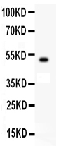
Western blot analysis of C-Kit using anti-C-Kit antibody (PB9258).
Electrophoresis was performed on a 5-20% SDS-PAGE gel at 70V (Stacking gel) / 90V (Resolving gel) for 2-3 hours.
Lane 1: Recombinan Human C-Kit Protein 0.5ng
After Electrophoresis, proteins were transferred to a Nitrocellulose membrane at 150mA for 50-90 minutes. Blocked the membrane with 5% Non-fat Milk/ TBS for 1.5 hour at RT. The membrane was incubated with rabbit anti-C-Kit antigen affinity purified polyclonal antibody (Catalog # PB9258) at 0.5 μg/mL overnight at 4°C, then washed with TBS-0.1%Tween 3 times with 5 minutes each and probed with a goat anti-rabbit IgG-HRP secondary antibody at a dilution of 1:10000 for 1.5 hour at RT. The signal is developed using an Enhanced Chemiluminescent detection (ECL) kit (Catalog # EK1002) with Tanon 5200 system. A specific band was detected for C-Kit at approximately
49KD. The expected band size for C-Kit is at 49KD.

Click image to see more details
Lumiflavin treatment reduces the development of DDP resistance of ovarian cancer OVCAR-3 cells line that is associated with cancer stem cells (CSCs). DDP resistance of ovarian cancer OVCAR-3 cell lines were induced by gradient concentration increment method ( ), and treated with 10, 20, 40, and 80 μm lumiflavin intervention simultaneously. The cell inhibition rate and resistance index of OVCAR-3cells to DDP were detected by CCK-8 assay kit. The proportion of CSCs (CD133+/CD177+ double positive cells) in OVCAR-3 cells was detected by flow cytometry. (A) Inhibitory curves of lumiflavin and DDP on OVCAR-3 and OVCAR-3/DDP cells. (B) Resistance index of OVCAR-3/DDP cells compared with OVCAR-3 cells. (C) Flow detection results of CD133+/CD117+ cells ratio of OVCAR-3 and OVCAR-3/DDP cells. (D) Statistical analysis graph of (C) . Mean ± SD ( n = 3). ** P < 0.01 difference between groups; * P < 0.05 difference between groups. DDP, cisplatin; OVCAR-3/DDP, DDP resistant OVCAR-3 cell lines; LUM, lumiflavin.
Index in PubMed under a CC BY license. PMID: 35669418

Click image to see more details
Effects of lumiflavin treatment on phenotypic differentiation of DDP-resistant cancer stem cells (CSCs/DDP). CSCs, CSCs/DDP were treated with 80 μM DDP, and CSCs/DDP were treated with 80 μM DDP and 20, 40, 80 μM lumiflavin for 72 h. Co-expression of CD133+/CD117+, CD44+/CD177, and CD44+/CD24– of CSCs/DDP were detected through flow cytometry. (A) Flow detection results of CD133+/CD117+, CD44+/CD177, and CD44+/CD24- cells of CSCs/DDP. (B) Statistical analysis graph of (A) . ** P < 0.01 between groups. Mean ± SD ( n = 3). CSCs/DDP, CSCs from DDP resistant OVCAR-3 cell lines; LUM, lumiflavin.
Index in PubMed under a CC BY license. PMID: 35669418

Click image to see more details
Western blot analysis of C-Kit using anti-C-Kit antibody (PB9258).
Electrophoresis was performed on a 5-20% SDS-PAGE gel at 70V (Stacking gel) / 90V (Resolving gel) for 2-3 hours.
Lane 1: HEPG2 Whole Cell Lysate
After Electrophoresis, proteins were transferred to a Nitrocellulose membrane at 150mA for 50-90 minutes. Blocked the membrane with 5% Non-fat Milk/ TBS for 1.5 hour at RT. The membrane was incubated with rabbit anti-C-Kit antigen affinity purified polyclonal antibody (Catalog # PB9258) at 0.5 μg/mL overnight at 4°C, then washed with TBS-0.1%Tween 3 times with 5 minutes each and probed with a goat anti-rabbit IgG-HRP secondary antibody at a dilution of 1:10000 for 1.5 hour at RT. The signal is developed using an Enhanced Chemiluminescent detection (ECL) kit (Catalog # EK1002) with Tanon 5200 system. A specific band was detected for C-Kit at approximately
109KD. The expected band size for C-Kit is at 109KD.

Click image to see more details
IHC analysis of C-Kit using anti-C-Kit antibody (PB9258).
C-Kit was detected in paraffin-embedded section of human intestinal cancer tissue. Heat mediated antigen retrieval was performed in citrate buffer (pH6, epitope retrieval solution) for 20 mins. The tissue section was blocked with 10% goat serum. The tissue section was then incubated with 1μg/ml rabbit anti-C-Kit Antibody (PB9258) overnight at 4°C. Biotinylated goat anti-rabbit IgG was used as secondary antibody and incubated for 30 minutes at 37°C. The tissue section was developed using Strepavidin-Biotin-Complex (SABC)(Catalog # SA1022) with DAB as the chromogen.

Click image to see more details
Flow Cytometry analysis of K562 cells using anti-C-Kit antibody (PB9258).
Overlay histogram showing K562 cells stained with PB9258 (Blue line). The cells were fixed with 4% paraformaldehyde and blocked with 10% normal goat serum. And then incubated with rabbit anti-C-Kit Antibody (PB9258,1μg/1x106 cells) for 30 min at 20°C. DyLight488 conjugated goat anti-rabbit IgG (BA1127, 5-10μg/1x106 cells) was used as secondary antibody for 30 minutes at 20°C. Isotype control antibody (Green line) was rabbit IgG (1μg/1x106) used under the same conditions. Unlabelled sample without incubation with primary antibody and secondary antibody (Red line) was used as a blank control.
Specific Publications For Anti-c-Kit Antibody Picoband® (PB9258)
Loading publications
Recommended Resources
Here are featured tools and databases that you might find useful.
- Boster's Pathways Library
- Protein Databases
- Bioscience Research Protocol Resources
- Data Processing & Analysis Software
- Photo Editing Software
- Scientific Literature Resources
- Research Paper Management Tools
- Molecular Biology Software
- Primer Design Tools
- Bioinformatics Tools
- Phylogenetic Tree Analysis
Customer Reviews
Have you used Anti-c-Kit Antibody Picoband®?
Share your experimental results or join a short interview to earn up to $1,000 in product credits or other rewards.
0 Reviews For Anti-c-Kit Antibody Picoband®
Customer Q&As
Have a question?
Find answers in Q&As, reviews.
Can't find your answer?
Submit your question
1 Customer Q&As for Anti-c-Kit Antibody Picoband®
Question
We are currently using anti-c-Kit antibody PB9258 for human tissue, and we are happy with the WB results. The species of reactivity given in the datasheet says human. Is it possible that the antibody can work on horse tissues as well?
C. Anderson
Verified customer
Asked: 2016-05-24
Answer
The anti-c-Kit antibody (PB9258) has not been validated for cross reactivity specifically with horse tissues, but there is a good chance of cross reactivity. We have an innovator award program that if you test this antibody and show it works in horse you can get your next antibody for free. Please contact me if I can help you with anything.
Boster Scientific Support
Answered: 2016-05-24
