Click image to see more details
-

-
-
-
-
+1
Product Info Summary
| SKU: | PB9741 |
|---|---|
| Size: | 100 μg/vial |
| Reactive Species: | Human, Mouse, Rat |
| Host: | Rabbit |
| Application: | IHC, WB |
Customers Who Bought This Also Bought

Product info
Product Name
Anti-c-Rel Antibody Picoband®
SKU/Catalog Number
PB9741
Size
100 μg/vial
Form
Lyophilized
Description
Boster Bio Anti-c-Rel Antibody Picoband® catalog # PB9741. Tested in IHC, WB applications. This antibody reacts with Human, Mouse, Rat. The brand Picoband indicates this is a premium antibody that guarantees superior quality, high affinity, and strong signals with minimal background in Western blot applications. Only our best-performing antibodies are designated as Picoband, ensuring unmatched performance.
Storage & Handling
Store at -20˚C for one year from date of receipt. After reconstitution, at 4˚C for one month. It can also be aliquotted and stored frozen at -20˚C for six months. Avoid repeated freeze-thaw cycles.
Cite This Product
Anti-c-Rel Antibody Picoband® (Boster Biological Technology, Pleasanton CA, USA, Catalog # PB9741)
Host
Rabbit
Contents
Each vial contains antibody formulated with stabilizing components, 0.9 mg NaCl, 0.2 mg Na2HPO4, and 0.05 mg NaN3.
*This antibody is supplied in a stabilized formulation.
Compatibility with conjugation reactions depends on the chemistry of the conjugation method used.
For conjugation methods that are not compatible with the stabilizing components present in this formulation, a carrier-free antibody format is required.
Clonality
Polyclonal
Isotype
Rabbit IgG
Immunogen
A synthetic peptide corresponding to a sequence in the middle region of mouse c-Rel, different from the related human sequence by four amino acids.
Cross-reactivity
No cross-reactivity with other proteins.
Reactive Species
PB9741 is reactive to Rel in Human, Mouse, Rat
Observed Molecular Weight
69 kDa
Calculated molecular weight
65.0 kDa
Background of Rel
The proto-oncogene c-Rel is a protein that in humans is encoded by the REL gene. This gene is mapped to chromosome 2p13-p12. The c-Rel protein is a member of the NF-κB family of transcription factors and contains a Rel homology domain (RHD) at its N-terminus and two C-terminal transactivation domains. c-Rel has an important role in B-cell survival and proliferation. The REL gene is amplified or mutated in several human B-cell lymphomas, including diffuse large B-cell lymphoma and Hodgkin's lymphoma.
Antibody Validation
Boster validates all antibodies on WB, IHC, ICC, Immunofluorescence, and ELISA with known positive control and negative samples to ensure specificity and high affinity, including thorough antibody incubations.
Application & Images
Applications
PB9741 is guaranteed for IHC, WB Boster Guarantee
Recommend Dilution
| Application | Dilution | Species |
|---|---|---|
| Western blot | 0.1-0.5μg/ml | Human, Mouse, Rat |
| Immunohistochemistry (Paraffin-embedded Section) | 0.5-1μg/ml | Human, Mouse, Rat |
Tested application
Suggested blocking solution with 5% non-fat milk or BSA; (*)Recommended protein loading: 20-40 µg per lane
Use TE buffer pH 9.0 for antigen retrieval; (*) citrate buffer pH 6.0 is an alternative.
Validation Images & Assay Conditions
Click image to see more details
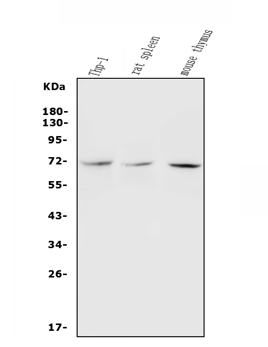
Western blot analysis of c-Rel using anti-c-Rel antibody (PB9741).
Electrophoresis was performed on a 5-20% SDS-PAGE gel at 70V (Stacking gel) / 90V (Resolving gel) for 2-3 hours. The sample well of each lane was loaded with 30 ug of sample under reducing conditions.
Lane 1: human THP-1 whole cell lysates,
Lane 2: rat spleen tissue lysates,
Lane 3: mouse thymus tissue lysates.
After electrophoresis, proteins were transferred to a nitrocellulose membrane at 150 mA for 50-90 minutes. Blocked the membrane with 5% non-fat milk/TBS for 1.5 hour at RT. The membrane was incubated with rabbit anti-c-Rel antigen affinity purified polyclonal antibody (Catalog # PB9741) at 0.5 μg/mL overnight at 4°C, then washed with TBS-0.1%Tween 3 times with 5 minutes each and probed with a goat anti-rabbit IgG-HRP secondary antibody at a dilution of 1:5000 for 1.5 hour at RT. The signal is developed using an Enhanced Chemiluminescent detection (ECL) kit (Catalog # EK1002) with Tanon 5200 system. A specific band was detected for c-Rel at approximately 69 kDa. The expected band size for c-Rel is at 65 kDa.

Click image to see more details
IHC analysis of c-Rel using anti-c-Rel antibody (PB9741).
c-Rel was detected in a paraffin-embedded section of mouse Intestine tissue. Heat mediated antigen retrieval was performed in EDTA buffer (pH 8.0, epitope retrieval solution). The tissue section was blocked with 10% goat serum. The tissue section was then incubated with 1 μg/ml rabbit anti-c-Rel Antibody (PB9741) overnight at 4°C. Biotinylated goat anti-rabbit IgG was used as secondary antibody and incubated for 30 minutes at 37°C. The tissue section was developed using Strepavidin-Biotin-Complex (SABC) (Catalog # SA1022) with DAB as the chromogen.

Click image to see more details
IL-25 inhibited LPS-induced translocation of c-Rel in STAT3-dependent manner in pulmonary macrophages. ( A ) The level of STAT3 phosphorylation was detected using Western blot. ( B ) Quantitative analysis of STAT3 phosphorylation in primary culture of pulmonary macrophages using ImageJ. Values are expressed in arbitrary units (a.u.). ( C - D ) Western blot was performed to measure the protein levels of c-Rel in the cytoplasm ( C ) and nucleus ( D ) of pulmonary macrophages. ( E - F ) Quantitative analysis of c-Rel expresion in cytoplasm ( E ) and nucleus ( F ) of pulmonary macrophages using ImageJ. ( G ) Representative images of c-Rel immunofluorescence staining in primary culture of macrophage from lung. Scale bar, 50 μm. ( H ) The protein levels of IL-12 A and IL-23 A of pulmonary macrophages was measured using Western blot. ( I - J ) Quantitative analysis of IL-12 A and IL-23 A in pulmonary macrophages using ImageJ. Values are expressed in arbitrary units (a.u.). ( K ) The graphical abstract of this study, which was drawn by Figdraw. The experiment was repeated 3 times independently, and the similar trend was obtained. One way ANOVA was used for statistical analysis (* P < 0.05; ** P < 0.01; *** P < 0.001)
Index in PubMed under a CC BY license. PMID: 37898756

Click image to see more details
IHC analysis of c-Rel using anti-c-Rel antibody (PB9741).
c-Rel was detected in a paraffin-embedded section of rat Intestine tissue. Heat mediated antigen retrieval was performed in EDTA buffer (pH 8.0, epitope retrieval solution). The tissue section was blocked with 10% goat serum. The tissue section was then incubated with 1 μg/ml rabbit anti-c-Rel Antibody (PB9741) overnight at 4°C. Biotinylated goat anti-rabbit IgG was used as secondary antibody and incubated for 30 minutes at 37°C. The tissue section was developed using Strepavidin-Biotin-Complex (SABC) (Catalog # SA1022) with DAB as the chromogen.

Click image to see more details
IHC analysis of c-Rel using anti-c-Rel antibody (PB9741).
c-Rel was detected in a paraffin-embedded section of human mammary cancer tissue. Heat mediated antigen retrieval was performed in EDTA buffer (pH 8.0, epitope retrieval solution). The tissue section was blocked with 10% goat serum. The tissue section was then incubated with 1 μg/ml rabbit anti-c-Rel Antibody (PB9741) overnight at 4°C. Biotinylated goat anti-rabbit IgG was used as secondary antibody and incubated for 30 minutes at 37°C. The tissue section was developed using Strepavidin-Biotin-Complex (SABC) (Catalog # SA1022) with DAB as the chromogen.
Specific Publications For Anti-c-Rel Antibody Picoband® (PB9741)
Loading publications
Recommended Resources
Here are featured tools and databases that you might find useful.
- Boster's Pathways Library
- Protein Databases
- Bioscience Research Protocol Resources
- Data Processing & Analysis Software
- Photo Editing Software
- Scientific Literature Resources
- Research Paper Management Tools
- Molecular Biology Software
- Primer Design Tools
- Bioinformatics Tools
- Phylogenetic Tree Analysis
Customer Reviews
Have you used Anti-c-Rel Antibody Picoband®?
Share your experimental results or join a short interview to earn up to $1,000 in product credits or other rewards.
0 Reviews For Anti-c-Rel Antibody Picoband®
Customer Q&As
Have a question?
Find answers in Q&As, reviews.
Can't find your answer?
Submit your question
1 Customer Q&As for Anti-c-Rel Antibody Picoband®
Question
We are currently using anti-c-Rel antibody PB9741 for rat tissue, and we are satisfied with the WB results. The species of reactivity given in the datasheet says human, mouse, rat. Is it possible that the antibody can work on primate tissues as well?
T. Banerjee
Verified customer
Asked: 2015-05-26
Answer
The anti-c-Rel antibody (PB9741) has not been tested for cross reactivity specifically with primate tissues, though there is a good chance of cross reactivity. We have an innovator award program that if you test this antibody and show it works in primate you can get your next antibody for free. Please contact me if I can help you with anything.
Boster Scientific Support
Answered: 2015-05-26
