Click image to see more details
Product Info Summary
| SKU: | A00048-1 |
|---|---|
| Size: | 100 µg/vial |
| Reactive Species: | Human |
| Host: | Rabbit |
| Application: | ELISA, WB |
Customers Who Bought This Also Bought

Product info
Product Name
Anti-Caspase 1(p20)/CASP1 Antibody Picoband®
SKU/Catalog Number
A00048-1
Size
100 µg/vial
Form
Lyophilized
Description
Boster Bio Anti-Caspase 1(p20)/CASP1 Antibody Picoband® catalog # A00048-1. Tested in ELISA, WB applications. This antibody reacts with Human. The brand Picoband indicates this is a premium antibody that guarantees superior quality, high affinity, and strong signals with minimal background in Western blot applications. Only our best-performing antibodies are designated as Picoband, ensuring unmatched performance.
Storage & Handling
At -20°C for one year from date of receipt. After reconstitution, at 4°C for one month. It can also be aliquotted and stored frozen at -20°C for six months. Avoid repeated freezing and thawing.
Cite This Product
Anti-Caspase 1(p20)/CASP1 Antibody Picoband® (Boster Biological Technology, Pleasanton CA, USA, Catalog # A00048-1)
Host
Rabbit
Contents
Each vial contains 4 mg Trehalose, 0.9 mg NaCl, 0.2 mg Na2HPO4.
Clonality
Polyclonal
Isotype
Rabbit IgG
Immunogen
E.coli-derived human Caspase 1(p20)/CASP1 recombinant protein (Position: N132-H404).
Cross-reactivity
No cross-reactivity with other proteins.
Reactive Species
A00048-1 is reactive to CASP1 in Human
Observed Molecular Weight
47 kDa
Calculated molecular weight
45.2 kDa
Background of CASP1
Caspase 1, apoptosis-related cysteine protease, is a cysteine protease that regulates inflammatory processes through its capacity to process and activate the interleukin-1-beta, IL18, and IL33 precursor proteins. Caspase 1 was purified ICE from the cytosol of the THP. human monocytic cell line and found that the active protease was made up of 2 peptides, which they called p20 and p10 based on their apparent molecular masses by SDS-PAGE. It belongs to a family of cysteine proteases known as caspases that always cleave proteins following an aspartic acid residue. The Caspase1 gene consists of 10 exons spanning at least 10.6 kb. The Caspase 1 gene is mapped to 11q23, a site frequently involved in rearrangement in human cancers, including a number of leukemias and lymphomas, by Southern DNA blot analysis of rodent-human hybrids and by in situ hybridization to normal human metaphase chromosomes. Caspase 1 has been shown to induce cell necrosis or pyroptosis and may function in various developmental stages.
Antibody Validation
Boster validates all antibodies on WB, IHC, ICC, Immunofluorescence, and ELISA with known positive control and negative samples to ensure specificity and high affinity, including thorough antibody incubations.
Application & Images
Applications
A00048-1 is guaranteed for ELISA, WB Boster Guarantee
Recommend Dilution
| Application | Dilution | Species |
|---|---|---|
| Western blot | 0.25-0.5 μg/ml | Human |
| ELISA | 0.1-0.5 μg/ml | - |
Tested application
Suggested blocking solution with 5% non-fat milk or BSA; (*)Recommended protein loading: 20-40 µg per lane
Validation Images & Assay Conditions
Click image to see more details
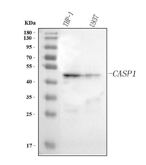
Western blot analysis of Caspase 1(P20)/CASP1 using anti-Caspase 1(P20)/CASP1 antibody (A00048-1).
Electrophoresis was performed on a 5-20% SDS-PAGE gel at 70V (Stacking gel) / 90V (Resolving gel) for 2-3 hours. The sample well of each lane was loaded with 30 ug of sample under reducing conditions.
Lane 1: human THP-2 whole cell lysates,
Lane 2: human U937 whole cell lysates.
After electrophoresis, proteins were transferred to a nitrocellulose membrane at 150 mA for 50-90 minutes. Blocked the membrane with 5% non-fat milk/TBS for 1.5 hour at RT. The membrane was incubated with rabbit anti-Caspase 1(P20)/CASP1 antigen affinity purified polyclonal antibody (Catalog # A00048-1) at 0.5 μg/mL overnight at 4°C, then washed with TBS-0.1%Tween 3 times with 5 minutes each and probed with a goat anti-rabbit IgG-HRP secondary antibody at a dilution of 1:5000 for 1.5 hour at RT. The signal is developed using an Enhanced Chemiluminescent detection (ECL) kit (Catalog # EK1002) with Tanon 5200 system. A specific band was detected for Caspase 1(P20)/CASP1 at approximately 47 kDa. The expected band size for Caspase 1(P20)/CASP1 is at 47 kDa.
Specific Publications For Anti-Caspase 1(p20)/CASP1 Antibody Picoband® (A00048-1)
Loading publications
Recommended Resources
Here are featured tools and databases that you might find useful.
- Boster's Pathways Library
- Protein Databases
- Bioscience Research Protocol Resources
- Data Processing & Analysis Software
- Photo Editing Software
- Scientific Literature Resources
- Research Paper Management Tools
- Molecular Biology Software
- Primer Design Tools
- Bioinformatics Tools
- Phylogenetic Tree Analysis
Customer Reviews
Have you used Anti-Caspase 1(p20)/CASP1 Antibody Picoband®?
Share your experimental results or join a short interview to earn up to $1,000 in product credits or other rewards.
0 Reviews For Anti-Caspase 1(p20)/CASP1 Antibody Picoband®
Customer Q&As
Have a question?
Find answers in Q&As, reviews.
Can't find your answer?
Submit your question
