Click image to see more details
Product Info Summary
| SKU: | A03693 |
|---|---|
| Size: | 100ug |
| Reactive Species: | Human |
| Host: | Rabbit |
| Application: | ELISA, IP, IHC, WB |
Customers Who Bought This Also Bought

Product info
Product Name
Anti-Cbl-c Antibody
SKU/Catalog Number
A03693
Size
100ug
Form
Liquid (sterile filtered)
Description
Boster Bio Anti-Cbl-c Antibody (Catalog # A03693). Tested in ELISA, IHC, IP, WB applications. This antibody reacts with Human.
Storage & Handling
Store vial at -20°C prior to opening. Aliquot contents and freeze at -20°C or below for extended storage. Avoid cycles of freezing and thawing. Centrifuge product if not completely clear after standing at room temperature. This product is stable for several weeks at 4°C as an undiluted liquid. Dilute only prior to immediate use. Expiration date is one (1) year from date of opening. (Ship on dry ice.)
Cite This Product
Anti-Cbl-c Antibody (Boster Biological Technology, Pleasanton CA, USA, Catalog # A03693)
Host
Rabbit
Contents
0.02 M Potassium Phosphate, 0.15 M Sodium Chloride, pH 7.2, 0.01% (w/v) Sodium Azide
Clonality
Polyclonal
Isotype
IgG
Immunogen
This affinity-purified antibody was prepared from whole rabbit serum produced by repeated immunizations with a synthetic peptide corresponding to the C-Terminal portion of Human Cbl-c.
Reactive Species
A03693 is reactive to CBLC in Human
Observed Molecular Weight
42 kDa
Calculated molecular weight
52.5 kDa
Background of CBLC
This antibody is suitable for Cancer, Immunology and Nuclear Signaling research. Cbl-c is also known as signal transduction protein CBL-C, SH3-binding protein CBL-C, CBL-3, and RING finger protein 57. Cbl proteins are a family of ubiquitin protein ligases (E3s) that negatively regulate signaling by targeting activated tyrosine kinases for degradation. Cbl-c (a.k.a. Cbl-3) is the most recently cloned member of the Cbl proteins and is expressed only in epithelial cells (the other Cbl proteins are ubiquitously expressed). Cbl-c, like the other mammalian Cbl proteins, can ubiquitinate the activated EGFR and target it for degradation. Cbl-c knock out mice show no obvious phenotype. Thus, the physiological role of Cbl-c is not known.
Antibody Validation
Boster validates all antibodies on WB, IHC, ICC, Immunofluorescence, and ELISA with known positive control and negative samples to ensure specificity and high affinity, including thorough antibody incubations.
Application & Images
Applications
A03693 is guaranteed for ELISA, IP, IHC, WB Boster Guarantee
Recommend Dilution
| Application | Dilution | Species |
|---|---|---|
| ELISA: 1:10 | 000 - 1:50 | 000 |
| IHC: 1:500 - 1:3 | 000 | |
| WB: 1:500 - 1:3 | 000 | |
| This affinity purified antibody has been tested for use in ELISA | immunohistochemistry | immunoprecipitation and western blot. Specific conditions for reactivity should be optimized by the end user. Expect a band at ~52 kDa in size corresponding to Cbl-c by western blotting in the appropriate cell lysate or extract. |
Validation Images & Assay Conditions
Click image to see more details
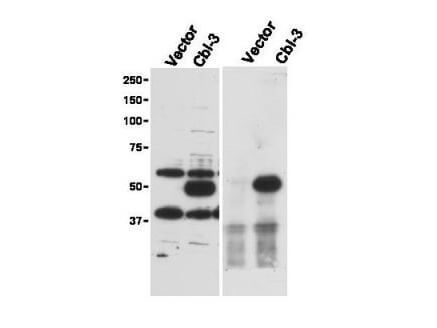
Immunoprecipitation (right) and western blot (left) using Boster's Affinity Purified anti-Cbl-c antibody shows detection of a predominant band at ~52 kDa corresponding to Cbl-c. Lysates used are from HEK293T cells transfected with empty vector or with Cbl-c and western blotting (left panel). The predicted size of Cbl-c is 52 kDa. Size markers in kDa are shown to the left of the panel. The (right panel) shows immunoprecipitation with Rabbit anti-Cbl-c followed by western blotting using a Goat anti-Cbl-c antibody. Personal Communication. Stan Lipkowitz, NCI, NIH, Bethesda, MD.

Click image to see more details
Boster's affinity purified anti-Cbl-c antibody was used at 5 µg/ml to detect signal in a variety of tissues including multi-human, multi-brain and multi-cancer slides. This image shows moderate intracellular positive staining of human pancreatic acinar epithelium at 40X. Tissue was formalin-fixed and paraffin embedded. The image shows localization of the antibody as the precipitated red signal, with a hematoxylin purple nuclear counterstain. Personal Communication, Tina Roush, LifeSpanBiosciences, Seattle, WA.

Click image to see more details
Boster's Affinity Purified anti-Cbl-c antibody shows strong nuclear and cytoplasmic staining of cells in tubuli in human kidney tissue. Tissue was formalin-fixed and paraffin embedded. Brown color indicates presence of protein, blue color shows cell nuclei. Personal Communication, Kenneth Wester, www.proteinatlas.org, Uppsala, Sweden.
Specific Publications For Anti-Cbl-c Antibody (A03693)
Loading publications
Recommended Resources
Here are featured tools and databases that you might find useful.
- Boster's Pathways Library
- Protein Databases
- Bioscience Research Protocol Resources
- Data Processing & Analysis Software
- Photo Editing Software
- Scientific Literature Resources
- Research Paper Management Tools
- Molecular Biology Software
- Primer Design Tools
- Bioinformatics Tools
- Phylogenetic Tree Analysis
Customer Reviews
Have you used Anti-Cbl-c Antibody?
Share your experimental results or join a short interview to earn up to $1,000 in product credits or other rewards.
0 Reviews For Anti-Cbl-c Antibody
Customer Q&As
Have a question?
Find answers in Q&As, reviews.
Can't find your answer?
Submit your question
3 Customer Q&As for Anti-Cbl-c Antibody
Question
I was wanting to use your anti-Cbl-c antibody for IHC for human skin on frozen tissues, but I want to know if it has been validated for this particular application. Has this antibody been validated and is this antibody a good choice for human skin identification?
Verified Customer
Verified customer
Asked: 2020-02-12
Answer
As indicated on the product datasheet, A03693 anti-Cbl-c antibody has been tested for IHC, WB on human tissues. We have an innovator award program that if you test this antibody and show it works in human skin in IHC-frozen, you can get your next antibody for free.
Boster Scientific Support
Answered: 2020-02-12
Question
Is a blocking peptide available for product anti-Cbl-c antibody (A03693)?
S. Thomas
Verified customer
Asked: 2015-04-16
Answer
We do provide the blocking peptide for product anti-Cbl-c antibody (A03693). If you would like to place an order for it please contact support@bosterbio.com and make a special request.
Boster Scientific Support
Answered: 2015-04-16
Question
We are currently using anti-Cbl-c antibody A03693 for human tissue, and we are satisfied with the IHC results. The species of reactivity given in the datasheet says human. Is it likely that the antibody can work on bovine tissues as well?
C. Moore
Verified customer
Asked: 2013-09-20
Answer
The anti-Cbl-c antibody (A03693) has not been validated for cross reactivity specifically with bovine tissues, though there is a good chance of cross reactivity. We have an innovator award program that if you test this antibody and show it works in bovine you can get your next antibody for free. Please contact me if I can help you with anything.
Boster Scientific Support
Answered: 2013-09-20
