Click image to see more details
-

-
-
-
-
+3
Product Info Summary
| SKU: | A04185-1 |
|---|---|
| Size: | 100 μg/vial |
| Reactive Species: | Human, Mouse, Rat |
| Host: | Rabbit |
| Application: | ELISA, IHC, WB |
Customers Who Bought This Also Bought

Product info
Product Name
Anti-CCXCR1/XCR1 Antibody Picoband®
SKU/Catalog Number
A04185-1
Size
100 μg/vial
Form
Lyophilized
Description
Boster Bio Anti-CCXCR1/XCR1 Antibody Picoband® catalog # A04185-1. Tested in ELISA, IHC, WB applications. This antibody reacts with Human, Mouse, Rat. The brand Picoband indicates this is a premium antibody that guarantees superior quality, high affinity, and strong signals with minimal background in Western blot applications. Only our best-performing antibodies are designated as Picoband, ensuring unmatched performance.
Storage & Handling
At -20°C for one year from date of receipt. After reconstitution, at 4°C for one month. It can also be aliquotted and stored frozen at -20°C for six months. Avoid repeated freezing and thawing.
Cite This Product
Anti-CCXCR1/XCR1 Antibody Picoband® (Boster Biological Technology, Pleasanton CA, USA, Catalog # A04185-1)
Host
Rabbit
Contents
Each vial contains 4 mg Trehalose, 0.9 mg NaCl, 0.2 mg Na2HPO4.
Clonality
Polyclonal
Isotype
Rabbit IgG
Immunogen
E.coli-derived human CCXCR1/XCR1 recombinant protein (Position: F246-N274).
Cross-reactivity
No cross-reactivity with other proteins.
Reactive Species
A04185-1 is reactive to XCR1 in Human, Mouse, Rat
Observed Molecular Weight
42 kDa
Calculated molecular weight
38.5 kDa
Background of XCR1
The protein encoded by this gene is a chemokine receptor belonging to the G protein-coupled receptor superfamily. The family members are characterized by the presence of 7 transmembrane domains and numerous conserved amino acids. This receptor is most closely related to RBS11 and the MIP1-alpha/RANTES receptor. It transduces a signal by increasing the intracellular calcium ions level. The viral macrophage inflammatory protein-II is an antagonist of this receptor and blocks signaling. Several alternatively spliced transcript variants encoding the same protein have been found for this gene.
Antibody Validation
Boster validates all antibodies on WB, IHC, ICC, Immunofluorescence, and ELISA with known positive control and negative samples to ensure specificity and high affinity, including thorough antibody incubations.
Application & Images
Applications
A04185-1 is guaranteed for ELISA, IHC, WB Boster Guarantee
Recommend Dilution
| Application | Dilution | Species |
|---|---|---|
| Western blot | 0.25-0.5 μg/ml | Human, Mouse, Rat |
| Immunohistochemistry(Paraffin-embedded Section) | 2-5 μg/ml | Human, Mouse, Rat |
| ELISA | 0.1-0.5 μg/ml | - |
Tested application
Suggested blocking solution with 5% non-fat milk or BSA; (*)Recommended protein loading: 20-40 µg per lane
Use TE buffer pH 9.0 for antigen retrieval; (*) citrate buffer pH 6.0 is an alternative.
Validation Images & Assay Conditions
Click image to see more details
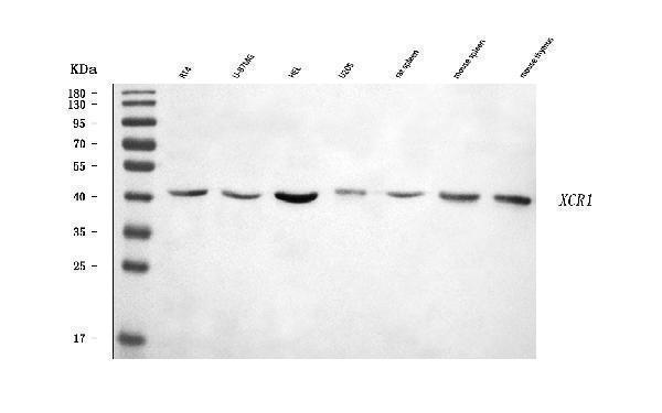
Western blot analysis of CCXCR1/XCR1 using anti-CCXCR1/XCR1 antibody (A04185-1).
Electrophoresis was performed on a 5-20% SDS-PAGE gel at 70V (Stacking gel) / 90V (Resolving gel) for 2-3 hours. The sample well of each lane was loaded with 30 ug of sample under reducing conditions.
Lane 1: human RT4 whole cell lysates,
Lane 2: human U-87MG whole cell lysates,
Lane 3: human HEL whole cell lysates,
Lane 4: human U20S whole cell lysates,
Lane 5: rat spleen tissue lysates,
Lane 6: mouse spleen tissue lysates,
Lane 7: mouse thymus tissue lysates.
After electrophoresis, proteins were transferred to a nitrocellulose membrane at 150 mA for 50-90 minutes. Blocked the membrane with 5% non-fat milk/TBS for 1.5 hour at RT. The membrane was incubated with rabbit anti-CCXCR1/XCR1 antigen affinity purified polyclonal antibody (Catalog # A04185-1) at 0.5 μg/mL overnight at 4°C, then washed with TBS-0.1%Tween 3 times with 5 minutes each and probed with a goat anti-rabbit IgG-HRP secondary antibody at a dilution of 1:5000 for 1.5 hour at RT. The signal is developed using an Enhanced Chemiluminescent detection (ECL) kit (Catalog # EK1002) with Tanon 5200 system. A specific band was detected for CCXCR1/XCR1 at approximately 42 kDa. The expected band size for CCXCR1/XCR1 is at 39 kDa.

Click image to see more details
cDC1s in SCI patients and mouse model of SCI. A PBMCs were collected from 10 healthy volunteers and 10 SCI patients. Identification of cDC1s and cDC2s subsets was performed by flow cytometry. CD11C + HLA-DR + represents cDCs, while CD141 + represents sDC1s and CD1c + represents cDC2 cells. Difference was calculated by Mann–Whitney U test. ** P < 0.01 vs. healthy group. B Mice were divided into sham ( n = 6) and SCI ( n = 6) groups. BMS score detection was performed. Difference was calculated by two-way ANOVA test. ** P < 0.01 vs. sham group. C–D The positive expressions of MHCII and XCR1 in spinal cord tissues were detected by IF assay. The difference was calculated by unpaired two-tailed T-test. ** P < 0.01 vs. sham group. SCI : spinal cord injury; dpi : days post-injury; cDC : conventional dendritic cell
Index in PubMed under a CC BY license. PMID: 39901071

Click image to see more details
cDC1 depletion reduces CD8 + T cell infiltration after SCI injury. A Mice were divided into SCI ( n = 6) and SCI + Qu ( n = 6) groups. Qu administration was started on day 14 after surgery. B BMS score was determined. The difference was calculated by unpaired two-tailed T-test. ** P < 0.01 vs. SCI group. C Positive expression of XCR1 in spinal cord tissues was detected by IF assay. The difference was calculated by unpaired two-tailed T-test. ** P < 0.01. D The results of flow cytometry of XCR1 + cDCs in lymph nodes and blood. The cDC population was expressed as CD11C and I-A/I-E (MHCII) double positive, and XCR1 + represents XCR1 + cDCs. E The proportion of CD8 + T cells in spinal cord tissues was determined by flow cytometry. CD3 + T cells were circled with CD3 gate, and further CD3 + CD8 + T cells were analyzed. The difference was calculated by unpaired two-tailed T-test. ** P < 0.01 vs. SCI group. N = 6. F ELISA was used to detect IFN-γ levels in spinal cord tissues. The difference was calculated by unpaired two-tailed T-test. ** P < 0.01 vs. SCI group. N = 6. dpi : days post-injury; Qu : Quizartinib
Index in PubMed under a CC BY license. PMID: 39901071

Click image to see more details
Excess cDC1s in non-lymphoid tissues in LNs of SCI mice is due to migration of cDC1s from the spinal cord. A Leukocyte (CD45 + ) and pre-DC subsets in bone marrow of sham and SCI groups were detected by flow cytometry. Immune cells were circled by CD45 + and murine pre-DCs were further detected by CD135 + CD11c + . The difference was calculated by unpaired two-tailed T-test. N = 6. B–C Non-lymphoid tissue cDC1 (CD11C + I-A/I-E + CD103 + ) proportion and lymphoid-resident cDC1 (CD11C + I-A/I-E + CD8α + ) subsets in dcLN and LLN of sham and SCI groups were detected by flow cytometry. The difference was calculated by unpaired two-tailed T-test. ** P < 0.01 vs. SCI group. N = 3. D cDC1s (CD11C + I-A/I-E + CD103 + ) and cDC2s (CD11C + I-A/I-E + CD11b + ) subsets in the blood of SCI-3 dpi and SCI-42 dpi groups were detected by flow cytometry. The difference was calculated by unpaired two-tailed T-test. ** P < 0.01 vs. SCI group. N = 6. E cDC1 subsets (CD45 + XCR1 + CD103 + ) in spinal cord tissues of SCI-3 dpi and SCI-42 dpi groups were detected by flow cytometry. The difference was calculated by unpaired two-tailed T-test. ** P < 0.01 vs. SCI group. N = 6. F The positive expressions of CD11C and LYVE-1 in the spinal cord tissues of SCI group mice were detected by IF assay. BM : bone marrow; dcLN : deep cervical lymph nodes; LLN : lumbar lymph nodes
Index in PubMed under a CC BY license. PMID: 39901071

Click image to see more details
IHC analysis of CCXCR1/XCR1 using anti-CCXCR1/XCR1 antibody (A04185-1).
CCXCR1/XCR1 was detected in a paraffin-embedded section of human spleen tissue. Heat mediated antigen retrieval was performed in EDTA buffer (pH 8.0, epitope retrieval solution). The tissue section was blocked with 10% goat serum. The tissue section was then incubated with 2 μg/ml rabbit anti-CCXCR1/XCR1 Antibody (A04185-1) overnight at 4°C. Peroxidase Conjugated Goat Anti-rabbit IgG was used as secondary antibody and incubated for 30 minutes at 37°C. The tissue section was developed using HRP Conjugated Rabbit IgG Super Vision Assay Kit (Catalog # SV0002) with DAB as the chromogen.

Click image to see more details
IHC analysis of CCXCR1/XCR1 using anti-CCXCR1/XCR1 antibody (A04185-1).
CCXCR1/XCR1 was detected in a paraffin-embedded section of mouse thymus tissue. Heat mediated antigen retrieval was performed in EDTA buffer (pH 8.0, epitope retrieval solution). The tissue section was blocked with 10% goat serum. The tissue section was then incubated with 2 μg/ml rabbit anti-CCXCR1/XCR1 Antibody (A04185-1) overnight at 4°C. Peroxidase Conjugated Goat Anti-rabbit IgG was used as secondary antibody and incubated for 30 minutes at 37°C. The tissue section was developed using HRP Conjugated Rabbit IgG Super Vision Assay Kit (Catalog # SV0002) with DAB as the chromogen.

Click image to see more details
IHC analysis of CCXCR1/XCR1 using anti-CCXCR1/XCR1 antibody (A04185-1).
CCXCR1/XCR1 was detected in a paraffin-embedded section of rat thymus tissue. Heat mediated antigen retrieval was performed in EDTA buffer (pH 8.0, epitope retrieval solution). The tissue section was blocked with 10% goat serum. The tissue section was then incubated with 2 μg/ml rabbit anti-CCXCR1/XCR1 Antibody (A04185-1) overnight at 4°C. Peroxidase Conjugated Goat Anti-rabbit IgG was used as secondary antibody and incubated for 30 minutes at 37°C. The tissue section was developed using HRP Conjugated Rabbit IgG Super Vision Assay Kit (Catalog # SV0002) with DAB as the chromogen.
Specific Publications For Anti-CCXCR1/XCR1 Antibody Picoband® (A04185-1)
Loading publications
Recommended Resources
Here are featured tools and databases that you might find useful.
- Boster's Pathways Library
- Protein Databases
- Bioscience Research Protocol Resources
- Data Processing & Analysis Software
- Photo Editing Software
- Scientific Literature Resources
- Research Paper Management Tools
- Molecular Biology Software
- Primer Design Tools
- Bioinformatics Tools
- Phylogenetic Tree Analysis
Customer Reviews
Have you used Anti-CCXCR1/XCR1 Antibody Picoband®?
Share your experimental results or join a short interview to earn up to $1,000 in product credits or other rewards.
0 Reviews For Anti-CCXCR1/XCR1 Antibody Picoband®
Customer Q&As
Have a question?
Find answers in Q&As, reviews.
Can't find your answer?
Submit your question
