Click image to see more details
-

-
-
-
-
+1
Product Info Summary
| SKU: | PB9050 |
|---|---|
| Size: | 100 μg/vial |
| Reactive Species: | Human |
| Host: | Rabbit |
| Application: | Flow Cytometry, IF, IHC, IHC-F, ICC, WB |
Customers Who Bought This Also Bought

Product info
Product Name
Anti-CD20/MS4A1 Antibody Picoband®
SKU/Catalog Number
PB9050
Size
100 μg/vial
Form
Lyophilized
Description
Boster Bio Anti-CD20/MS4A1 Antibody Picoband® catalog # PB9050. Tested in Flow Cytometry, IF, IHC, IHC-F, ICC, WB applications. This antibody reacts with Human. The brand Picoband indicates this is a premium antibody that guarantees superior quality, high affinity, and strong signals with minimal background in Western blot applications. Only our best-performing antibodies are designated as Picoband, ensuring unmatched performance.
Storage & Handling
Store at -20˚C for one year from date of receipt. After reconstitution, at 4˚C for one month. It can also be aliquotted and stored frozen at -20˚C for six months. Avoid repeated freeze-thaw cycles.
Cite This Product
Anti-CD20/MS4A1 Antibody Picoband® (Boster Biological Technology, Pleasanton CA, USA, Catalog # PB9050)
Host
Rabbit
Contents
Each vial contains antibody formulated with stabilizing components, 0.9 mg NaCl, 0.2 mg Na2HPO4, and 0.05 mg NaN3.
*This antibody is supplied in a stabilized formulation.
Compatibility with conjugation reactions depends on the chemistry of the conjugation method used.
For conjugation methods that are not compatible with the stabilizing components present in this formulation, a carrier-free antibody format is required.
Clonality
Polyclonal
Isotype
Rabbit IgG
Immunogen
E.coli-derived human CD20 recombinant protein (Position: M1-D261). Human CD20 shares 75% amino acid (aa) sequence identity with mouse CD20.
Cross-reactivity
No cross-reactivity with other proteins
Reactive Species
PB9050 is reactive to MS4A1 in Human
Observed Molecular Weight
35 kDa
Calculated molecular weight
33.1 kDa
Background of MS4A1
CD20, also known as MS4A1, is an activated-glycosylated phosphoprotein expressed on the surface of all B-cells beginning at the pro-B phase (CD45R+, CD117+) and progressively increasing in concentration until maturity. It is mapped to 11q12.2. This gene encodes a member of the membrane-spanning 4A gene family. The function of CD20 is to enable optimal B-cell immune response, specifically against T-independent antigens. It is suspected that CD20 acts as a calcium channel in the cell membrane. What’s more, this protein may be involved in the regulation of B-cell activation and proliferation.
Antibody Validation
Boster validates all antibodies on WB, IHC, ICC, Immunofluorescence, and ELISA with known positive control and negative samples to ensure specificity and high affinity, including thorough antibody incubations.
Application & Images
Applications
PB9050 is guaranteed for Flow Cytometry, IF, IHC, IHC-F, ICC, WB Boster Guarantee
Recommend Dilution
| Application | Dilution | Species |
|---|---|---|
| Western blot | 0.1-0.5μg/ml | |
| Immunohistochemistry (Paraffin-embedded Section) | 0.5-1μg/ml | |
| Immunohistochemistry (Frozen Section) | 0.5-1μg/ml | |
| Immunocytochemistry | 0.5-1μg/ml | |
| Immunofluorescence | 2μg/ml | Human |
| Flow Cytometry (Fixed) | 1-3μg/1x106cells |
Tested application
Suggested blocking solution with 5% non-fat milk or BSA; (*)Recommended protein loading: 20-40 µg per lane
Use TE buffer pH 9.0 for antigen retrieval; (*) citrate buffer pH 6.0 is an alternative.
Validation Images & Assay Conditions
Click image to see more details
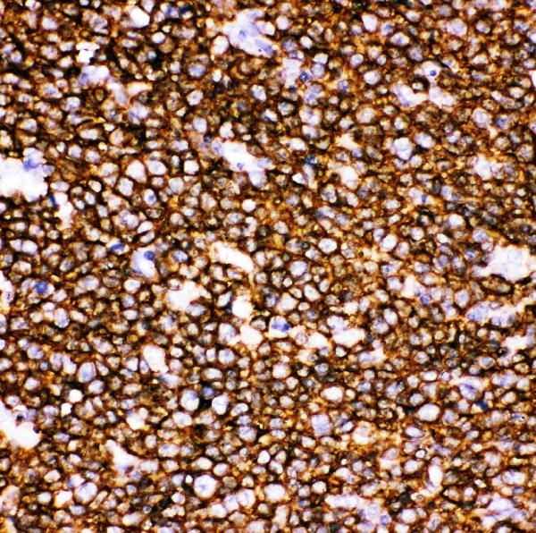
IHC analysis of CD20 using anti-CD20 antibody (PB9050).
CD20 was detected in paraffin-embedded section of Human Tonsil Tissue. Heat mediated antigen retrieval was performed in citrate buffer (pH6, epitope retrieval solution) for 20 mins. The tissue section was blocked with 10% goat serum. The tissue section was then incubated with 1μg/ml rabbit anti-CD20 Antibody (PB9050) overnight at 4°C. Biotinylated goat anti-rabbit IgG was used as secondary antibody and incubated for 30 minutes at 37°C. The tissue section was developed using Strepavidin-Biotin-Complex (SABC)(Catalog # SA1022) with DAB as the chromogen.

Click image to see more details
Western blot analysis of CD20 using anti-CD20 antibody (PB9050).
Electrophoresis was performed on a 5-20% SDS-PAGE gel at 70V (Stacking gel) / 90V (Resolving gel) for 2-3 hours. The sample well of each lane was loaded with 30 ug of sample under reducing conditions.
Lane 1: human Daudi whole cell lysates.
After electrophoresis, proteins were transferred to a nitrocellulose membrane at 150 mA for 50-90 minutes. Blocked the membrane with 5% non-fat milk/TBS for 1.5 hour at RT. The membrane was incubated with rabbit anti-CD20 antigen affinity purified polyclonal antibody (Catalog # PB9050) at 0.5 μg/mL overnight at 4°C, then washed with TBS-0.1%Tween 3 times with 5 minutes each and probed with a goat anti-rabbit IgG-HRP secondary antibody at a dilution of 1:5000 for 1.5 hour at RT. The signal is developed using an Enhanced Chemiluminescent detection (ECL) kit (Catalog # EK1002) with Tanon 5200 system. A specific band was detected for CD20 at approximately 35 kDa. The expected band size for CD20 is at 33 kDa.

Click image to see more details
IHC analysis of CD20 using anti-CD20 antibody (PB9050).
CD20 was detected in paraffin-embedded section of Human Tonsil Tissue. Heat mediated antigen retrieval was performed in citrate buffer (pH6, epitope retrieval solution) for 20 mins. The tissue section was blocked with 10% goat serum. The tissue section was then incubated with 1μg/ml rabbit anti-CD20 Antibody (PB9050) overnight at 4°C. Biotinylated goat anti-rabbit IgG was used as secondary antibody and incubated for 30 minutes at 37°C. The tissue section was developed using Strepavidin-Biotin-Complex (SABC)(Catalog # SA1022) with DAB as the chromogen.

Click image to see more details
IF analysis of CD20 using anti-CD20 antibody (PB9050)
CD20 was detected in paraffin-embedded section of human B lymphocytic tumor tissues. Heat mediated antigen retrieval was performed in citrate buffer (pH6, epitope retrieval solution ) for 20 mins. The tissue section was blocked with 10% goat serum. The tissue section was then incubated with 1μg/mL rabbit anti-CD20 Antibody (PB9050) overnight at 4°C. DyLight®488 Conjugated Goat Anti-Rabbit IgG (BA1127) was used as secondary antibody at 1:100 dilution and incubated for 30 minutes at 37°C. The section was counterstained with DAPI. Visualize using a fluorescence microscope and filter sets appropriate for the label used.

Click image to see more details
Flow Cytometry analysis of A431 cells using anti-CD20 antibody (PB9050).
Overlay histogram showing A431 cells stained with PB9050 (Blue line). To facilitate intracellular staining, cells were fixed with 4% paraformaldehyde and permeabilized with permeabilization buffer. The cells were blocked with 10% normal goat serum. And then incubated with rabbit anti-CD20 Antibody (PB9050,1μg/1x106 cells) for 30 min at 20°C. DyLight®488 conjugated goat anti-rabbit IgG (BA1127, 5-10μg/1x106 cells) was used as secondary antibody for 30 minutes at 20°C. Isotype control antibody (Green line) was rabbit IgG (1μg/1x106) used under the same conditions. Unlabelled sample without incubation with primary antibody and secondary antibody (Red line) was used as a blank control.
Specific Publications For Anti-CD20/MS4A1 Antibody Picoband® (PB9050)
Loading publications
Recommended Resources
Here are featured tools and databases that you might find useful.
- Boster's Pathways Library
- Protein Databases
- Bioscience Research Protocol Resources
- Data Processing & Analysis Software
- Photo Editing Software
- Scientific Literature Resources
- Research Paper Management Tools
- Molecular Biology Software
- Primer Design Tools
- Bioinformatics Tools
- Phylogenetic Tree Analysis
Customer Reviews
Have you used Anti-CD20/MS4A1 Antibody Picoband®?
Share your experimental results or join a short interview to earn up to $1,000 in product credits or other rewards.
0 Reviews For Anti-CD20/MS4A1 Antibody Picoband®
Customer Q&As
Have a question?
Find answers in Q&As, reviews.
Can't find your answer?
Submit your question
5 Customer Q&As for Anti-CD20/MS4A1 Antibody Picoband®
Question
We are currently using anti-CD20/MS4A1 antibody PB9050 for human tissue, and we are content with the Flow Cytometry results. The species of reactivity given in the datasheet says human. Is it possible that the antibody can work on bovine tissues as well?
Verified Customer
Verified customer
Asked: 2019-12-23
Answer
The anti-CD20/MS4A1 antibody (PB9050) has not been tested for cross reactivity specifically with bovine tissues, though there is a good chance of cross reactivity. We have an innovator award program that if you test this antibody and show it works in bovine you can get your next antibody for free. Please contact me if I can help you with anything.
Boster Scientific Support
Answered: 2019-12-23
Question
We have seen staining in human t-cell. Are there any suggestions? Is anti-CD20/MS4A1 antibody supposed to stain t-cell positively?
Verified Customer
Verified customer
Asked: 2019-08-07
Answer
From what I have seen in literature t-cell does express MS4A1. From what I have seen in Uniprot.org, MS4A1 is expressed in blood, lymph, t-cell, b-cell, among other tissues. Regarding which tissues have MS4A1 expression, here are a few articles citing expression in various tissues:
B-cell, Pubmed ID: 22615937
Lymph, Pubmed ID: 15489334
T-cell, Pubmed ID: 19367720
Boster Scientific Support
Answered: 2019-08-07
Question
Our team were happy with the WB result of your anti-CD20/MS4A1 antibody. However we have been able to see positive staining in t-cell cell membrane using this antibody. Is that expected? Could you tell me where is MS4A1 supposed to be expressed?
Verified Customer
Verified customer
Asked: 2018-11-06
Answer
From what I have seen in literature, t-cell does express MS4A1. Generally MS4A1 expresses in cell membrane. Regarding which tissues have MS4A1 expression, here are a few articles citing expression in various tissues:
B-cell, Pubmed ID: 22615937
Lymph, Pubmed ID: 15489334
T-cell, Pubmed ID: 19367720
Boster Scientific Support
Answered: 2018-11-06
Question
We ordered your anti-CD20/MS4A1 antibody for WB on blood in a previous experiment. I am using human, and I plan to use the antibody for Flow Cytometry next. I am interested in examining blood as well as lymph in our next experiment. Could you please give me some suggestion on which antibody would work the best for Flow Cytometry?
V. Banerjee
Verified customer
Asked: 2017-08-25
Answer
I viewed the website and datasheets of our anti-CD20/MS4A1 antibody and it appears that PB9050 has been validated on human in both WB and Flow Cytometry. Thus PB9050 should work for your application. Our Boster satisfaction guarantee will cover this product for Flow Cytometry in human even if the specific tissue type has not been validated. We do have a comprehensive range of products for Flow Cytometry detection and you can check out our website bosterbio.com to find out more information about them.
Boster Scientific Support
Answered: 2017-08-25
Question
Our lab want to know about using your anti-CD20/MS4A1 antibody for humoral immune response studies. Has this antibody been tested with western blotting on raji whole cell lysate? We would like to see some validation images before ordering.
A. Anderson
Verified customer
Asked: 2017-05-22
Answer
Thank you for your inquiry. This PB9050 anti-CD20/MS4A1 antibody is validated on human tonsil tissue, raji whole cell lysate, cem whole cell lysate, u937 cells, a431 cells. It is guaranteed to work for Flow Cytometry, IF, IHC-P, IHC-F, ICC, WB in human. Our Boster guarantee will cover your intended experiment even if the sample type has not been be directly tested.
Boster Scientific Support
Answered: 2017-05-22
