Click image to see more details
-

-
-
-
-
+2
Product Info Summary
| SKU: | PB9933 |
|---|---|
| Size: | 100 μg/vial |
| Reactive Species: | Human, Mouse, Rat |
| Host: | Rabbit |
| Application: | Flow Cytometry, IHC, WB |
Customers Who Bought This Also Bought

Product info
Product Name
Anti-CDCP1 Antibody Picoband®
SKU/Catalog Number
PB9933
PB0978 is an alternative SKU for this antibody, used in previous lots.
Size
100 μg/vial
Form
Lyophilized
Description
Boster Bio Anti-CDCP1 Antibody Picoband® catalog # PB9933. Tested in Flow Cytometry, IHC, WB applications. This antibody reacts with Human, Mouse, Rat. The brand Picoband indicates this is a premium antibody that guarantees superior quality, high affinity, and strong signals with minimal background in Western blot applications. Only our best-performing antibodies are designated as Picoband, ensuring unmatched performance.
Storage & Handling
Store at -20˚C for one year from date of receipt. After reconstitution, at 4˚C for one month. It can also be aliquotted and stored frozen at -20˚C for six months. Avoid repeated freeze-thaw cycles.
Cite This Product
Anti-CDCP1 Antibody Picoband® (Boster Biological Technology, Pleasanton CA, USA, Catalog # PB9933)
Host
Rabbit
Contents
Each vial contains antibody formulated with stabilizing components, 0.9 mg NaCl, 0.2 mg Na2HPO4, and 0.05 mg NaN3.
*This antibody is supplied in a stabilized formulation.
Compatibility with conjugation reactions depends on the chemistry of the conjugation method used.
For conjugation methods that are not compatible with the stabilizing components present in this formulation, a carrier-free antibody format is required.
Clonality
Polyclonal
Isotype
Rabbit IgG
Immunogen
E. coli-derived human CDCP1 recombinant protein (Position: R582-T667). Human CDCP1 shares 84.5% amino acid (aa) sequence identity with mouse CDCP1.
Cross-reactivity
No cross-reactivity with other proteins
Reactive Species
PB9933 is reactive to CDCP1 in Human, Mouse, Rat
Observed Molecular Weight
130 kDa
Calculated molecular weight
92.9 kDa
Background of CDCP1
CUB domain-containing protein 1 (CDCP1) is a protein that in humans is encoded by the CDCP1 gene. It has also been designated as CD318 (cluster of differentiation 318) and Trask (Transmembrane and associated with src kinases). CDCP1/Trask is a 140 kD transmembrane glycoprotein with a large extracellular domain (ECD) containing two CUB domains, and a smaller intracellular domain (ICD) containing five tyrosines. The tyrosine phosphorylation of Trask is tightly regulated and reciprocally linked with the state of cell adhesion. The tyrosine phosphorylation of CDCP1 in cultured cells occurs when cells are induced to detach by trypsin or EDTA, or seen spontaneously during mitotic detachment. The overexpression of CDCP1 leads to the loss of cell adhesion and a detached phenotype. CDCP1 is widely expressed in human epithelial tissues, but its phosphorylation is only seen in mitotically detached or shedding cells, consistent with its role in the negative regulation of cell adhesion.
Antibody Validation
Boster validates all antibodies on WB, IHC, ICC, Immunofluorescence, and ELISA with known positive control and negative samples to ensure specificity and high affinity, including thorough antibody incubations.
Application & Images
Applications
PB9933 is guaranteed for Flow Cytometry, IHC, WB Boster Guarantee
Recommend Dilution
| Application | Dilution | Species |
|---|---|---|
| Western blot | 0.1-0.5μg/ml | Human |
| Immunohistochemistry (Paraffin-embedded Section) | 0.5-1μg/ml | Human, Mouse, Rat |
| Flow Cytometry (Fixed) | 1-3μg/1x106 cells | Human |
Tested application
Suggested blocking solution with 5% non-fat milk or BSA; (*)Recommended protein loading: 20-40 µg per lane
Use TE buffer pH 9.0 for antigen retrieval; (*) citrate buffer pH 6.0 is an alternative.
Validation Images & Assay Conditions
Click image to see more details
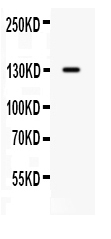
Western blot analysis of CDCP1 using anti-CDCP1 antibody (PB9933).
Electrophoresis was performed on a 5-20% SDS-PAGE gel at 70V (Stacking gel) / 90V (Resolving gel) for 2-3 hours. The sample well of each lane was loaded with 30 ug of sample under reducing conditions.
Lane 1: SW620 whole cell lysates.
After electrophoresis, proteins were transferred to a nitrocellulose membrane at 150 mA for 50-90 minutes. Blocked the membrane with 5% non-fat milk/TBS for 1.5 hour at RT. The membrane was incubated with rabbit anti-CDCP1 antigen affinity purified polyclonal antibody (Catalog # PB9933) at 0.5 μg/mL overnight at 4°C, then washed with TBS-0.1%Tween 3 times with 5 minutes each and probed with a goat anti-rabbit IgG-HRP secondary antibody at a dilution of 1:5000 for 1.5 hour at RT. The signal is developed using an Enhanced Chemiluminescent detection (ECL) kit (Catalog # EK1002) with Tanon 5200 system. A specific band was detected for CDCP1 at approximately 130 kDa. The expected band size for CDCP1 is at 93 kDa.

Click image to see more details
IHC analysis of CDCP1 using anti-CDCP1 antibody (PB9933).
CDCP1 was detected in a paraffin-embedded section of mouse intestine tissue. Heat mediated antigen retrieval was performed in EDTA buffer (pH 8.0, epitope retrieval solution). The tissue section was blocked with 10% goat serum. The tissue section was then incubated with 1 μg/ml rabbit anti-CDCP1 Antibody (PB9933) overnight at 4°C. Biotinylated goat anti-rabbit IgG was used as secondary antibody and incubated for 30 minutes at 37°C. The tissue section was developed using Strepavidin-Biotin-Complex (SABC) (Catalog # SA1022) with DAB as the chromogen.

Click image to see more details
Recruitment of immune cells and evidence of local spread. Paraffin-embedded sections prepared from the intestinal or colonic tumors isolated from untreated (Un), DSS-treated or AOM+DSS-treated Apc1638N/+ mice were subjected to F4/80 (upper panel), CD3/β-catenin co-localization (middle panel) and CD110 and CDCP1 (lower panel) staining, respectively. DAPI was used as nuclear stain. Scale bars = 150-250μm; n = 3 independent experiments.
Index in PubMed under a CC BY license. PMID: 31040926

Click image to see more details
IHC analysis of CDCP1 using anti-CDCP1 antibody (PB9933).
CDCP1 was detected in a paraffin-embedded section of rat intestine tissue. Heat mediated antigen retrieval was performed in EDTA buffer (pH 8.0, epitope retrieval solution). The tissue section was blocked with 10% goat serum. The tissue section was then incubated with 1 μg/ml rabbit anti-CDCP1 Antibody (PB9933) overnight at 4°C. Biotinylated goat anti-rabbit IgG was used as secondary antibody and incubated for 30 minutes at 37°C. The tissue section was developed using Strepavidin-Biotin-Complex (SABC) (Catalog # SA1022) with DAB as the chromogen.

Click image to see more details
IHC analysis of CDCP1 using anti-CDCP1 antibody (PB9933).
CDCP1 was detected in a paraffin-embedded section of human placenta tissue. Heat mediated antigen retrieval was performed in EDTA buffer (pH 8.0, epitope retrieval solution). The tissue section was blocked with 10% goat serum. The tissue section was then incubated with 1 μg/ml rabbit anti-CDCP1 Antibody (PB9933) overnight at 4°C. Biotinylated goat anti-rabbit IgG was used as secondary antibody and incubated for 30 minutes at 37°C. The tissue section was developed using Strepavidin-Biotin-Complex (SABC) (Catalog # SA1022) with DAB as the chromogen.

Click image to see more details
Flow Cytometry analysis of PC-3 cells using anti-CDCP1 antibody (PB9933).
Overlay histogram showing PC-3 cells stained with PB9933 (Blue line). To facilitate intracellular staining, cells were fixed with 4% paraformaldehyde and permeabilized with permeabilization buffer. The cells were blocked with 10% normal goat serum. And then incubated with rabbit anti-CDCP1 Antibody (PB9933,1μg/1x106 cells) for 30 min at 20°C. DyLight488 conjugated goat anti-rabbit IgG (BA1127, 5-10μg/1x106 cells) was used as secondary antibody for 30 minutes at 20°C. Isotype control antibody (Green line) was rabbit IgG (1μg/1x106) used under the same conditions. Unlabelled sample without incubation with primary antibody and secondary antibody (Red line) was used as a blank control.
Specific Publications For Anti-CDCP1 Antibody Picoband® (PB9933)
Loading publications
Recommended Resources
Here are featured tools and databases that you might find useful.
- Boster's Pathways Library
- Protein Databases
- Bioscience Research Protocol Resources
- Data Processing & Analysis Software
- Photo Editing Software
- Scientific Literature Resources
- Research Paper Management Tools
- Molecular Biology Software
- Primer Design Tools
- Bioinformatics Tools
- Phylogenetic Tree Analysis
Customer Reviews
Have you used Anti-CDCP1 Antibody Picoband®?
Share your experimental results or join a short interview to earn up to $1,000 in product credits or other rewards.
0 Reviews For Anti-CDCP1 Antibody Picoband®
Customer Q&As
Have a question?
Find answers in Q&As, reviews.
Can't find your answer?
Submit your question
4 Customer Q&As for Anti-CDCP1 Antibody Picoband®
Question
Does anti-CDCP1 antibody PB9933 work on canine Flow Cytometry with placenta?
Verified Customer
Verified customer
Asked: 2020-03-18
Answer
Our lab technicians have not tested anti-CDCP1 antibody PB9933 on canine. You can run a BLAST between canine and the immunogen sequence of anti-CDCP1 antibody PB9933 to see if they may cross-react. If the sequence homology is close, then you can perform a pilot test. Keep in mind that since we have not validated canine samples, this use of the antibody is not covered by our guarantee. However we have an innovator award program that if you test this antibody and show it works in canine placenta in Flow Cytometry, you can get your next antibody for free.
Boster Scientific Support
Answered: 2020-03-18
Question
I see that the anti-CDCP1 antibody PB9933 works with IHC-P, what is the protocol used to produce the result images on the product page?
Verified Customer
Verified customer
Asked: 2020-02-26
Answer
You can find protocols for IHC-P on the "support/technical resources" section of our navigation menu. If you have any further questions, please send an email to support@bosterbio.com
Boster Scientific Support
Answered: 2020-02-26
Question
We are currently using anti-CDCP1 antibody PB9933 for human tissue, and we are satisfied with the IHC-P results. The species of reactivity given in the datasheet says human, mouse, rat. Is it likely that the antibody can work on bovine tissues as well?
J. Wu
Verified customer
Asked: 2019-07-09
Answer
The anti-CDCP1 antibody (PB9933) has not been tested for cross reactivity specifically with bovine tissues, though there is a good chance of cross reactivity. We have an innovator award program that if you test this antibody and show it works in bovine you can get your next antibody for free. Please contact me if I can help you with anything.
Boster Scientific Support
Answered: 2019-07-09
Question
Will PB9933 anti-CDCP1 antibody work on parafin embedded sections? If so, which fixation method do you recommend we use (PFA, paraformaldehyde, other)?
Verified Customer
Verified customer
Asked: 2019-07-03
Answer
You can see on the product datasheet, PB9933 anti-CDCP1 antibody as been validated on IHC-P. It is best to use PFA for fixation because it has better tissue penetration ability. PFA needs to be prepared fresh before use. Long term stored PFA turns into formalin, as the PFA molecules congregate and become formalin.
Boster Scientific Support
Answered: 2019-07-03
