Click image to see more details
-

-
-
-
-
+1
Product Info Summary
| SKU: | A00386-1 |
|---|---|
| Size: | 100 μg/vial |
| Reactive Species: | Human |
| Host: | Rabbit |
| Application: | ELISA, Flow Cytometry, IF, ICC, WB |
Customers Who Bought This Also Bought

Product info
Product Name
Anti-CEBP Alpha/CEBPA Antibody Picoband®
SKU/Catalog Number
A00386-1
Size
100 μg/vial
Form
Lyophilized
Description
Boster Bio Anti-CEBP Alpha/CEBPA Antibody Picoband® catalog # A00386-1. Tested in ELISA, Flow Cytometry, IF, ICC, WB applications. This antibody reacts with Human. The brand Picoband indicates this is a premium antibody that guarantees superior quality, high affinity, and strong signals with minimal background in Western blot applications. Only our best-performing antibodies are designated as Picoband, ensuring unmatched performance.
Storage & Handling
Store at -20˚C for one year from date of receipt. After reconstitution, at 4˚C for one month. It can also be aliquotted and stored frozen at -20˚C for six months. Avoid repeated freeze-thaw cycles.
Cite This Product
Anti-CEBP Alpha/CEBPA Antibody Picoband® (Boster Biological Technology, Pleasanton CA, USA, Catalog # A00386-1)
Host
Rabbit
Contents
Each vial contains 4mg Trehalose, 0.9mg NaCl, 0.2mg Na2HPO4, 0.05mg NaN3.
Clonality
Polyclonal
Isotype
Rabbit IgG
Immunogen
E.coli-derived human CEBP Alpha/CEBPA recombinant protein (Position: Y138-Q221).
Cross-reactivity
No cross-reactivity with other proteins.
Reactive Species
A00386-1 is reactive to CEBPA in Human
Observed Molecular Weight
42 kDa
Calculated molecular weight
37.6 kDa
Background of CEBPA
CEBPA, CCAAT/enhancer-binding protein alpha is a protein that in humans is encoded by the CEBPA gene. The CEBPA gene is intronless. Using human/hamster somatic cell hybrids containing restricted fragments of human chromosome 19, the CEBPA gene is mapped to chromosome 19q13.1, between the GPI and TGFB1 genes. The protein encoded by this intronless gene is a bZIP transcription factor which can bind as a homodimer to certain promoters and enhancers. It can also form heterodimers with the related proteins CEBP-beta and CEBP-gamma. The encoded protein has been shown to bind to the promoter and modulate the expression of the gene encoding leptin, a protein that plays an important role in body weight homeostasis.
Antibody Validation
Boster validates all antibodies on WB, IHC, ICC, Immunofluorescence, and ELISA with known positive control and negative samples to ensure specificity and high affinity, including thorough antibody incubations.
Application & Images
Applications
A00386-1 is guaranteed for ELISA, Flow Cytometry, IF, ICC, WB Boster Guarantee
Assay Dilutions Recommendation
The recommendations below provide a starting point for assay optimization. The actual working concentration varies and should be decided by the user.
Western blot, 0.25-0.5μg/ml, Human
Immunocytochemistry/Immunofluorescence, 5μg/ml, Human
Flow Cytometry (Fixed), 1-3μg/1x106 cells, Human
ELISA, 0.1-0.5μg/ml, -
Positive Control
WB: human THP-1 whole cell, human U-937 whole cell, human HepG2 whole cell
ICC/IF: HepG2 cell
FCM: CACO-2 cell
Validation Images & Assay Conditions
Click image to see more details
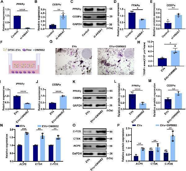
ANXA1 mediated PPARγ-CEBPα pathway to regulate osteoclast differentiation (A) The mRNA level of PPARγ in RAW264.7 cultured with siANXA1-EVs. (B) The mRNA level of CEBPα in RAW264.7 cultured with siANXA1-EVs. (C) The protein level of PPARγ and CEBPα in RAW264.7 cultured with siANXA1-EVs. (D) Quantitative analysis of PPARγ protein expression. (E) Quantitative analysis of CEBPα protein expression. (F) Schematic illustration of PPARγ inhibited RAW264.7 and DFSC-EVs co-culture system. (G) Representative images of TRAP staining. Scale bar = 200 μm. (H) Quantitative analysis of TRAP-positive area. (I) PPARγ inhibited RAW264.7 construction. (J) The mRNA level of CEBPα in PPARγ inhibited RAW264.7. (K) The protein level of PPARγ and CEBPα in PPARγ inhibited RAW264.7. (L) Quantitative analysis of PPARγ protein expression. (M) Quantitative analysis of CEBPα protein expression. (N) The mRNA level of ACP5 , CTSK and CFOS in PPARγ inhibited RAW264.7. (O) The protein level of ACP5, CTSK and CFOS in PPARγ inhibited RAW264.7. (P) Western blotting quantification. * p < 0.05, ** p < 0.01, *** p < 0.001, **** p < 0.0001. n = 3.
Index in PubMed under a CC BY license. PMID: 39834384

Click image to see more details
DFSCs-EVs/ANXA1 regulating tooth eruption by affecting osteoclast differentiation. (A) Representative micro-CT images of detecting tooth eruption distance. (B) Analysis of tooth eruption distance based on micro-CT. (C) Representative H&E staining images of the first mandibular molar area. (D) Analysis of tooth eruption distance based on H&E staining. (E) Representative images of TRAP staining. (F) Quantitative analysis of TRAP-positive area. (G) Representative immunohistochemistry staining images of PPARγ expression in the first mandibular molar area. (H) Quantitative analysis of PPARγ expression in the first mandibular molar area. (I) Representative immunohistochemistry staining images of CEBPα expression in the first mandibular molar area. (J) Quantitative analysis of CEBPα expression in the first mandibular molar area. ns, not significant. Scale bar = 1 mm ** p < 0. 01. n = 3.
Index in PubMed under a CC BY license. PMID: 39834384

Click image to see more details
Flow Cytometry analysis of CACO-2 cells using anti-CEBP Alpha/CEBPA antibody (A00386-1).
Overlay histogram showing CACO-2 cells stained with A00386-1 (Blue line). To facilitate intracellular staining, cells were fixed with 4% paraformaldehyde and permeabilized with permeabilization buffer. The cells were blocked with 10% normal goat serum. And then incubated with rabbit anti-CEBP Alpha/CEBPA Antibody (A00386-1, 1μg/1x106 cells) for 30 min at 20°C. DyLight®488 conjugated goat anti-rabbit IgG (BA1127, 5-10μg/1x106 cells) was used as secondary antibody for 30 minutes at 20°C. Isotype control antibody (Green line) was rabbit IgG (1μg/1x106) used under the same conditions. Unlabelled sample without incubation with primary antibody and secondary antibody (Red line) was used as a blank control.

Click image to see more details
Western blot analysis of CEBP using anti-CEBP antibody (A00386-1).
Electrophoresis was performed on a 5-20% SDS-PAGE gel at 70V (Stacking gel) / 90V (Resolving gel) for 2-3 hours. The sample well of each lane was loaded with 30 ug of sample under reducing conditions.
Lane 1: human THP-1 whole cell lysates,
Lane 2: human U-937 whole cell lysates,
Lane 3: human HepG2 whole cell lysates.
After electrophoresis, proteins were transferred to a nitrocellulose membrane at 150 mA for 50-90 minutes. Blocked the membrane with 5% non-fat milk/TBS for 1.5 hour at RT. The membrane was incubated with rabbit anti-CEBP antigen affinity purified polyclonal antibody (Catalog # A00386-1) at 0.5 μg/mL overnight at 4°C, then washed with TBS-0.1%Tween 3 times with 5 minutes each and probed with a goat anti-rabbit IgG-HRP secondary antibody at a dilution of 1:5000 for 1.5 hour at RT. The signal is developed using an Enhanced Chemiluminescent detection (ECL) kit (Catalog # EK1002) with Tanon 5200 system. A specific band was detected for CEBP at approximately 42 kDa. The expected band size for CEBP is at 37 kDa.

Click image to see more details
IF analysis of CEBP Alpha/CEBPA using anti- CEBP Alpha/CEBPA antibody (A00386-1).
CEBP Alpha/CEBPA was detected in immunocytochemical section of HepG2 cells. Enzyme antigen retrieval was performed using IHC enzyme antigen retrieval reagent (AR0022) for 15 mins. The cells were blocked with 10% goat serum. And then incubated with 5μg/mL rabbit anti- CEBP Alpha/CEBPA Antibody (A00386-1) overnight at 4°C. DyLight®488 Conjugated Goat Anti-Rabbit IgG (BA1127) was used as secondary antibody at 1:100 dilution and incubated for 30 minutes at 37°C. The section was counterstained with DAPI. Visualize using a fluorescence microscope and filter sets appropriate for the label used.
Specific Publications For Anti-CEBP Alpha/CEBPA Antibody Picoband® (A00386-1)
Loading publications
Recommended Resources
Here are featured tools and databases that you might find useful.
- Boster's Pathways Library
- Protein Databases
- Bioscience Research Protocol Resources
- Data Processing & Analysis Software
- Photo Editing Software
- Scientific Literature Resources
- Research Paper Management Tools
- Molecular Biology Software
- Primer Design Tools
- Bioinformatics Tools
- Phylogenetic Tree Analysis
Customer Reviews
Have you used Anti-CEBP Alpha/CEBPA Antibody Picoband®?
Share your experimental results or join a short interview to earn up to $1,000 in product credits or other rewards.
0 Reviews For Anti-CEBP Alpha/CEBPA Antibody Picoband®
Customer Q&As
Have a question?
Find answers in Q&As, reviews.
Can't find your answer?
Submit your question
5 Customer Q&As for Anti-CEBP Alpha/CEBPA Antibody Picoband®
Question
Our lab were content with the WB result of your anti-CEBP Alpha/CEBPA antibody. However we have been able to see positive staining in liver isoform 4: nucleus using this antibody. Is that expected? Could you tell me where is CEBPA supposed to be expressed?
Verified Customer
Verified customer
Asked: 2019-07-25
Answer
According to literature, liver does express CEBPA. Generally CEBPA expresses in nucleus., isoform 4: nucleus, nucleolus. Regarding which tissues have CEBPA expression, here are a few articles citing expression in various tissues:
Liver, Pubmed ID: 15057824
Pancreas, Pubmed ID: 15489334
Umbilical cord, Pubmed ID: 7575576
Boster Scientific Support
Answered: 2019-07-25
Question
We are currently using anti-CEBP Alpha/CEBPA antibody A00386-1 for mouse tissue, and we are content with the ELISA results. The species of reactivity given in the datasheet says human, mouse, rat. Is it possible that the antibody can work on zebrafish tissues as well?
Verified Customer
Verified customer
Asked: 2019-05-24
Answer
The anti-CEBP Alpha/CEBPA antibody (A00386-1) has not been tested for cross reactivity specifically with zebrafish tissues, though there is a good chance of cross reactivity. We have an innovator award program that if you test this antibody and show it works in zebrafish you can get your next antibody for free. Please contact me if I can help you with anything.
Boster Scientific Support
Answered: 2019-05-24
Question
We have tried in the past anti-CEBP Alpha/CEBPA antibody for WB on adipose tissue of abdominal region a few months ago. I am using human, and We want to use the antibody for ELISA next. My lab would like examining adipose tissue of abdominal region as well as umbilical cord in our next experiment. Could you please give me some suggestion on which antibody would work the best for ELISA?
Verified Customer
Verified customer
Asked: 2018-10-11
Answer
I viewed the website and datasheets of our anti-CEBP Alpha/CEBPA antibody and it appears that A00386-1 has been tested on human in both WB and ELISA. Thus A00386-1 should work for your application. Our Boster satisfaction guarantee will cover this product for ELISA in human even if the specific tissue type has not been validated. We do have a comprehensive range of products for ELISA detection and you can check out our website bosterbio.com to find out more information about them.
Boster Scientific Support
Answered: 2018-10-11
Question
I am interested in using your anti-CEBP Alpha/CEBPA antibody for transcriptional regulation of white adipocyte differentiation studies. Has this antibody been tested with western blotting on mouse lung tissue? We would like to see some validation images before ordering.
Verified Customer
Verified customer
Asked: 2018-09-19
Answer
We appreciate your inquiry. This A00386-1 anti-CEBP Alpha/CEBPA antibody is tested on human hepg2 whole cell lysates, rat lung tissue, mouse lung tissue. It is guaranteed to work for ELISA, WB in human, mouse, rat. Our Boster guarantee will cover your intended experiment even if the sample type has not been be directly tested.
Boster Scientific Support
Answered: 2018-09-19
Question
We have observed staining in rat pancreas. What should we do? Is anti-CEBP Alpha/CEBPA antibody supposed to stain pancreas positively?
Verified Customer
Verified customer
Asked: 2017-10-13
Answer
From literature pancreas does express CEBPA. From Uniprot.org, CEBPA is expressed in adipose tissue of abdominal region, umbilical cord, liver, pancreas, among other tissues. Regarding which tissues have CEBPA expression, here are a few articles citing expression in various tissues:
Liver, Pubmed ID: 15057824
Pancreas, Pubmed ID: 15489334
Umbilical cord, Pubmed ID: 7575576
Boster Scientific Support
Answered: 2017-10-13
