Click image to see more details
Product Info Summary
| SKU: | PA1202 |
|---|---|
| Size: | 100 μg/vial |
| Reactive Species: | Human, Mouse, Rat |
| Host: | Rabbit |
| Application: | WB |
Customers Who Bought This Also Bought

Product info
Product Name
Anti-Chk2/CHEK2 Antibody Picoband®
SKU/Catalog Number
PA1202
BA0741-2 is an alternative SKU for this antibody, used in previous lots.
Size
100 μg/vial
Form
Lyophilized
Description
Boster Bio Anti-Chk2/CHEK2 Antibody catalog # PA1202. Tested in WB applications. This antibody reacts with Human, Mouse, Rat. The brand Picoband indicates this is a premium antibody that guarantees superior quality, high affinity, and strong signals with minimal background in Western blot applications. Only our best-performing antibodies are designated as Picoband, ensuring unmatched performance.
Storage & Handling
Store at -20˚C for one year from date of receipt. After reconstitution, at 4˚C for one month. It can also be aliquotted and stored frozen at -20˚C for six months. Avoid repeated freeze-thaw cycles.
Cite This Product
Anti-Chk2/CHEK2 Antibody Picoband® (Boster Biological Technology, Pleasanton CA, USA, Catalog # PA1202)
Host
Rabbit
Contents
Each vial contains antibody formulated with stabilizing components, 0.9mg NaCl, 0.2mg Na2HPO4, 0.05mg Thimerosal, 0.05mg NaN3.
*This antibody is supplied in a stabilized formulation.
Compatibility with conjugation reactions depends on the chemistry of the conjugation method used.
For conjugation methods that are not compatible with the stabilizing components present in this formulation, a carrier-free antibody format is required.
Clonality
Polyclonal
Isotype
Rabbit IgG
Immunogen
A synthetic peptide corresponding to a sequence at the C-terminus of human Chk2, different from the related rat and mouse sequences by one amino acid.
Cross-reactivity
No cross-reactivity with other proteins
Reactive Species
PA1202 is reactive to CHEK2 in Human, Mouse, Rat
Observed Molecular Weight
69 kDa
Calculated molecular weight
60.9 kDa
Background of CHEK2
CHK2, a protein kinase that is activated in response to DNA damage, is involved in cell cycle arrest. Mapped on 22q12.1, CHK2 has a potential regulatory region rich in SQ and TQ amino acid pairs. It regulates BRCA1 function after DNA damage by phosphorylating serine-988 of BRCA1. Additionally, CHK2 can be modified by phosphorylation and activated in response to ionizing radiation, and can be also modified in response to hydroxyurea treatment. Furthermore, oligomerization of CHEK2 increases the efficiency of transautophosphorylation, resulting in the release of active CHEK2 monomers that proceed to enforce checkpoint control in irradiated cells. Morever, CHK2 is a tumor suppressor gene conferring predisposition to sarcoma, breast cancer, and brain tumors, and that their observations provided a link between the central role of p53 inactivation in human cancer and the well-defined G2 checkpoint in yeast. There is a wide expression of small amounts of CHK2 mRNA with larger amounts in human testis, spleen, colon, and peripheral blood leukocytes.
Antibody Validation
Boster validates all antibodies on WB, IHC, ICC, Immunofluorescence, and ELISA with known positive control and negative samples to ensure specificity and high affinity, including thorough antibody incubations.
Application & Images
Applications
PA1202 is guaranteed for WB Boster Guarantee
Recommend Dilution
| Application | Dilution | Species |
|---|---|---|
| Western blot | 0.1-0.5μg/ml | Human, Rat, Mouse |
Tested application
Suggested blocking solution with 5% non-fat milk or BSA; (*)Recommended protein loading: 20-40 µg per lane
Validation Images & Assay Conditions
Click image to see more details
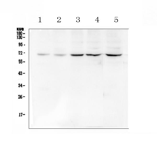
Western blot analysis of Chk2 using anti-Chk2 antibody (PA1202).
Electrophoresis was performed on a 5-20% SDS-PAGE gel at 70V (Stacking gel) / 90V (Resolving gel) for 2-3 hours. The sample well of each lane was loaded with 50ug of sample under reducing conditions.
Lane 1: human MDA-MB-231 whole cell lysates,
Lane 2: human MDA-MB-453 whole cell lysates,
Lane 3: human HepG2 whole cell lysates,
Lane 4: human K562 whole cell lysates,
Lane 5: human Caco-2 whole cell lysates,
After Electrophoresis, proteins were transferred to a Nitrocellulose membrane at 150mA for 50-90 minutes. Blocked the membrane with 5% Non-fat Milk/ TBS for 1.5 hour at RT. The membrane was incubated with rabbit anti-Chk2 antigen affinity purified polyclonal antibody (Catalog # PA1202) at 0.5 μg/mL overnight at 4°C, then washed with TBS-0.1%Tween 3 times with 5 minutes each and probed with a goat anti-rabbit IgG-HRP secondary antibody at a dilution of 1:10000 for 1.5 hour at RT. The signal is developed using an Enhanced Chemiluminescent detection (ECL) kit (Catalog # EK1002) with Tanon 5200 system. A specific band was detected for Chk2 at approximately 69KD. The expected band size for Chk2 is at 61KD.
Specific Publications For Anti-Chk2/CHEK2 Antibody Picoband® (PA1202)
Loading publications
Recommended Resources
Here are featured tools and databases that you might find useful.
- Boster's Pathways Library
- Protein Databases
- Bioscience Research Protocol Resources
- Data Processing & Analysis Software
- Photo Editing Software
- Scientific Literature Resources
- Research Paper Management Tools
- Molecular Biology Software
- Primer Design Tools
- Bioinformatics Tools
- Phylogenetic Tree Analysis
Customer Reviews
Have you used Anti-Chk2/CHEK2 Antibody Picoband®?
Share your experimental results or join a short interview to earn up to $1,000 in product credits or other rewards.
0 Reviews For Anti-Chk2/CHEK2 Antibody Picoband®
Customer Q&As
Have a question?
Find answers in Q&As, reviews.
Can't find your answer?
Submit your question
4 Customer Q&As for Anti-Chk2/CHEK2 Antibody Picoband®
Question
My boss were happy with the WB result of your anti-Chk2/CHEK2 antibody. However we have been able to see positive staining in t-cell isoform 12: nucleus. using this antibody. Is that expected? Could you tell me where is CHEK2 supposed to be expressed?
Verified Customer
Verified customer
Asked: 2020-04-10
Answer
From literature, t-cell does express CHEK2. Generally CHEK2 expresses in isoform 2: nucleus. note=isoform 10 is, isoform 4: nucleus., isoform 7: nucleus., isoform 9: nucleus., isoform 12: nucleus., nucleus, pml body. nucleus, nucleoplasm. Regarding which tissues have CHEK2 expression, here are a few articles citing expression in various tissues:
Mammary gland, Pubmed ID: 15361853
Muscle, Pubmed ID: 15489334
T-cell, Pubmed ID: 14702039
Boster Scientific Support
Answered: 2020-04-10
Question
I am looking for using your anti-Chk2/CHEK2 antibody for regulation of tp53 activity through methylation studies. Has this antibody been tested with western blotting on human k562? We would like to see some validation images before ordering.
Verified Customer
Verified customer
Asked: 2020-02-24
Answer
Thank you for your inquiry. This PA1202 anti-Chk2/CHEK2 antibody is tested on human k562, hepg2 whole cell lysates, k562 whole cell lysates. It is guaranteed to work for WB in human, mouse, rat. Our Boster guarantee will cover your intended experiment even if the sample type has not been be directly tested.
Boster Scientific Support
Answered: 2020-02-24
Question
We are currently using anti-Chk2/CHEK2 antibody PA1202 for rat tissue, and we are content with the WB results. The species of reactivity given in the datasheet says human, mouse, rat. Is it possible that the antibody can work on monkey tissues as well?
Verified Customer
Verified customer
Asked: 2019-07-11
Answer
The anti-Chk2/CHEK2 antibody (PA1202) has not been tested for cross reactivity specifically with monkey tissues, though there is a good chance of cross reactivity. We have an innovator award program that if you test this antibody and show it works in monkey you can get your next antibody for free. Please contact me if I can help you with anything.
Boster Scientific Support
Answered: 2019-07-11
Question
We have been able to see staining in mouse t-cell. What should we do? Is anti-Chk2/CHEK2 antibody supposed to stain t-cell positively?
Verified Customer
Verified customer
Asked: 2017-07-10
Answer
From literature t-cell does express CHEK2. From Uniprot.org, CHEK2 is expressed in tibial nerve, mammary gland, colon carcinoma, t-cell, muscle, among other tissues. Regarding which tissues have CHEK2 expression, here are a few articles citing expression in various tissues:
Mammary gland, Pubmed ID: 15361853
Muscle, Pubmed ID: 15489334
T-cell, Pubmed ID: 14702039
Boster Scientific Support
Answered: 2017-07-10
