Click image to see more details
-

-
-
-
-
+2
Product Info Summary
| SKU: | A07404-1 |
|---|---|
| Size: | 0.1 mg |
| Reactive Species: | Human, Mouse, Rat |
| Host: | Rabbit |
| Application: | ELISA, IF, WB |
Customers Who Bought This Also Bought

Product info
Product Name
Anti-CIDE-B Antibody
SKU/Catalog Number
A07404-1
Size
0.1 mg
Form
Liquid
Description
Boster Bio Anti-CIDE-B Antibody (Catalog # A07404-1). Tested in ELISA, WB, IF applications. This antibody reacts with Human, Mouse, Rat.
Storage & Handling
CIDE-B antibody can be stored at 4°C for three months and -20°C, stable for up to one year. Avoid repeated freeze-thaw cycles. Antibodies should not be exposed to prolonged high temperatures.
Cite This Product
Anti-CIDE-B Antibody (Boster Biological Technology, Pleasanton CA, USA, Catalog # A07404-1)
Host
Rabbit
Contents
CIDE-B Antibody is supplied in PBS containing 0.02% sodium azide.
Clonality
Polyclonal
Isotype
IgG
Immunogen
CIDE-B antibody was raised against a peptide corresponding to 15 amino acids near the carboxy terminus of human CIDE-B. The immunogen is located within the last 50 amino acids of CIDE-B.
Cross-reactivity
CIDE-B antibody has no cross activity to CIDE-A.
Reactive Species
A07404-1 is reactive to CIDEB in Human, Mouse, Rat
Observed Molecular Weight
26 kDa
Calculated molecular weight
24.7 kDa
Background of CIDEB
Apoptosis is related to many diseases and induced by a family of cell death receptors and their ligands. Cell death signals are transduced by death domain containing adapter molecules and members of the caspase family of proteases. These death signals finally cause the degradation of chromosomal DNA by activated DNase. DFF45/ICAD has been identified as inhibitor of caspase activated DNase DFF40/CAD. DFF45 related proteins CIDE-A and CIDE-B (for cell death-inducing DFF-like effector A and B) were recently identified. CIDE contains a new type of domain termed CIDE-N, which has high homology with the regulatory domains of DFF45/ICAD and DFF40/CAD. Expression of CIDE-B induces apoptosis, which is inhibited by DFF45. CIDE-B is a DFF45-inhibitable effector that promotes cell death and DNA fragmentation. CIDE-B is expressed mainly in liver and at lower levels in spleen, kidney, peripheral blood lymphocytes and bone marrow.
Antibody Validation
Boster validates all antibodies on WB, IHC, ICC, Immunofluorescence, and ELISA with known positive control and negative samples to ensure specificity and high affinity, including thorough antibody incubations.
Application & Images
Applications
A07404-1 is guaranteed for ELISA, IF, WB Boster Guarantee
Recommend Dilution
| Application | Dilution | Species |
|---|---|---|
| Antibody validated: Western Blot in human | mouse and rat samples; Immunofluorescence in the human | mouse and rat samples. All other applications and species not yet tested. |
Validation Images & Assay Conditions
Click image to see more details
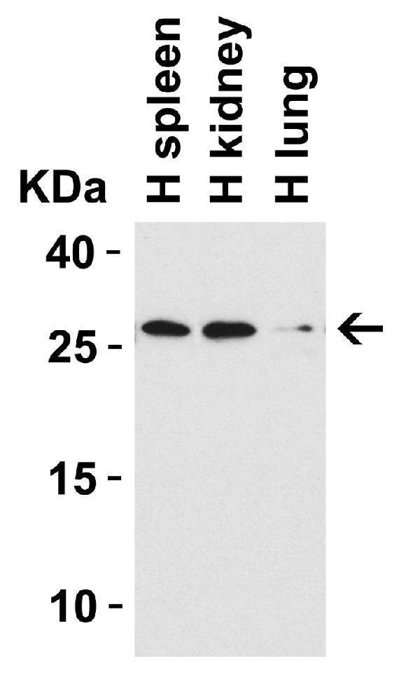
WB Validation in Human Tissues
Loading: 15 μg of lysate Antibodies: CIDE-B, A07404-1, 1 μ g/mL , 1 h incubation at RT in 5% NFDM/TBST. Secondary: Goat Anti-Rabbit IgG HRP conjugate at 1:10000 dilution.

Click image to see more details
WB Validation in Mouse Small Intestine
Loading: 15 μg of lysate Antibodies: CIDE-B, A07404-1, 1 μ g/mL , 1 h incubation at RT in 5% NFDM/TBST. Secondary: Goat Anti-Rabbit IgG HRP conjugate at 1:10000

Click image to see more details
WB Validation in Rat Small Intestine
Loading: 15 μg of lysate Antibodies: CIDE-B, A07404-1, 2 μg/mL , 1 h incubation at RT in 5% NFDM/TBST. Secondary: Goat Anti-Rabbit IgG HRP conjugate at 1:10000 dilution.

Click image to see more details
Immunofluorescence Validation of CIDE-B in Human Small Intestine Immunofluorescent analysis of 4% paraformaldehyde-fixed human small intestine tissue labeling CIDE-B with A07404-1 at 10 μg/mL, followed by goat anti-rabbit IgG secondary antibody at 1/500 dilution (red) and DAPI staining (blue).

Click image to see more details
Immunofluorescence Validation of CIDE-B in Mouse Small Intestine Immunofluorescent analysis of 4% paraformaldehyde-fixed mouse small intestine tissue labeling CIDE-B with A07404-1 at 10 μg/mL, followed by goat anti-rabbit IgG secondary antibody at 1/500 dilution (red) and DAPI staining (blue).

Click image to see more details
Immunofluorescence Validation of CIDE-B in Rat Liver Immunofluorescent analysis of 4% paraformaldehyde-fixed rat liver tissue labeling CIDE-B with A07404-1 at 20 μg/mL, followed by goat anti-rabbit IgG secondary antibody at 1/500 dilution (red) and DAPI staining (blue).
Specific Publications For Anti-CIDE-B Antibody (A07404-1)
Loading publications
Recommended Resources
Here are featured tools and databases that you might find useful.
- Boster's Pathways Library
- Protein Databases
- Bioscience Research Protocol Resources
- Data Processing & Analysis Software
- Photo Editing Software
- Scientific Literature Resources
- Research Paper Management Tools
- Molecular Biology Software
- Primer Design Tools
- Bioinformatics Tools
- Phylogenetic Tree Analysis
Customer Reviews
Have you used Anti-CIDE-B Antibody?
Share your experimental results or join a short interview to earn up to $1,000 in product credits or other rewards.
0 Reviews For Anti-CIDE-B Antibody
Customer Q&As
Have a question?
Find answers in Q&As, reviews.
Can't find your answer?
Submit your question
