Click image to see more details
Product Info Summary
| SKU: | PB9493 |
|---|---|
| Size: | 100 μg/vial |
| Reactive Species: | Human, Mouse, Rat |
| Host: | Rabbit |
| Application: | Flow Cytometry, IF, ICC, WB |
Customers Who Bought This Also Bought

Product info
Product Name
Anti-CTCF Antibody Picoband®
SKU/Catalog Number
PB9493
PB0514 is an alternative SKU for this antibody, used in previous lots.
Size
100 μg/vial
Form
Lyophilized
Description
Boster Bio Anti-CTCF Antibody Picoband® catalog # PB9493. Tested in Flow Cytometry, IF, ICC, WB applications. This antibody reacts with Human, Mouse, Rat. The brand Picoband indicates this is a premium antibody that guarantees superior quality, high affinity, and strong signals with minimal background in Western blot applications. Only our best-performing antibodies are designated as Picoband, ensuring unmatched performance.
Storage & Handling
Store at -20˚C for one year from date of receipt. After reconstitution, at 4˚C for one month. It can also be aliquotted and stored frozen at -20˚C for six months. Avoid repeated freeze-thaw cycles.
Cite This Product
Anti-CTCF Antibody Picoband® (Boster Biological Technology, Pleasanton CA, USA, Catalog # PB9493)
Host
Rabbit
Contents
Each vial contains 4 mg Trehalose, 0.9 mg NaCl and 0.2 mg Na2HPO4.
Clonality
Polyclonal
Isotype
Rabbit IgG
Immunogen
E.coli-derived human CTCF recombinant protein (Position: K521-R727). Human CTCF shares 94% and 93.1% amino acid (aa) sequence identity with mouse and rat CTCF, respectively.
Cross-reactivity
No cross-reactivity with other proteins
Reactive Species
PB9493 is reactive to CTCF in Human, Mouse, Rat
Observed Molecular Weight
150 kDa
Calculated molecular weight
82.8 kDa
Background of CTCF
Transcriptional repressor CTCF also known as 11-zinc finger protein or CCCTC-binding factor is a transcription factor that in humans is encoded by the CTCF gene. This gene is a member of the BORIS + CTCF gene family and encodes a transcriptional regulator protein with 11 highly conserved zinc finger (ZF) domains. And this nuclear protein is able to use different combinations of the ZF domains to bind different DNA target sequences and proteins. Depending upon the context of the site, the protein can bind a histone acetyltransferase (HAT)-containing complex and function as a transcriptional activator or bind a histone deacetylase (HDAC)-containing complex and function as a transcriptional repressor. If the protein is bound to a transcriptional insulator element, it can block communication between enhancers and upstream promoters, thereby regulating imprinted expression. Mutations in this gene have been associated with invasive breast cancers, prostate cancers, and Wilms' tumors. Alternatively spliced transcript variants encoding different isoforms have been found for this gene.
Antibody Validation
Boster validates all antibodies on WB, IHC, ICC, Immunofluorescence, and ELISA with known positive control and negative samples to ensure specificity and high affinity, including thorough antibody incubations.
Application & Images
Applications
PB9493 is guaranteed for Flow Cytometry, IF, ICC, WB Boster Guarantee
Recommend Dilution
| Application | Dilution | Species |
|---|---|---|
| Western blot | 0.1-0.5μg/ml | Human, Mouse, Rat |
| Immunocytochemistry/Immunofluorescence | 5 μg/ml | Human |
| Flow Cytometry(Fixed) | 1-3 μg/1x106 cells | Human |
Tested application
Suggested blocking solution with 5% non-fat milk or BSA; (*)Recommended protein loading: 20-40 µg per lane
Validation Images & Assay Conditions
Click image to see more details
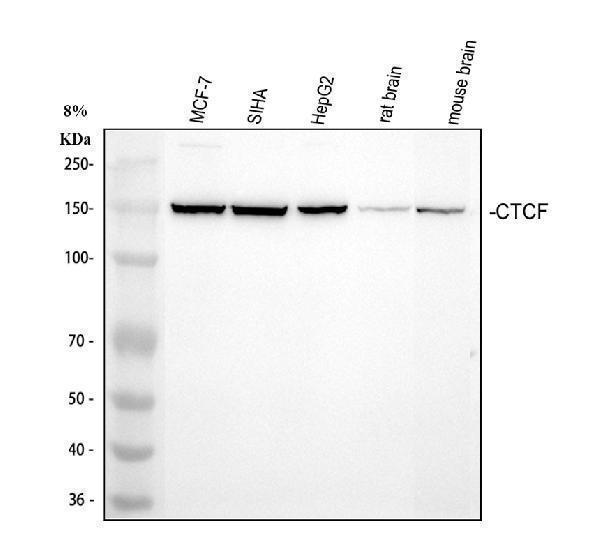
Western blot analysis of CTCF using anti-CTCF antibody (PB9493).
Electrophoresis was performed on a 8% SDS-PAGE gel at 80V (Stacking gel) / 120V (Resolving gel) for 2 hours. The sample well of each lane was loaded with 30 ug of sample under reducing conditions.
Lane 1: human MCF-7 whole cell lysates,
Lane 2: human SiHa whole cell lysates,
Lane 3: human HepG2 whole cell lysates,
Lane 4: rat brain tissue lysates,
Lane 5: mouse brain tissue lysates.
After electrophoresis, proteins were transferred to a nitrocellulose membrane at 150 mA for 50-90 minutes. Blocked the membrane with 5% non-fat milk/TBS for 1.5 hour at RT. The membrane was incubated with rabbit anti-CTCF antigen affinity purified polyclonal antibody (PB9493) at 0.5 μg/mL overnight at 4°C, then washed with TBS-0.1%Tween 3 times with 5 minutes each and probed with a goat anti-rabbit IgG-HRP secondary antibody (Catalog # BA1054) at a dilution of 1:5000 for 1.5 hour at RT. The signal is developed using an ECL Plus Western Blotting Substrate (Catalog # AR1196-200) with Tanon 5200 system. A specific band was detected for CTCF at approximately 150 kDa. The expected band size for CTCF is at 83 kDa.

Click image to see more details
IF analysis of CTCF using anti-CTCF antibody (PB9493) and anti-Tubulin Alpha antibody (M03989-3).
CTCF was detected in immunocytochemical section of U2OS cell. Enzyme antigen retrieval was performed using IHC enzyme antigen retrieval reagent (AR0022) for 15 mins. The cells were blocked with 10% goat serum. And then incubated with 5 μg/mL rabbit anti-CTCF Antibody (PB9493) and mouse anti-Tubulin Alpha antibody (M03989-3) overnight at 4°C. DyLight®488 Conjugated Goat Anti-Rabbit IgG (BA1127) and Cy3 Conjugated Goat Anti-Mouse IgG (BA1031) were used as secondary antibody at 1:500 dilution and incubated for 30 minutes at 37°C. Visualize using a fluorescence microscope and filter sets appropriate for the label used.

Click image to see more details
Flow Cytometry analysis of MCF-7 cells using anti-CTCF antibody (PB9493).
Overlay histogram showing MCF-7 cells stained with A04887-1 (Blue line). To facilitate intracellular staining, cells were fixed with 4% paraformaldehyde and permeabilized with permeabilization buffer. The cells were blocked with 10% normal goat serum. And then incubated with rabbit anti-CTCF Antibody (PB9493, 1 μg/1x106 cells) for 30 min at 20°C. DyLight®488 conjugated goat anti-rabbit IgG (BA1127, 5-10 μg/1x106 cells) was used as secondary antibody for 30 minutes at 20°C. Isotype control antibody (Green line) was rabbit IgG (1 μg/1x106) used under the same conditions. Unlabelled sample without incubation with primary antibody and secondary antibody (Red line) was used as a blank control.
Specific Publications For Anti-CTCF Antibody Picoband® (PB9493)
Loading publications
Recommended Resources
Here are featured tools and databases that you might find useful.
- Boster's Pathways Library
- Protein Databases
- Bioscience Research Protocol Resources
- Data Processing & Analysis Software
- Photo Editing Software
- Scientific Literature Resources
- Research Paper Management Tools
- Molecular Biology Software
- Primer Design Tools
- Bioinformatics Tools
- Phylogenetic Tree Analysis
Customer Reviews
Have you used Anti-CTCF Antibody Picoband®?
Share your experimental results or join a short interview to earn up to $1,000 in product credits or other rewards.
0 Reviews For Anti-CTCF Antibody Picoband®
Customer Q&As
Have a question?
Find answers in Q&As, reviews.
Can't find your answer?
Submit your question
5 Customer Q&As for Anti-CTCF Antibody Picoband®
Question
We have observed staining in mouse amniotic fluid. Any tips? Is anti-CTCF antibody supposed to stain amniotic fluid positively?
Verified Customer
Verified customer
Asked: 2019-11-26
Answer
From what I have seen in literature amniotic fluid does express CTCF. From what I have seen in Uniprot.org, CTCF is expressed in amniotic fluid, brain, placenta, cervix carcinoma, leukemic t-cell, among other tissues. Regarding which tissues have CTCF expression, here are a few articles citing expression in various tissues:
Brain, Pubmed ID: 15616553
Cervix carcinoma, Pubmed ID: 18669648, 20068231
Leukemic T-cell, Pubmed ID: 19690332
Placenta, Pubmed ID: 15489334
Boster Scientific Support
Answered: 2019-11-26
Question
you antibody using your anti-CTCF antibody for negative regulation of cell population proliferation studies. Has this antibody been tested with western blotting on rat intestine tissue? We would like to see some validation images before ordering.
Verified Customer
Verified customer
Asked: 2019-08-21
Answer
We appreciate your inquiry. This PB9493 anti-CTCF antibody is validated on mouse intestine tissue, rat intestine tissue, lung cancer tissue. It is guaranteed to work for IHC, WB in human, mouse, rat. Our Boster guarantee will cover your intended experiment even if the sample type has not been be directly tested.
Boster Scientific Support
Answered: 2019-08-21
Question
Our lab used your anti-CTCF antibody for WB on cervix carcinoma in a previous project. I am using human, and I plan to use the antibody for IHC next. My question regards examining cervix carcinoma as well as amniotic fluid in our next experiment. Do you have any suggestion on which antibody would work the best for IHC?
Verified Customer
Verified customer
Asked: 2019-06-03
Answer
I took a look at the website and datasheets of our anti-CTCF antibody and it appears that PB9493 has been tested on human in both WB and IHC. Thus PB9493 should work for your application. Our Boster satisfaction guarantee will cover this product for IHC in human even if the specific tissue type has not been validated. We do have a comprehensive range of products for IHC detection and you can check out our website bosterbio.com to find out more information about them.
Boster Scientific Support
Answered: 2019-06-03
Question
We were content with the WB result of your anti-CTCF antibody. However we have been able to see positive staining in brain nucleus using this antibody. Is that expected? Could you tell me where is CTCF supposed to be expressed?
Verified Customer
Verified customer
Asked: 2019-05-27
Answer
From literature, brain does express CTCF. Generally CTCF expresses in nucleus, nucleoplasm. Regarding which tissues have CTCF expression, here are a few articles citing expression in various tissues:
Brain, Pubmed ID: 15616553
Cervix carcinoma, Pubmed ID: 18669648, 20068231
Leukemic T-cell, Pubmed ID: 19690332
Placenta, Pubmed ID: 15489334
Boster Scientific Support
Answered: 2019-05-27
Question
We are currently using anti-CTCF antibody PB9493 for rat tissue, and we are satisfied with the WB results. The species of reactivity given in the datasheet says human, mouse, rat. Is it true that the antibody can work on dog tissues as well?
Verified Customer
Verified customer
Asked: 2019-05-14
Answer
The anti-CTCF antibody (PB9493) has not been tested for cross reactivity specifically with dog tissues, though there is a good chance of cross reactivity. We have an innovator award program that if you test this antibody and show it works in dog you can get your next antibody for free. Please contact me if I can help you with anything.
Boster Scientific Support
Answered: 2019-05-14
