Click image to see more details
Product Info Summary
| SKU: | A00745 |
|---|---|
| Size: | 100uL |
| Reactive Species: | Human |
| Host: | Rabbit |
| Application: | ELISA, IP, IHC, WB |
Customers Who Bought This Also Bought

Product info
Product Name
Anti-Cyclin B1 CCNB1 Antibody
SKU/Catalog Number
A00745
Size
100uL
Form
Liquid (sterile filtered)
Description
Boster Bio Anti-Cyclin B1 CCNB1 Antibody (Catalog # A00745). Tested in IHC, WB applications. This antibody reacts with Human.
Storage & Handling
Store vial at -20°C prior to opening. Aliquot contents and freeze at -20°C or below for extended storage. Avoid cycles of freezing and thawing. Centrifuge product if not completely clear after standing at room temperature. This product is stable for several weeks at 4°C as an undiluted liquid. Dilute only prior to immediate use. Expiration date is one (1) year from date of opening. (Ship on dry ice.)
Cite This Product
Anti-Cyclin B1 CCNB1 Antibody (Boster Biological Technology, Pleasanton CA, USA, Catalog # A00745)
Host
Rabbit
Contents
0.01% (w/v) Sodium Azide
Clonality
Polyclonal
Isotype
Antiserum
Immunogen
Anti-Cyclin B1 antibody was produced by repeated immunizations of full length fusion protein corresponding to the human gene.
Reactive Species
A00745 is reactive to CCNB1 in Human
Observed Molecular Weight
39 kDa
Calculated molecular weight
48.3 kDa
Background of CCNB1
The protein encoded by this gene is a regulatory protein involved in mitosis. The gene product forms a complex with p34 (cdc2) to form the maturation-promoting factor (MPF). Two alternative transcripts have been found, a constitutively expressed transcript and a cell cycle-regulated transcript that is expressed predominantly during G2/M phase. The different transcripts result from the use of alternate transcription initiation sites.
Antibody Validation
Boster validates all antibodies on WB, IHC, ICC, Immunofluorescence, and ELISA with known positive control and negative samples to ensure specificity and high affinity, including thorough antibody incubations.
Application & Images
Applications
A00745 is guaranteed for ELISA, IP, IHC, WB Boster Guarantee
Recommend Dilution
| Application | Dilution | Species |
|---|---|---|
| ELISA: 1:2 | 000 - 1:10 | 000 |
| WB: 1:500 - 1:1 | 000 | |
| Anti-Cyclin B1 antibody has been tested by western blot and immunohistochemistry and is suitable for use in ELISA | immunoblotting | immunoprecipitation, immunohistochemistry, and other immunological methods requiring high titer and specificity. Specific conditions for reactivity and detection of Cyclin B1 should be optimized by the end user. Expect a band approximately 55-66 kDa in size corresponding to Cyclin B1 by western blotting in the appropriate cell lysate or extract. H23 cells may be used as a positive control. |
Validation Images & Assay Conditions
Click image to see more details
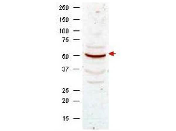
Western blot analysis using Boster's anti-Cyclin B1 antibody shows detection of Cyclin B1 present in asynchronous HeLa cell lysates. Comparison to a molecular weight marker indicates a band of ~55 kDa corresponding to human Cyclin B1 (arrowhead). Approximately 50 µg of lysate was loaded on to a 7% SDS-PAGE gel for separation. After transfer to nitrocellulose, the blot was incubated with a 1:500 dilution of the antibody for 1 h at room temperature. Detection occurred using a 1:10,000 of HRP conjugated Goat-a-Rabbit IgG . Personal communication, Luca D'Agostino, Temple University, Philadelphia, PA.

Click image to see more details
Boster's anti-Cyclin B1 antibody was diluted 1:500 to detect Cyclin B1 in human brain cerebellum tissue. Tissue was formalin fixed and paraffin embedded. No pre-treatment of sample was required. The image shows the localization of antibody as the precipitated red signal, with a hematoxylin purple nuclear counter stain.

Click image to see more details
Western blot analysis using Boster's Anti-Cyclin B1 antibody shows detection of human Cyclin B1 present in asynchronous HN30 cell lysates. HN30 cells are from head and neck cancer tumors that over express cyclin B1 and D1. Comparison to a molecular weight marker indicates a band of ~62 kDa corresponding to the expected molecular weight for the protein. The blot was incubated with a 1:500 dilution of the antibody for 1 h at room temperature. Detection occurred using a 1:10,000 of HRP conjugated Goat-a-Rabbit IgG 611-103-122 and chemiluminescence reagent with a 1-min exposure time. Other detection systems will yield similar results. Personal communication, Luca Cote, Temple University, Philadelphia, PA.
Specific Publications For Anti-Cyclin B1 CCNB1 Antibody (A00745)
Loading publications
Recommended Resources
Here are featured tools and databases that you might find useful.
- Boster's Pathways Library
- Protein Databases
- Bioscience Research Protocol Resources
- Data Processing & Analysis Software
- Photo Editing Software
- Scientific Literature Resources
- Research Paper Management Tools
- Molecular Biology Software
- Primer Design Tools
- Bioinformatics Tools
- Phylogenetic Tree Analysis
Customer Reviews
Have you used Anti-Cyclin B1 CCNB1 Antibody?
Share your experimental results or join a short interview to earn up to $1,000 in product credits or other rewards.
0 Reviews For Anti-Cyclin B1 CCNB1 Antibody
Customer Q&As
Have a question?
Find answers in Q&As, reviews.
Can't find your answer?
Submit your question
