This website uses cookies to ensure you get the best experience on our website.
- Table of Contents
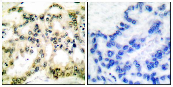
10 Citations 1 Q&As


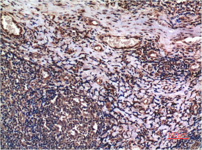
4 Citations


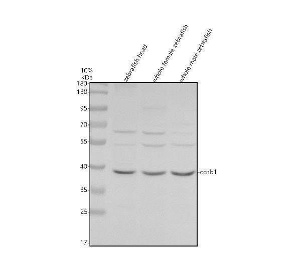
9 Citations
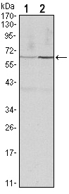
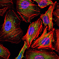
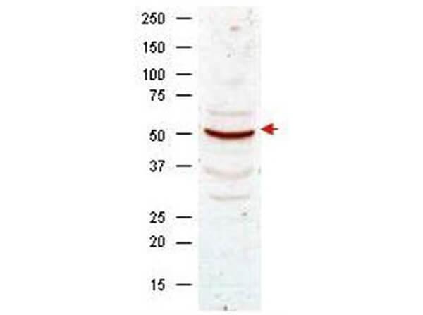
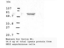
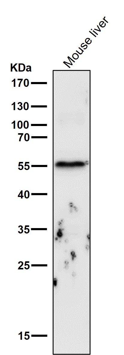
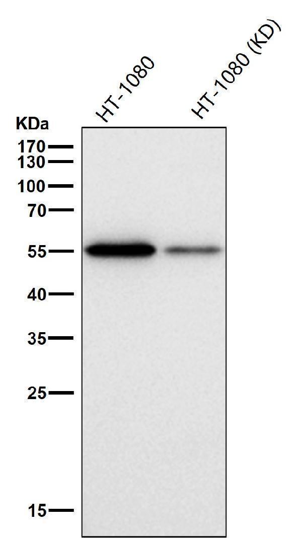
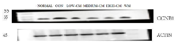
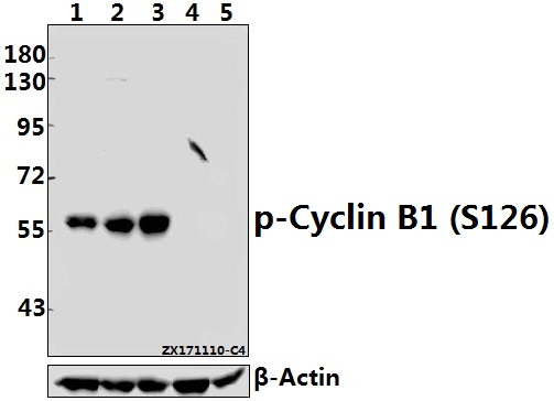
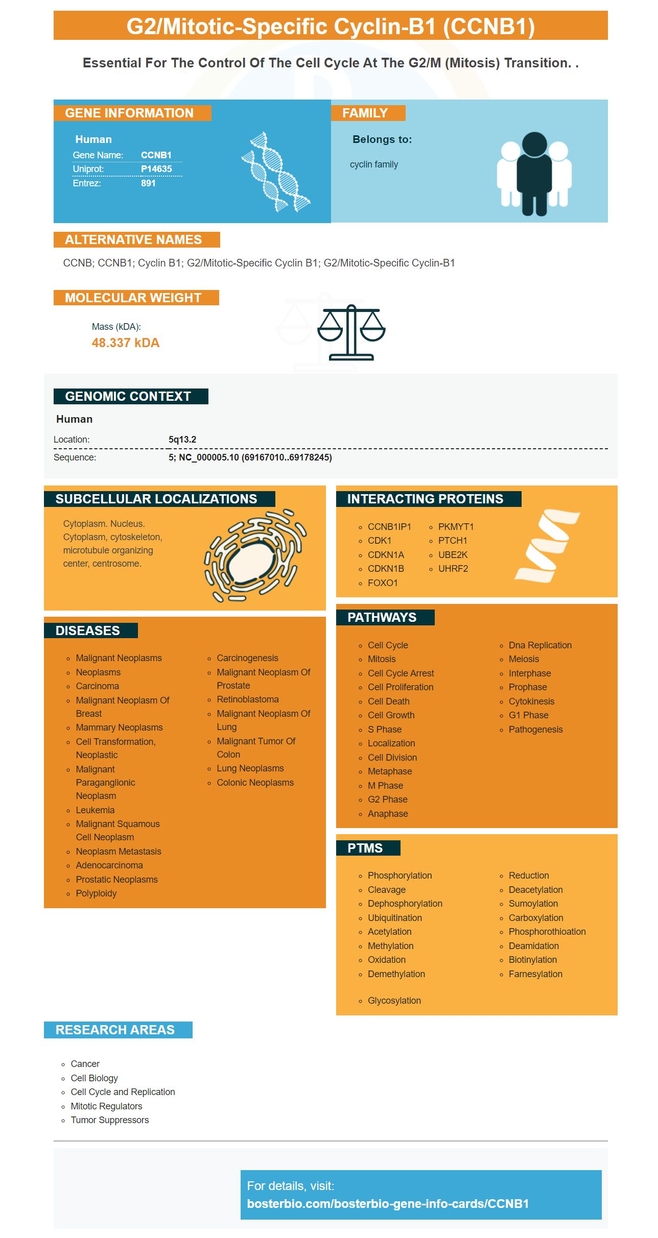
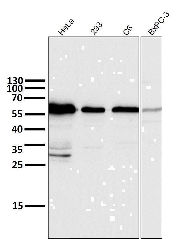
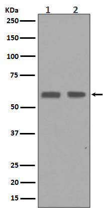
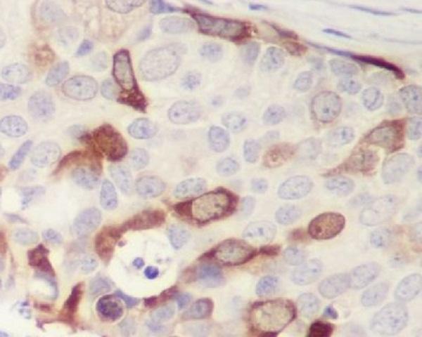
Facts about G2/mitotic-specific cyclin-B1.

| Human | |
|---|---|
| Gene Name: | CCNB1 |
| Uniprot: | P14635 |
| Entrez: | 891 |

| Belongs to: |
|---|
| cyclin family |

CCNB; CCNB1; Cyclin B1; G2/mitotic-specific cyclin B1; G2/mitotic-specific cyclin-B1
Mass (kDA):
48.337 kDA

| Human | |
|---|---|
| Location: | 5q13.2 |
| Sequence: | 5; NC_000005.10 (69167010..69178245) |
Cytoplasm. Nucleus. Cytoplasm, cytoskeleton, microtubule organizing center, centrosome.


PMID: 2570636 by Pines J., et al. Isolation of a human cyclin cDNA: evidence for cyclin mRNA and protein regulation in the cell cycle and for interaction with p34cdc2.
PMID: 7843284 by Piaggio G., et al. Structure and growth-dependent regulation of the human cyclin B1 promoter.
*Showing only the more recent 20. More publications can be found for each product on its corresponding product page