Click image to see more details
-

-
-
-
-
+8
Product Info Summary
| SKU: | A00254 |
|---|---|
| Size: | 100 μg/vial |
| Reactive Species: | Human, Mouse, Rat |
| Host: | Rabbit |
| Application: | ELISA, IHC |
Customers Who Bought This Also Bought

Product info
Product Name
Anti-Ki67/Mki67 Antibody
SKU/Catalog Number
A00254
Size
100 μg/vial
Form
Lyophilized
Description
Boster Bio Anti-Ki67/Mki67 Antibody catalog # A00254. Tested in ELISA, IHC applications. This antibody reacts with Human, Mouse, Rat.
Storage & Handling
Store at -20˚C for one year from date of receipt. After reconstitution, at 4˚C for one month. It can also be aliquotted and stored frozen at -20˚C for six months. Avoid repeated freeze-thaw cycles.
Cite This Product
Anti-Ki67/Mki67 Antibody (Boster Biological Technology, Pleasanton CA, USA, Catalog # A00254)
Host
Rabbit
Contents
Each vial contains 4mg Trehalose, 0.9mg NaCl, 0.2mg Na2HPO4.
Clonality
Polyclonal
Isotype
Rabbit IgG
Immunogen
E.coli-derived mouse Mki67 recombinant protein (Position: F29-S3177).
Cross-reactivity
No cross-reactivity with other proteins.
Reactive Species
A00254 is reactive to Mki67 in Human, Mouse, Rat
Calculated molecular weight
350.9 kDa
Background of Mki67
Ki-67 (Proliferation-related Ki-67 antigen), also known as MKI67 or KIA, is a protein that in humans is encoded by the MKI67 gene. From study of a panel of human-rodent somatic cell hybrids, it has been demonstrated that a gene involved in the expression of the MKI67 antigen is located on chromosome 10. By in situ hybridization, Fonatsch et al. (1991) regionalized the MKI67 gene to chromosome 10q25-qter. By FISH, Traut et al. (1998) mapped the mouse Mki67 gene to chromosome 7F3-F5. Antigen KI-67 is a nuclear protein that is associated with and may be necessary for cellular proliferation. Furthermore it is associated with ribosomal RNA transcription. Inactivation of antigen KI-67 leads to inhibition of ribosomal RNA synthesis.
Antibody Validation
Boster validates all antibodies on WB, IHC, ICC, Immunofluorescence, and ELISA with known positive control and negative samples to ensure specificity and high affinity, including thorough antibody incubations.
Application & Images
Applications
A00254 is guaranteed for ELISA, IHC Boster Guarantee
Recommend Dilution
| Application | Dilution | Species |
|---|---|---|
| Immunohistochemistry (Paraffin-embedded Section) | 2-5μg/ml | Human, Mouse, Rat |
| ELISA | 0.1-0.5μg/ml | - |
Tested application
Use TE buffer pH 9.0 for antigen retrieval; (*) citrate buffer pH 6.0 is an alternative.
Validation Images & Assay Conditions
Click image to see more details
IHC analysis of Ki67/Mki67 using anti-Ki67/Mki67 antibody (A00254).
Ki67/Mki67 was detected in a paraffin-embedded section of human tonsil tissue. Heat mediated antigen retrieval was performed in EDTA buffer (pH 8.0, epitope retrieval solution). The tissue section was blocked with 10% goat serum. The tissue section was then incubated with 2 μg/ml rabbit anti-Ki67/Mki67 Antibody (A00254) overnight at 4°C. Peroxidase Conjugated Goat Anti-rabbit IgG was used as secondary antibody and incubated for 30 minutes at 37°C. The tissue section was developed using HRP Conjugated Rabbit IgG Super Vision Assay Kit (Catalog # SV0002) with DAB as the chromogen.

Click image to see more details
IHC analysis of Ki67/Mki67 using anti-Ki67/Mki67 antibody (A00254).
Ki67/Mki67 was detected in a paraffin-embedded section of human lung cancer tissue. Heat mediated antigen retrieval was performed in EDTA buffer (pH 8.0, epitope retrieval solution). The tissue section was blocked with 10% goat serum. The tissue section was then incubated with 2 μg/ml rabbit anti-Ki67/Mki67 Antibody (A00254) overnight at 4°C. Peroxidase Conjugated Goat Anti-rabbit IgG was used as secondary antibody and incubated for 30 minutes at 37°C. The tissue section was developed using HRP Conjugated Rabbit IgG Super Vision Assay Kit (Catalog # SV0002) with DAB as the chromogen.

Click image to see more details
IHC analysis of Ki67/Mki67 using anti-Ki67/Mki67 antibody (A00254).
Ki67/Mki67 was detected in a paraffin-embedded section of human thyroid cancer tissue. Heat mediated antigen retrieval was performed in EDTA buffer (pH 8.0, epitope retrieval solution). The tissue section was blocked with 10% goat serum. The tissue section was then incubated with 2 μg/ml rabbit anti-Ki67/Mki67 Antibody (A00254) overnight at 4°C. Peroxidase Conjugated Goat Anti-rabbit IgG was used as secondary antibody and incubated for 30 minutes at 37°C. The tissue section was developed using HRP Conjugated Rabbit IgG Super Vision Assay Kit (Catalog # SV0002) with DAB as the chromogen.

Click image to see more details
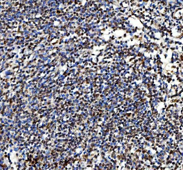
IHC analysis of Ki67/Mki67 using anti-Ki67/Mki67 antibody (A00254).
Ki67/Mki67 was detected in a paraffin-embedded section of mouse thymus tissue. Heat mediated antigen retrieval was performed in EDTA buffer (pH 8.0, epitope retrieval solution). The tissue section was blocked with 10% goat serum. The tissue section was then incubated with 2 μg/ml rabbit anti-Ki67/Mki67 Antibody (A00254) overnight at 4°C. Peroxidase Conjugated Goat Anti-rabbit IgG was used as secondary antibody and incubated for 30 minutes at 37°C. The tissue section was developed using HRP Conjugated Rabbit IgG Super Vision Assay Kit (Catalog # SV0002) with DAB as the chromogen.

Click image to see more details
IHC analysis of Ki67/Mki67 using anti-Ki67/Mki67 antibody (A00254).
Ki67/Mki67 was detected in a paraffin-embedded section of rat thymus tissue. Heat mediated antigen retrieval was performed in EDTA buffer (pH 8.0, epitope retrieval solution). The tissue section was blocked with 10% goat serum. The tissue section was then incubated with 2 μg/ml rabbit anti-Ki67/Mki67 Antibody (A00254) overnight at 4°C. Peroxidase Conjugated Goat Anti-rabbit IgG was used as secondary antibody and incubated for 30 minutes at 37°C. The tissue section was developed using HRP Conjugated Rabbit IgG Super Vision Assay Kit (Catalog # SV0002) with DAB as the chromogen.

Click image to see more details
miR-150 suppresses tumor growth and metastasis in vivo . A. Transduced MHCC97-H cells were injected subcutaneously into nude mice (n=5). After 28 days, the mice were euthanized, and the tumors were excised. Representative tumors are shown. B. Growth curve of tumor volumes. C. Tumor weight. D. Representative H&E and immunohistochemical staining for Ki-67, GAB1 and phospho-ERK1/2 in tumors are shown. E. Representative bioluminescence images 35 days after injection are shown. F. The luminescence intensity of lung metastases from MHCC97-H-luc-miR-150 cells was weaker than that of lung metastases from MHCC97-H-luc-miR-NC cells. G. miR-150 reduced the incidence of lung metastases. H. miR-150 overexpression significantly reduced the number of lung metastatic nodules. I. Representative H&E and immunohistochemical staining for Ki-67, GAB1 and phospho-ERK1/2 in lung metastatic nodules are shown. * P <0.05.
Index in PubMed under a CC BY license. PMID: 26871477

Click image to see more details
Kaplan-Meier analysis of overall survival in HCC patients according to Nek7 and Ki-67 expression. A. Higher Nek7 expression was closely correlated with poor overall survival ( p < 0.001). B. The HCC group with Ki-67 higher expression indicated poorer overall survival ( p < 0.001).
Index in PubMed under a CC BY license. PMID: 26921196

Click image to see more details
Nek7 gene silencing suppressed growth of SMMC7721 xenograft tumors. A. Tumors isolated from mice of different groups are shown. B. Growth curves of xenograft tumors from the experiments with BALB/c nude mice. Changes in tumor volumes measured at the indicated days are shown. C. Therapeutic lenti-shNek7-1 reduced the tumor weight. Real-time PCR and Western blot assays to determine the mRNA and protein levels of cell growth associated genes, Bcl-2, cyclinD1 and cyclinB1 in HCC cells D. and dissected tumors E, F. Immunohistochemical staining was used to detect the Nek7 and Ki-67 expression pattern in xenograft tumors from different groups. β-actin was used as internal control for Western blot and Real-time PCR. Experiments were repeated three times. (* p < 0.05).
Index in PubMed under a CC BY license. PMID: 26921196

Click image to see more details
Immunohistochemistry of Nek7 and Ki-67 on HCC specimens. Representative immunohistochemical staining of HCC specimens with different histologic grade, as determined using anti-Nek7 and anti-Ki-67 antibodies. A. The expression status of Nek7 in low-grade HCC tissue. B. An example of moderate-differentiated HCC with Nek7 expression. C. Nek7 expression in high-grade HCC tissue. D. Ki-67 expression status in low-grade HCC tissue. E. Example of moderate-differentiated HCC with Ki-67 expression. F. Ki-67 expression in high-grade HCC tissue.
Index in PubMed under a CC BY license. PMID: 26921196

Click image to see more details
Immunohistochemistry staining for YAP, KI-67 and Bcl-2 protein in xenograft tumour model and pulmonary metastasis model. Cytoplasmic staining was considered to be positive for YAP, KI-67 and Bcl-2. b & f Higher YAP expression in both xenograft tumour and pulmonary models. c & d High YAP expression mainly in the nucleus of LSCC. e & f Low YAP expression in LSCC in both the cytoplasm and nucleus of tumour cells. g & h Low expression of YAP protein mainly in the nucleus of LSCC. [A, a, E, e] × 40; [B, b, C, c, D, d, F, f, G, g, H, h] × 100
Index in PubMed under a CC BY license. PMID: 31269911

Click image to see more details
LINC00326 downregulation accelerates tumor growth in vivo. a Appearance of tumors four weeks after injection, showing the blank control, negative control, LINC00326 overexpression, shCTRL, and LINC00326 knockdown. b Tumor volumes in the five groups (mean ± SD) at time of sacrifice. c , d LINC00326 and miR-657 mRNA expression in tumors from blank, OE-CTRL, OE- LINC00326, shCTRL, and shLINC00326 groups. e Wnt1, Wnt3A, and β-catenin protein levels in tumors from blank, OE-CTRL, OE- LINC00326, shCTRL, and shLINC00326 groups. f–h Ki67 and TUNEL staining in blank, OE-CTRL, OE- LINC00326, shCTRL, and shLINC00326 groups, respectively (× 200, scale bars, 100 µm); n = 3. Data are presented as mean ± SEM; *P < 0.05; **P < 0.01; ***P < 0.001
Index in PubMed under a CC BY license. PMID: 36747258

Click image to see more details
Targeting MUS81 sensitizes the anticancer effect of WEE1 inhibitor MK1775 in gastric cancer in vitro and in vivo. a and b MTT assay was performed to examine the effect of MK1775 on proliferation of MUS81 knockdown and parental gastric cancer cells SGC7901 and BGC823. Data are reported as mean ± SD for five independent experiments. c Clonogenic assay was performed to detect the anticancer effect of MK1775 in MUS81 knockdown and parental gastric cancer cells SGC7901 (left) and BGC823 (right). Representative images are displayed. d Data of the clonogenic assay in SGC7901 (left) and BGC823 (right) cells are reported as mean ± SD for three independent experiments. e The image of tumors in the SGC7901 gastric cancer xenograft mice model (n = 6 for each group) after treatment for 16 d. f Tumor growth curve of indicated groups in the SGC7901 gastric cancer xenograft mice model. The data are presented as mean ± SD (n = 6 for each group). g Tumor volume on day 16 after treatment. Data are presented as mean ± SD (n = 6 for each group). h Representative images of immunohistochemical (IHC) staining of Ki67 in the gastric cancer xenograft mice model. Scale bar: 25 μm. i IHC staining analysis of Ki 67-positive cells in different groups. The data are presented as mean ± SD (n = 3). * P < 0.05; ** P < 0.01; *** P < 0.001; **** P < 0.0001; ns. , not significant
Index in PubMed under a CC BY license. PMID: 34625086
Specific Publications For Anti-Ki67/Mki67 Antibody (A00254)
Loading publications
Recommended Resources
Here are featured tools and databases that you might find useful.
- Boster's Pathways Library
- Protein Databases
- Bioscience Research Protocol Resources
- Data Processing & Analysis Software
- Photo Editing Software
- Scientific Literature Resources
- Research Paper Management Tools
- Molecular Biology Software
- Primer Design Tools
- Bioinformatics Tools
- Phylogenetic Tree Analysis
Customer Reviews
Have you used Anti-Ki67/Mki67 Antibody?
Share your experimental results or join a short interview to earn up to $1,000 in product credits or other rewards.
0 Reviews For Anti-Ki67/Mki67 Antibody
Customer Q&As
Have a question?
Find answers in Q&As, reviews.
Can't find your answer?
Submit your question
